
9.4: Pneumocystis jiroveci
- Page ID
- 123389
Pneumocystis jiroveci, (formerly called Pneumocystis carinii ), causes Pneumocystis pneumonia (PCP). It is seen almost exclusively in highly immunosuppressed individuals such as those with AIDS, late -stage malignancies, or leukemias. PCP is one of the more common infections associated with AIDS.
P. jiroveci can be found in 3 distinct morphologic stages:
- The trophozoite (trophic form), a haploid amoeboid form 1-4 µm in diameter that replicates by mitosis and binary fission. The trophic forms are irregular shaped and often appears in clusters. See Fig. \(\PageIndex{1A}\).
- A precystic form or early cyst. Haploid trophic forms conjugate and produce a diploid precyst form or sporocyte.
- The precyst form matures into a cyst form, which contains several intracystic bodies or spores are 5-8 µm in diameter. It has been postulated that in formation of the cyst form (late phase cyst), the zygote undergoes meiosis and subsequent mitosis to typically produce eight haploid ascospores (sporozoites) See Fig. \(\PageIndex{1B}\). As the haploid ascospores are released the cysts often collapse forming crescent-shaped bodies (see Fig. \(\PageIndex{1C}\)). P. jiroveci is usually transmitted by inhalation of the cyst form. Released ascospores then develop into replicating trophic forms that attach to the wall of the alveoli and replicate to fill the alveoli.
- A proposed life cycle for Pneumocystis jiroveci can be seen in Fig. \(\PageIndex{1D}\).
|
Fig \(\PageIndex{1A}\): Trophic Form of Pneumocystis jiroveci from the Lungs of a Person with PCP |
Fig. \(\PageIndex{1B}\): Cysts of Pneumocystis jiroveci from the Lungs of a Person with PCP |
Fig \(\PageIndex{1C}\): Cysts of Pneumocystis jiroveci in Smear from Bronchoalveolar Lavage |
Fig \(\PageIndex{1D}\): Proposed life cycle for Pneumocystis jiroveci |
|---|---|---|---|
 |
 |
 |
 |
| Cysts of Pneumocystis jiroveci from the Lungs of a Person with PCP | Cysts of Pneumocystis jiroveci in bronchoalveolar material, Giemsa stain method.The rounded cysts (arrows) are 4 to 7 µm in diameter and contain 6 to 8 intracystic bodies, whose nuclei are stained by the dye. The walls of the cysts are not stained. | Cysts of Pneumocystis jiroveci in lung tissue, Gomori methenamine silver stain method. The walls of the cysts are stained black and often appear crescent shaped or like crushed ping-pong balls. The intracystic bodies are not visible with this stain. |
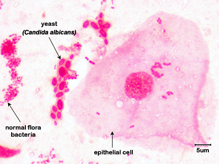
Note epithelial cells, rod-shaped bacteria, and Candida albicans in its hyphal form. |
| By Content Providers: CDC [Public domain]. Courtesy of the Centers for Disease Control and Prevention |
Copyright; Gary E. Kaiser, Ph.D. The Community College of Baltimore County, Catonsville Campus CC-BY-3.0 | By Content Providers: CDC [Public domain]. Courtesy of the Centers for Disease Control and Prevention |
By Content Providers: CDC [Public domain]. Courtesy of the Centers for Disease Control and Prevention |
In biopsies from lung tissue or in tracheobronchial aspirates, both a trophic form about 1-4 µm in diameter with a distinct nucleus and a cyst form between 5-8 µm in diameter with 6-8 intracystic bodies (ascospores) can be seen.
When viewing cysts of P. jiroveci in lung tissue after utilizing the Gomori methenamine silver stain method, the walls of the cysts are stained black and often appear crescent shaped or like crushed ping-pong balls, as seen in Fig. \(\PageIndex{8C}\) above.The intracystic bodies are not visible with this stain.
Contributors and Attributions
Dr. Gary Kaiser (COMMUNITY COLLEGE OF BALTIMORE COUNTY, CATONSVILLE CAMPUS)