
7.5: Examples of Bacterial Motility
- Page ID
- 123369
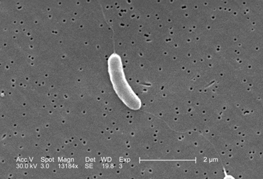
1. monotrichous - a single flagellum at one pole. (See Figs. \(\PageIndex{1A}\), \(\PageIndex{1B}\), and \(\PageIndex{1C}\),)
|
Fig. \(\PageIndex{1A}\): Flagella Stain Showing Monotrichous Arrangement of Flagella of a Vibrio species |
Fig. \(\PageIndex{1B}\): Flagella Stain of Vibrio cholerae showing Monotrichous Arrangement of Flagella |
Fig. \(\PageIndex{1C}\): Scanning Electron Micrograph Showing a Monotrichous Flagellum of Vibrio vulnificus |
|---|---|---|
 |
 |
 |
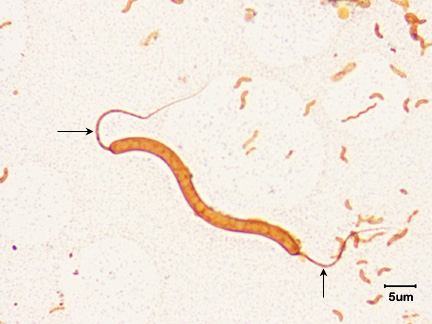
| Note single flagellum (arrow) | Note single flagellum at each pole (arrows). |
Aseptically pipette 0.1 ml of the 10-4 bacterial dilution onto a plate of TSA. This will give a 1/100,000 or 10-5 dilution of the sample. Using a bent-glass rod and a turntable, spread the bacteria out over the surface of the plate. Incubate the 3 plates at 37°C. |
| (Copyright; Gary E. Kaiser, Ph.D. The Community College of Baltimore County, Catonsville Campus CC-BY-3.0) | By Content Provider: CDC [Public domain] Courtesy of the Centers for Disease Control and Prevention. |
By Content Providers(s): CDC/Dr. Edwin P. Ewing, Jr [Public domain] Courtesy of the Centers for Disease Control and Prevention. |
2. amphitrichous - a single flagellum at both poles. (See Fig. \(\PageIndex{2}\).)
3. lophotrichous - two or more flagella at one or both poles of the cell. (See Fig. \(\PageIndex{3}\).)
4. peritrichous - completely surrounded by flagella. (See Fig. \(\PageIndex{4}\).)
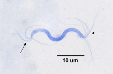
One group of bacteria, the spirochetes, has internally-located axial filaments. (see Fig. \(\PageIndex{5}\)) or endoflagella. Axial filaments wrap around the spirochete towards the middle from both ends. They are located above the peptidoglycan cell wall but underneath the outer membrane or sheath.
Some bacteria use motility to contact host cells and disseminate within a host. The mucosal surfaces of the respiratory tract, the intestinal tract, and the genitourinary tract constantly flush bacteria away in order to prevent colonization of host mucous membranes. Motile bacteria can use their motility and chemotaxis to swim through mucus towards mucosal epithelial cells. Many bacteria that can colonize the mucous membranes of the bladder and the intestines, in fact, are motile. Motility probably helps these bacteria move through the mucus between the mucin strands or in places where the mucus is less viscous.
Figure \(\PageIndex{6}\): Spirochete Axial Filaments. (Copyright; Gary E. Kaiser, Ph.D. The Community College of Baltimore County, Catonsville Campus CC-BY-3.0)
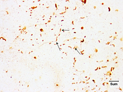
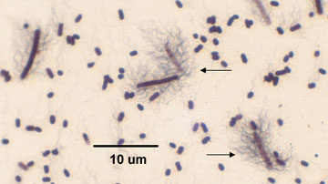
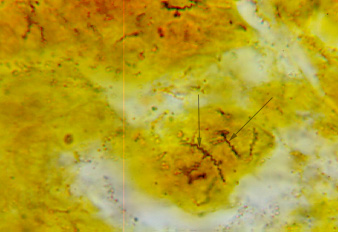
In addition, because of their thinness, their internal flagella (axial filaments), their corkscrew shape, and their motility, certain spirochetes are more readily able to penetrate host mucous membranes, skin abrasions, etc., and enter the body. Motility and penetration may also enable the spirochetes to penetrate deeper in tissue and enter the lymphatics and bloodstream and disseminate to other body sites. Spirochetes that infect humans include Treponema pallidum that causes syphilis, Leptospira that causes leptospirosis, and Borrelia burgdorferi that causes Lyme disease. (See Figs. \(\PageIndex{21A}\), \(\PageIndex{21B}\), and \(\PageIndex{21C}\).)
|
Fig. \(\PageIndex{7A}\): The Spirochete Treponema pallidum (arrows) in Tissue. |
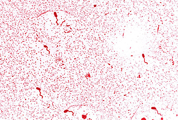
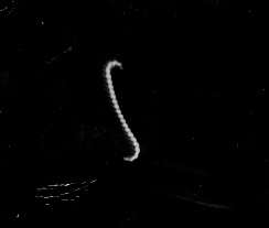
Fig. \(\PageIndex{7B}\): Darkfield Microscope Photomicrograph of the Spirochete Leptospira |
Fig. \(\PageIndex{7C}\): Borrelia burgdorferi |
|---|---|---|
 |
 |
_lores.jpg?revision=1) |
| Aseptically pipette 0.1 ml of the 10-6 bacterial dilution onto a plate of TSA. This will give a 1/10,000,000 or 10-7 dilution of the sample. Using a bent-glass rod and a turntable, spread the bacteria out over the surface of the plate. | Aseptically pipette 0.1 ml of the 10-5 bacterial dilution onto a plate of TSA. This will give a 1/1,000,000 or 10-6 dilution of the sample. Using a bent-glass rod and a turntable, spread the bacteria out over the surface of the plate. |
Aseptically pipette 0.1 ml of the 10-4 bacterial dilution onto a plate of TSA. This will give a 1/100,000 or 10-5 dilution of the sample. Using a bent-glass rod and a turntable, spread the bacteria out over the surface of the plate. Incubate the 3 plates at 37°C. |
| (Copyright; Gary E. Kaiser, Ph.D. The Community College of Baltimore County, Catonsville Campus CC-BY-3.0) | (Copyright; Gary E. Kaiser, Ph.D. The Community College of Baltimore County, Catonsville Campus CC-BY-3.0) | Photo Credit:Content Providers(s): CDC, Public domain, via Wikimedia Commons |
Contributors and Attributions
Dr. Gary Kaiser (COMMUNITY COLLEGE OF BALTIMORE COUNTY, CATONSVILLE CAMPUS)