
9.4: Pelvic Girdle
- Page ID
- 53652

\( \newcommand{\vecs}[1]{\overset { \scriptstyle \rightharpoonup} {\mathbf{#1}} } \)
\( \newcommand{\vecd}[1]{\overset{-\!-\!\rightharpoonup}{\vphantom{a}\smash {#1}}} \)
\( \newcommand{\dsum}{\displaystyle\sum\limits} \)
\( \newcommand{\dint}{\displaystyle\int\limits} \)
\( \newcommand{\dlim}{\displaystyle\lim\limits} \)
\( \newcommand{\id}{\mathrm{id}}\) \( \newcommand{\Span}{\mathrm{span}}\)
( \newcommand{\kernel}{\mathrm{null}\,}\) \( \newcommand{\range}{\mathrm{range}\,}\)
\( \newcommand{\RealPart}{\mathrm{Re}}\) \( \newcommand{\ImaginaryPart}{\mathrm{Im}}\)
\( \newcommand{\Argument}{\mathrm{Arg}}\) \( \newcommand{\norm}[1]{\| #1 \|}\)
\( \newcommand{\inner}[2]{\langle #1, #2 \rangle}\)
\( \newcommand{\Span}{\mathrm{span}}\)
\( \newcommand{\id}{\mathrm{id}}\)
\( \newcommand{\Span}{\mathrm{span}}\)
\( \newcommand{\kernel}{\mathrm{null}\,}\)
\( \newcommand{\range}{\mathrm{range}\,}\)
\( \newcommand{\RealPart}{\mathrm{Re}}\)
\( \newcommand{\ImaginaryPart}{\mathrm{Im}}\)
\( \newcommand{\Argument}{\mathrm{Arg}}\)
\( \newcommand{\norm}[1]{\| #1 \|}\)
\( \newcommand{\inner}[2]{\langle #1, #2 \rangle}\)
\( \newcommand{\Span}{\mathrm{span}}\) \( \newcommand{\AA}{\unicode[.8,0]{x212B}}\)
\( \newcommand{\vectorA}[1]{\vec{#1}} % arrow\)
\( \newcommand{\vectorAt}[1]{\vec{\text{#1}}} % arrow\)
\( \newcommand{\vectorB}[1]{\overset { \scriptstyle \rightharpoonup} {\mathbf{#1}} } \)
\( \newcommand{\vectorC}[1]{\textbf{#1}} \)
\( \newcommand{\vectorD}[1]{\overrightarrow{#1}} \)
\( \newcommand{\vectorDt}[1]{\overrightarrow{\text{#1}}} \)
\( \newcommand{\vectE}[1]{\overset{-\!-\!\rightharpoonup}{\vphantom{a}\smash{\mathbf {#1}}}} \)
\( \newcommand{\vecs}[1]{\overset { \scriptstyle \rightharpoonup} {\mathbf{#1}} } \)
\(\newcommand{\longvect}{\overrightarrow}\)
\( \newcommand{\vecd}[1]{\overset{-\!-\!\rightharpoonup}{\vphantom{a}\smash {#1}}} \)
\(\newcommand{\avec}{\mathbf a}\) \(\newcommand{\bvec}{\mathbf b}\) \(\newcommand{\cvec}{\mathbf c}\) \(\newcommand{\dvec}{\mathbf d}\) \(\newcommand{\dtil}{\widetilde{\mathbf d}}\) \(\newcommand{\evec}{\mathbf e}\) \(\newcommand{\fvec}{\mathbf f}\) \(\newcommand{\nvec}{\mathbf n}\) \(\newcommand{\pvec}{\mathbf p}\) \(\newcommand{\qvec}{\mathbf q}\) \(\newcommand{\svec}{\mathbf s}\) \(\newcommand{\tvec}{\mathbf t}\) \(\newcommand{\uvec}{\mathbf u}\) \(\newcommand{\vvec}{\mathbf v}\) \(\newcommand{\wvec}{\mathbf w}\) \(\newcommand{\xvec}{\mathbf x}\) \(\newcommand{\yvec}{\mathbf y}\) \(\newcommand{\zvec}{\mathbf z}\) \(\newcommand{\rvec}{\mathbf r}\) \(\newcommand{\mvec}{\mathbf m}\) \(\newcommand{\zerovec}{\mathbf 0}\) \(\newcommand{\onevec}{\mathbf 1}\) \(\newcommand{\real}{\mathbb R}\) \(\newcommand{\twovec}[2]{\left[\begin{array}{r}#1 \\ #2 \end{array}\right]}\) \(\newcommand{\ctwovec}[2]{\left[\begin{array}{c}#1 \\ #2 \end{array}\right]}\) \(\newcommand{\threevec}[3]{\left[\begin{array}{r}#1 \\ #2 \\ #3 \end{array}\right]}\) \(\newcommand{\cthreevec}[3]{\left[\begin{array}{c}#1 \\ #2 \\ #3 \end{array}\right]}\) \(\newcommand{\fourvec}[4]{\left[\begin{array}{r}#1 \\ #2 \\ #3 \\ #4 \end{array}\right]}\) \(\newcommand{\cfourvec}[4]{\left[\begin{array}{c}#1 \\ #2 \\ #3 \\ #4 \end{array}\right]}\) \(\newcommand{\fivevec}[5]{\left[\begin{array}{r}#1 \\ #2 \\ #3 \\ #4 \\ #5 \\ \end{array}\right]}\) \(\newcommand{\cfivevec}[5]{\left[\begin{array}{c}#1 \\ #2 \\ #3 \\ #4 \\ #5 \\ \end{array}\right]}\) \(\newcommand{\mattwo}[4]{\left[\begin{array}{rr}#1 \amp #2 \\ #3 \amp #4 \\ \end{array}\right]}\) \(\newcommand{\laspan}[1]{\text{Span}\{#1\}}\) \(\newcommand{\bcal}{\cal B}\) \(\newcommand{\ccal}{\cal C}\) \(\newcommand{\scal}{\cal S}\) \(\newcommand{\wcal}{\cal W}\) \(\newcommand{\ecal}{\cal E}\) \(\newcommand{\coords}[2]{\left\{#1\right\}_{#2}}\) \(\newcommand{\gray}[1]{\color{gray}{#1}}\) \(\newcommand{\lgray}[1]{\color{lightgray}{#1}}\) \(\newcommand{\rank}{\operatorname{rank}}\) \(\newcommand{\row}{\text{Row}}\) \(\newcommand{\col}{\text{Col}}\) \(\renewcommand{\row}{\text{Row}}\) \(\newcommand{\nul}{\text{Nul}}\) \(\newcommand{\var}{\text{Var}}\) \(\newcommand{\corr}{\text{corr}}\) \(\newcommand{\len}[1]{\left|#1\right|}\) \(\newcommand{\bbar}{\overline{\bvec}}\) \(\newcommand{\bhat}{\widehat{\bvec}}\) \(\newcommand{\bperp}{\bvec^\perp}\) \(\newcommand{\xhat}{\widehat{\xvec}}\) \(\newcommand{\vhat}{\widehat{\vvec}}\) \(\newcommand{\uhat}{\widehat{\uvec}}\) \(\newcommand{\what}{\widehat{\wvec}}\) \(\newcommand{\Sighat}{\widehat{\Sigma}}\) \(\newcommand{\lt}{<}\) \(\newcommand{\gt}{>}\) \(\newcommand{\amp}{&}\) \(\definecolor{fillinmathshade}{gray}{0.9}\)Pelvic Girdle
Coxal Bones (Os Coxae)
The pelvic girdle functions to connect and support the thighs to each side of the trunk of the body. The rest of the bones in the lower extremity form joints and regions of the lower limb (i.e. hip, thigh, knee, leg, ankle, and foot).
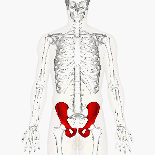
Above: Ox coxae highlighted in red.
The pelvic girdle is name given the left and right coxal bones. Colloquially, these are known as the “hip bones”. The pelvic girdle is just the two coxal bones, but the pelvis itself is the bowl-like structure created by the two coxal bones joined in the anterior by the sacrum, and coccyx. (“pelvis" comes from the Latin word for “basin”.)
Above: The three regions of the os coxae: ilium (top), ischium (middle), and pubis (bottom).
At birth, each coxal bone starts out as three separate bones – the ilium, (ILL-ee-um), the ischium, (ISH-ee-um) and the pubis (PYOO-bus) bones – joined by hyaline cartilage. The figure below shows what these bones look like initially. By the age of 25, these three bones have fully fused into a single coxal bone. We still subdivide the fully-formed coxal bone into three regions based on the positions of the three bones that fused to form it, each region named after the bone that gave rise to that region.

Above: Lateral view of coxal bone (os coxa) prior to fusion of the ilium, ischium, and pubis.

Above: Os coxae, or coxal bones. (A) Anterior view of the right and left coxal bones. (B) Posterior view of the right and left coxal bones. (C) Lateral view of the left coxal bone. (D) Lateral view of the right coxal bone. Ilium is colored pink in all images, pubis is colored blue in all images, and ischium is colored yellow in all images.
In a fully fused coxal bone, the ilium is the most superior portion, forming the “wing” that makes up the most prominent part of the coxal bone. The interior-facing side of this “wing” is called the iliac fossa. The ilium is where the sacrum attaches to each coxal bone to complete the pelvic bowl. This attachment point is alternatively called the sacroiliac joint, the sacroiliac articulation, or the iliac tuberosity, and is a rough surface. In anatomical position, the rough sacroiliac joint is always facing anterior.
In anatomical position, the ischium is posterior to the pubis. You “sit on the ischium” portion of the coxal bone. It is thicker and stronger than the pubis, allowing it to support your weight.
The large socket of the coxal bone is called the acetabulum (“ah-set-TAB-you-lum”). It faces laterally and is where the ball-like head of the femur bone articulates with the pelvis. Its name is derived from the Latin for vinegar cup, because of its cup-like shape. Inferior to the acetabulum is a large opening called the obturator foramen (“OB-tur-aye-tor for-AY-men”).
The Pelvis (Coxal Bones + Sacrum + Coccyx)
The pelvis is the bowl-shaped structure generated by the two coxal bones articulated with the sacrum and coccyx bones. On the anterior side of the pelvis, the pubis portions of the two coxal bones do not articulate with each other, but instead are joined with a small piece of cartilage called the pubic symphysis (“PYOO-bick SIM-fiss-is”)

Above: The pelvis is composed of the sacrum and coccyx of the axial skeleton and the coxal bones including their component bones: ilium, pubis, and ischium.
Anatomists divide the pelvis into two regions. The false pelvis is superior and is surrounded by iliac fossa portions of the coxal bones and the upper portion of the sacrum. The true pelvis is inferior and is surrounded by the pubis and ischium portions of the coxal bones, in addition to the lower sections of the ilium and the sacrum. In women, the true pelvis defines the space babies must squeeze through during childbirth.

Above: The false pelvis versus the true pelvis.

Above: Three features that distinguish male and female pelvises are (1) the pelvic inlet, (2) the pubic arch, and (3) sacrum concavity.
One of the few ways a skeleton stripped of all flesh can be reliably established as either male or female comes from examining the pelvis. The female pelvis can be distinguished from the male pelvis by a number or criterion, three of which are shown in the figure below.
Most of these anatomical differences between the pelvises of males and females reflect the fact that only female pelvises have to serve as part of the birth canal, and these sex differences are not as pronounced in the skeletons of children who have not finished puberty.
In female pelvises, both the pelvic inlet and the pelvic outlet are wider and more oval-shaped than those in male pelvises. The pelvic inlet in males tends to be more heart-shaped (narrower on the dorsal side) and the pelvic outlet tends to be more narrow. The pubic arch, found immediately inferior to the pubic symphysis, tends to form an angle closer to 90° in females, but forms an angle closer to 60° in males. The sacrum in female pelvises tends to be less curved; in male pelvises, the sacrum is more curved and tends to impinge upon the space of the pubic outlet.
Attributions
- "Anatomy 204L: Laboratory Manual (Second Edition)" by Ethan Snow, University of North Dakota is licensed under CC BY-NC 4.0
- "BodyParts3D/Anatomography" by The Database Center for Life Science is licensed under CC BY-SA 2.1
- "BIOL 250 Human Anatomy Lab Manual SU 19" by Yancy Aquino, Skyline College is licensed under CC BY-NC-SA 4.0
- "Gray237.png" by Henry Vandyke Carter is in the Public Domain
- "Hip bone animation.gif" by Anatomography is licensed under CC BY-SA 2.1
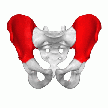
- "Ilium 03 animation.gif" by BodyParts3D is made by DBCLS is licensed under CC BY-SA 2.1
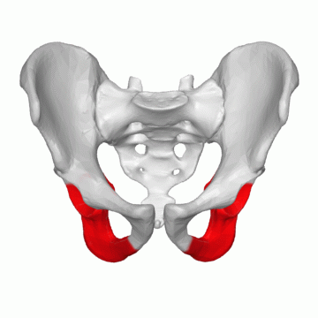
- "Ischium 03 animation.gif" by BodyParts3D is made by DBCLS is licensed under CC BY-SA 2.1
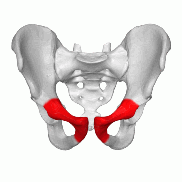
- "Pubis 03 animation.gif" by BodyParts3D is made by DBCLS is licensed under CC BY-SA 2.1
Updated 2026.