9.5: Lower Limb
- Page ID
- 53653
Lower Limb
For anatomists, the lower limb consists of the thigh (hip to knee), the leg (knee to ankle), and the foot. The thigh consists of a single bone, the femur. The leg consists of two long bones, the tibia and fibula, and the sesamoid bone, the patella that serves as the knee cap. The foot consists of 26 bones, which are grouped into the tarsals, metatarsals, and phalanges.
Above: Diagram showing the components of a lower limb: 1 femur, 1 patella, 1 tibia, 1 fibula, 7 tarsal bones, 5 metatarsal bones, and 14 phalanges.
Above: The right and left femur (A) anterior view and (B) posterior view.
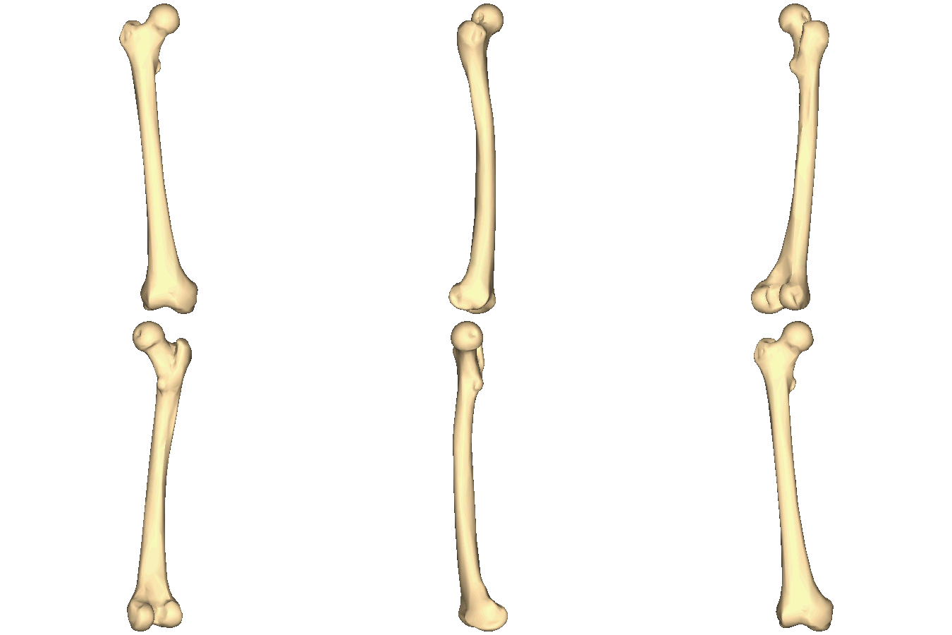
The femur, the thigh bone, is the strongest and heaviest bone in the human body. The head of the femur articulates with the acetabulum of a coxal bone. A dimple-like depression on the head of the femur, fovea capitis, is the site of attachment of a ligament securing the femur in the acetabulum (ligamentum teres). Although the femur is very strong, its weakest point is its neck, the most common site of femur fractures and usually called a "broken hip." The greater trochanter, lesser trochanter, and gluteal tuberosity are all markings of the femur where muscles attach.

Above: Right femur.
Above: Left patella (left) anterior view and (right) posterior view.
At the distal end of the femur are two condyles, a medial condyle and a lateral condyle, that articulate with the tibia at the articulation surface of the medial condyle and the articulation surface of the lateral condyle, respectively, forming the knee joint. A smooth surface called the patellar surface between the two condyles of the femur on the anterior aspect serves as a location where the patella, the knee cap, articulates with the femur in the knee joint.
Above: Articulation of the condyles of the femur with the articular surfaces of the tibia in the left knee joint. Articulation of the patella and fibula are also shown with some of the ligaments and cartilage of the knee joint.
Above: Right and left tibia and fibula (A) anterior view and (B) posterior view.
The bones of the foot are shown in below. The calcaneus is the heel bone, and the talus bone forms the ankle joint with the tibia and fibula. The calcaneus and tarsus are two of the seven tarsal bones that are posterior to the first long bones of the foot, the metatarsal bones. The bones of the toes are phalanges, the same name used for finger bones.
Above: The bones of the right and left feet, superior view. There are seven tarsal (ankle) bones: calcaneus (heel), talus, navicular, medial cuneiform, intermediate cuneiform, lateral cuneiform, and cuboid. The bones of the feet are metatarsals numbered one (medial) through five (lateral). The phalanges of the foot have the same numbering system as the metatarsals and also get designations of proximal, middle, and distal based on their positions. Phalanx #1 only has proximal and distal phalanges (no middle phalanx).
Above: The tarsal bones and their articulation with the tibial and fibula on the (left) left and (right) right foot viewed from their lateral aspects.
Attributions
- "BodyParts3D/Anatomography" by The Database Center for Life Science is licensed under CC BY-SA 2.1
- "Human knee joint FE model.png" by Hamid nbe is licensed under CC BY-SA 4.0
- "BIOL 250 Human Anatomy Lab Manual SU 19" by Yancy Aquino, Skyline College is licensed under CC BY-NC-SA 4.0
- "Femur - animation.gif" by Anatomography is licensed under CC BY-SA 2.1
- "Right femur - close-up - animation.gif" by Anatomography is licensed under CC BY-SA 2.1


