

1.9: Mitosis
- Page ID
- 222595
\( \newcommand{\vecs}[1]{\overset { \scriptstyle \rightharpoonup} {\mathbf{#1}} } \)
\( \newcommand{\vecd}[1]{\overset{-\!-\!\rightharpoonup}{\vphantom{a}\smash {#1}}} \)
\( \newcommand{\dsum}{\displaystyle\sum\limits} \)
\( \newcommand{\dint}{\displaystyle\int\limits} \)
\( \newcommand{\dlim}{\displaystyle\lim\limits} \)
\( \newcommand{\id}{\mathrm{id}}\) \( \newcommand{\Span}{\mathrm{span}}\)
( \newcommand{\kernel}{\mathrm{null}\,}\) \( \newcommand{\range}{\mathrm{range}\,}\)
\( \newcommand{\RealPart}{\mathrm{Re}}\) \( \newcommand{\ImaginaryPart}{\mathrm{Im}}\)
\( \newcommand{\Argument}{\mathrm{Arg}}\) \( \newcommand{\norm}[1]{\| #1 \|}\)
\( \newcommand{\inner}[2]{\langle #1, #2 \rangle}\)
\( \newcommand{\Span}{\mathrm{span}}\)
\( \newcommand{\id}{\mathrm{id}}\)
\( \newcommand{\Span}{\mathrm{span}}\)
\( \newcommand{\kernel}{\mathrm{null}\,}\)
\( \newcommand{\range}{\mathrm{range}\,}\)
\( \newcommand{\RealPart}{\mathrm{Re}}\)
\( \newcommand{\ImaginaryPart}{\mathrm{Im}}\)
\( \newcommand{\Argument}{\mathrm{Arg}}\)
\( \newcommand{\norm}[1]{\| #1 \|}\)
\( \newcommand{\inner}[2]{\langle #1, #2 \rangle}\)
\( \newcommand{\Span}{\mathrm{span}}\) \( \newcommand{\AA}{\unicode[.8,0]{x212B}}\)
\( \newcommand{\vectorA}[1]{\vec{#1}} % arrow\)
\( \newcommand{\vectorAt}[1]{\vec{\text{#1}}} % arrow\)
\( \newcommand{\vectorB}[1]{\overset { \scriptstyle \rightharpoonup} {\mathbf{#1}} } \)
\( \newcommand{\vectorC}[1]{\textbf{#1}} \)
\( \newcommand{\vectorD}[1]{\overrightarrow{#1}} \)
\( \newcommand{\vectorDt}[1]{\overrightarrow{\text{#1}}} \)
\( \newcommand{\vectE}[1]{\overset{-\!-\!\rightharpoonup}{\vphantom{a}\smash{\mathbf {#1}}}} \)
\( \newcommand{\vecs}[1]{\overset { \scriptstyle \rightharpoonup} {\mathbf{#1}} } \)
\(\newcommand{\longvect}{\overrightarrow}\)
\( \newcommand{\vecd}[1]{\overset{-\!-\!\rightharpoonup}{\vphantom{a}\smash {#1}}} \)
\(\newcommand{\ket}[1]{\left| #1 \right>}\)
\(\newcommand{\bra}[1]{\left< #1 \right|}\)
\(\newcommand{\braket}[2]{\left< #1 \vphantom{#2} \right| \left. #2 \vphantom{#1} \right>}\)
\(\newcommand{\braopket}[3]{\left< #1 \vphantom{#2}\vphantom{#3} \right| #2 \vphantom{#1}\vphantom{#3} \left| #3 \vphantom{#1}\vphantom{#2} \right>}\)
\(\newcommand{\qmvec}[1]{\mathbf{\vec{#1}}}\)
\(\newcommand{\op}[1]{\hat{\mathbf{#1}}}\)
\(\newcommand{\expect}[1]{\langle #1 \rangle}\)
\(\newcommand{\dfn}[1]{\emph{\textbf{#1}}}\)
Introduction
- Describe the phases of the cell cycle
- Identify and explain each of the four phases of mitosis
Early in the semester we learned about cell theory. One of the components of cell theory is that cells must come from other cells. How does that happen though? There are actually several ways to replicate cells. Which process a cell uses depends on what type of organism the cell is and the reason why the cell is replicating.
One such method is binary fission -- a replication process used by simple organisms such as bacteria and archaea. In binary fission, the bacterium’s single circular chromosome is replicated with each copy moving to opposite sides of the cell. Then proteins begin pinching the cell in the middle, a septum forms, and the cell splits into two. This process is both simple and fast, meaning bacteria can replicate very quickly.
Another method is mitosis. This process is used by the more complex eukaryotic cells. In multicellular organisms like ourselves, mitosis is used for growth and repair. Consider how big you were when you were born. Are you the same size? Hopefully, no. But rather than your cells getting larger, your body has simply made more cells to create the growth you’ve experienced over the years. You’ve also probably experienced a few injuries before. But if you get a cut, it heals. This healing you see is because the cells that were killed and damaged by whatever cut your skin were replaced. Nearby healthy cells used mitosis to replace them.
Cell Cycle
In eukaryotic organisms like plants and animals, cell replication is a tightly regulated cycle. Cells must receive various signals and meet certain criteria before being able to divide. These checks and balances ensure that the cell has enough building material, that the DNA replicates correctly, and that the chromosomes are aligned properly to divide. This helps ensure that the organism is keeping up with normal rates of cell death and not wasting resources. When this system fails, things like cancer can result.
The cell cycle can be broken down into two main parts: interphase and the mitotic phase. Let’s look at interphase first. In interphase, there are three distinct phases: G1, S, and G2. In the G1 phase, the cell is running its daily activities and is gathering needed materials for the next steps. Then the cell enters the S phase. S phase is unique in that this is when a cell replicates its entire genome (the complete set of DNA) of the cell. After all of the DNA has been replicated, the cell then enters the G2 phase. In the G2 phase, the cell makes final preparations to divide.

Figure 1: Stages of Cell Cycle. (Modified by Karen Marks, from OpenStax, CC BY 4.0, via Wikimedia Commons)
Looking at the figure above, do you think a cell spends more time in interphase or mitotic phase?
Mitosis in Plant and Animal Cells
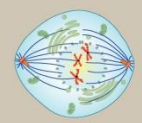
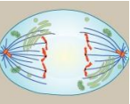
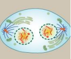
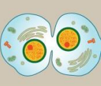
Mitosis has five phases: prophase, prometaphase, metaphase, anaphase, and telophase. Once the cell has completed these phases, it will split in two via cytokinesis.
| PHASE | DESCRIPTION | APPEARANCE |
|---|---|---|
| Prophase |
|
 |
| Prometaphase |
|
 |
| Metaphase |
|
 |
| Anaphase |
|
 |
| Telophase |
|
 |
| Cytokinesis |
|
 |
Observe the phases of Mitosis in Plant Cells
Exercise 1: Observing the Phases of Mitosis in the Onion Root Tip
Materials:
- Prepared slide of the onion root tip
- Compound light microscope
Procedure:
- Examine a slide of a longitudinal section of an onion root tip. Adjust the slide to view the region just above the root cap, where there are likely to be dividing cells.
- Focus on the dividing cells using the 4x scanning objective lens, then switch to the 10x objective and then the 40x objective.
- Survey the slide to find a cell in each phase of mitosis. Draw a cell for each phase in the boxes provided.
| Magnification | Interphase | Prophase | Metaphase | Anaphase | Telophase/Cytokineses |
|---|---|---|---|---|---|
|
4x Objective |
|||||
|
10x Objective |
|||||
|
40x Objective |
Interphase
DNA is uncondensed and in the form of chromatin. Individual chromosomes are not visible. The nuclear membrane is intact. The nucleolus is visible.
Figure 2: Interphase in plant cells.
Prophase
Chromatin begins to condense into distinguishable chromosomes. These “puffy” structures are seen throughout the nucleus. Nucleoli begin to disappear. In late prophase (often called prometaphase) the nuclear membrane is no longer visible.
Figure 3: Prophase in plant cells.
Metaphase
The chromosomes line up in the middle of the cell. Spindle fibers attach to kinetochores at the centromere and extend to the poles of the cell.
Figure 4: Metaphase in plant cells.
Anaphase
Centromeres split, separating each former chromatid into two individual chromosomes. The chromosomes move toward opposite poles.
Figure 5: Anaphase in plant cells.
Telophase and Cytokinesis
Chromosomes reach the poles. The nuclear envelopes begin to reform. The formation of a cell plate forms between the two cells to carry out cytokinesis.
Figure 6: Telophase in plant cells.
Figure 7: Cytokinesis in plant cells.
Observe the phases of Mitosis in Animal Cells
Exercise 2: Observing the Phases of Mitosis in the Whitefish Blastula
Materials:
- Prepared slide of whitefish blastula
- Compound light microscope
Procedure:
The blastula is an early embryonic stage where many of the cells are dividing at any one time.
- Focus on the dividing cells using the 4x scanning objective lens, then switch to the 10x objective and then the 40x objective.
- Survey the slide to find a cell in each phase of mitosis. Draw a cell for each phase in the boxes provided.
| Magnification | Interphase | Prophase | Metaphase | Anaphase | Telophase/Cytokineses |
|---|---|---|---|---|---|
|
4x Objective |
|||||
|
10x Objective |
|||||
|
40x Objective |
Interphase
The DNA is uncondensed and in the form of chromatin. Individual chromosomes are not visible. The nuclear membrane is intact. The nucleolus is visible.
Figure 8: Interphase in animal cells.
Prophase
Chromatin begins to condense and chromosomes are distinguishable. These “puffy” structures are seen throughout the nucleus. The nucleoli begin to disappear. In late prophase (often called prometaphase) the nuclear membrane is no longer visible.
Figure 9: Prophase in animal cells.
Metaphase
The chromosomes line up in the middle of the cell. Spindle fibers attach to kinetochores at the centromere and extend to the poles of the cell.
Figure 10: Metaphase in animal cells.
Anaphase
Centromeres split, separating each former chromatid into two individual chromosomes. The chromosomes move toward opposite poles.
Figure 11: Anaphase in animal cells.
Telophase and Cytokinesis
Chromosomes reach the poles. The nuclear envelopes begin to reform. A cleavage furrow forms between the two cells to carry out cytokinesis.
Figure 12: Telophase in animal cells.
Figure 13: Cytokinesis in animal cells.
- Why would the method of cytokinesis in animal cells not work in plant cells? Explain.
__________________________________________________________________
How Long Does a Cell Spend in Each Phase of the Cell Cycle?
Exercise 3: Determining Time Spent in Different Phases of the Cell Cycle (Optional)
Materials:
- Laptop
- Calculator
Procedure:
- Count ~ 30 cells in total per field of view using the onion root tip slide.
- Use the formula given below and record your results in the table.
- The duration of each stage of mitosis can be determined by using the following formula. Compute the length of time for each stage and place your calculations in the table below.
(Number of cells in a stage ÷ Total number of cells) x 1440 (min in a day) = minutes a cell spends in each stage in one day.
|
Number of cells in each stage |
Interphase ______ |
Prophase ______ |
Metaphase ______ |
Anaphase _______ |
Telophase _______ |
Total 36 |
|---|---|---|---|---|---|---|
|
Percent of cells (# of cells / Total) |
100% |
|||||
|
Time (in minutes) spent in Stage – use calculation above |
Modeling Mitosis
Now that we’ve studied cells at the various stages of mitosis, it’s your group’s turn to model mitosis in the lab. For this exercise, your group will use pop beads to make two pairs of homologous chromosomes. Each pair of homologous chromosomes must be in a different color. Then consider the desk as a cell and arrange the chromosomes to indicate phases of mitosis on the lab bench and explain what happens during each phase to your instructor.
Example of chromosomes made with pop beads:

Replicated chromosomes:

In your explanations, you must follow these "rules":
- Everyone in the group must participate
- You must use the proper terminology to explain something
- You cannot read steps directly from a book or other resource
- Be prepared before calling the instructor over
Group Members:
Verified complete:_______________________________ (Instructor signature)
Licensing and Attributions
- 14: Mitosis is shared under a CC BY 4.0 license and was authored, remixed, and/or curated by Darcy Ernst, May Chen, Katie Foltz, and Bridget Greuel (Open Educational Resource Initiative at Evergreen Valley College)
- 14.1: Introduction is shared under a CC BY 4.0 license and was authored, remixed, and/or curated by Darcy Ernst, May Chen, Katie Foltz, and Bridget Greuel (Open Educational Resource Initiative at Evergreen Valley College)
- 14.2: Exercise is shared under a CC BY 4.0 license and was authored, remixed, and/or curated by Darcy Ernst, May Chen, Katie Foltz, and Bridget Greuel (Open Educational Resource Initiative at Evergreen Valley College)
- 8: From One Cell Comes Many - Mitosis and Meiosis is shared under a CC BY-NC-SA 4.0 license and was authored, remixed, and/or curated by Karen Marks and Valeria Hochman Adler.
- 8.2: The Cell Cycle is shared under a CC BY-NC-SA 4.0 license and was authored, remixed, and/or curated by Karen Marks and Valeria Hochman Adler.
- 8.3: Mitosis in Plant and Animal Cells is shared under a CC BY-NC-SA 4.0 license and was authored, remixed, and/or curated by Karen Marks and Valeria Hochman Adler.
- 8.4: Modeling Mitosis is shared under a CC BY-NC-SA 4.0 license and was authored, remixed, and/or curated by Karen Marks and Valeria Hochman Adler.
- Belwood, Jacqueline; Rogers, Brandy; and Christian, Jason, Foundations of Biology Lab Manual (Georgia Highlands College). “Lab 10: Mitosis & Meiosis,” (2019). Biological Sciences Open Textbooks. 18. CC-BY (https://oer.galileo.usg.edu/biology-textbooks/18)