
Lab 7 Musculoskeletal Anatomy Part 3: Articulation and Kinematics of movement
- Page ID
- 53333
\( \newcommand{\vecs}[1]{\overset { \scriptstyle \rightharpoonup} {\mathbf{#1}} } \)
\( \newcommand{\vecd}[1]{\overset{-\!-\!\rightharpoonup}{\vphantom{a}\smash {#1}}} \)
\( \newcommand{\id}{\mathrm{id}}\) \( \newcommand{\Span}{\mathrm{span}}\)
( \newcommand{\kernel}{\mathrm{null}\,}\) \( \newcommand{\range}{\mathrm{range}\,}\)
\( \newcommand{\RealPart}{\mathrm{Re}}\) \( \newcommand{\ImaginaryPart}{\mathrm{Im}}\)
\( \newcommand{\Argument}{\mathrm{Arg}}\) \( \newcommand{\norm}[1]{\| #1 \|}\)
\( \newcommand{\inner}[2]{\langle #1, #2 \rangle}\)
\( \newcommand{\Span}{\mathrm{span}}\)
\( \newcommand{\id}{\mathrm{id}}\)
\( \newcommand{\Span}{\mathrm{span}}\)
\( \newcommand{\kernel}{\mathrm{null}\,}\)
\( \newcommand{\range}{\mathrm{range}\,}\)
\( \newcommand{\RealPart}{\mathrm{Re}}\)
\( \newcommand{\ImaginaryPart}{\mathrm{Im}}\)
\( \newcommand{\Argument}{\mathrm{Arg}}\)
\( \newcommand{\norm}[1]{\| #1 \|}\)
\( \newcommand{\inner}[2]{\langle #1, #2 \rangle}\)
\( \newcommand{\Span}{\mathrm{span}}\) \( \newcommand{\AA}{\unicode[.8,0]{x212B}}\)
\( \newcommand{\vectorA}[1]{\vec{#1}} % arrow\)
\( \newcommand{\vectorAt}[1]{\vec{\text{#1}}} % arrow\)
\( \newcommand{\vectorB}[1]{\overset { \scriptstyle \rightharpoonup} {\mathbf{#1}} } \)
\( \newcommand{\vectorC}[1]{\textbf{#1}} \)
\( \newcommand{\vectorD}[1]{\overrightarrow{#1}} \)
\( \newcommand{\vectorDt}[1]{\overrightarrow{\text{#1}}} \)
\( \newcommand{\vectE}[1]{\overset{-\!-\!\rightharpoonup}{\vphantom{a}\smash{\mathbf {#1}}}} \)
\( \newcommand{\vecs}[1]{\overset { \scriptstyle \rightharpoonup} {\mathbf{#1}} } \)
\( \newcommand{\vecd}[1]{\overset{-\!-\!\rightharpoonup}{\vphantom{a}\smash {#1}}} \)
\(\newcommand{\avec}{\mathbf a}\) \(\newcommand{\bvec}{\mathbf b}\) \(\newcommand{\cvec}{\mathbf c}\) \(\newcommand{\dvec}{\mathbf d}\) \(\newcommand{\dtil}{\widetilde{\mathbf d}}\) \(\newcommand{\evec}{\mathbf e}\) \(\newcommand{\fvec}{\mathbf f}\) \(\newcommand{\nvec}{\mathbf n}\) \(\newcommand{\pvec}{\mathbf p}\) \(\newcommand{\qvec}{\mathbf q}\) \(\newcommand{\svec}{\mathbf s}\) \(\newcommand{\tvec}{\mathbf t}\) \(\newcommand{\uvec}{\mathbf u}\) \(\newcommand{\vvec}{\mathbf v}\) \(\newcommand{\wvec}{\mathbf w}\) \(\newcommand{\xvec}{\mathbf x}\) \(\newcommand{\yvec}{\mathbf y}\) \(\newcommand{\zvec}{\mathbf z}\) \(\newcommand{\rvec}{\mathbf r}\) \(\newcommand{\mvec}{\mathbf m}\) \(\newcommand{\zerovec}{\mathbf 0}\) \(\newcommand{\onevec}{\mathbf 1}\) \(\newcommand{\real}{\mathbb R}\) \(\newcommand{\twovec}[2]{\left[\begin{array}{r}#1 \\ #2 \end{array}\right]}\) \(\newcommand{\ctwovec}[2]{\left[\begin{array}{c}#1 \\ #2 \end{array}\right]}\) \(\newcommand{\threevec}[3]{\left[\begin{array}{r}#1 \\ #2 \\ #3 \end{array}\right]}\) \(\newcommand{\cthreevec}[3]{\left[\begin{array}{c}#1 \\ #2 \\ #3 \end{array}\right]}\) \(\newcommand{\fourvec}[4]{\left[\begin{array}{r}#1 \\ #2 \\ #3 \\ #4 \end{array}\right]}\) \(\newcommand{\cfourvec}[4]{\left[\begin{array}{c}#1 \\ #2 \\ #3 \\ #4 \end{array}\right]}\) \(\newcommand{\fivevec}[5]{\left[\begin{array}{r}#1 \\ #2 \\ #3 \\ #4 \\ #5 \\ \end{array}\right]}\) \(\newcommand{\cfivevec}[5]{\left[\begin{array}{c}#1 \\ #2 \\ #3 \\ #4 \\ #5 \\ \end{array}\right]}\) \(\newcommand{\mattwo}[4]{\left[\begin{array}{rr}#1 \amp #2 \\ #3 \amp #4 \\ \end{array}\right]}\) \(\newcommand{\laspan}[1]{\text{Span}\{#1\}}\) \(\newcommand{\bcal}{\cal B}\) \(\newcommand{\ccal}{\cal C}\) \(\newcommand{\scal}{\cal S}\) \(\newcommand{\wcal}{\cal W}\) \(\newcommand{\ecal}{\cal E}\) \(\newcommand{\coords}[2]{\left\{#1\right\}_{#2}}\) \(\newcommand{\gray}[1]{\color{gray}{#1}}\) \(\newcommand{\lgray}[1]{\color{lightgray}{#1}}\) \(\newcommand{\rank}{\operatorname{rank}}\) \(\newcommand{\row}{\text{Row}}\) \(\newcommand{\col}{\text{Col}}\) \(\renewcommand{\row}{\text{Row}}\) \(\newcommand{\nul}{\text{Nul}}\) \(\newcommand{\var}{\text{Var}}\) \(\newcommand{\corr}{\text{corr}}\) \(\newcommand{\len}[1]{\left|#1\right|}\) \(\newcommand{\bbar}{\overline{\bvec}}\) \(\newcommand{\bhat}{\widehat{\bvec}}\) \(\newcommand{\bperp}{\bvec^\perp}\) \(\newcommand{\xhat}{\widehat{\xvec}}\) \(\newcommand{\vhat}{\widehat{\vvec}}\) \(\newcommand{\uhat}{\widehat{\uvec}}\) \(\newcommand{\what}{\widehat{\wvec}}\) \(\newcommand{\Sighat}{\widehat{\Sigma}}\) \(\newcommand{\lt}{<}\) \(\newcommand{\gt}{>}\) \(\newcommand{\amp}{&}\) \(\definecolor{fillinmathshade}{gray}{0.9}\)Objectives:
At the end of this lab, you will be able to…
- Differentiate between classifications of articulations
- Correctly describe the types of movements at a given articulation
- Describe what is meant by the kinematic chain
Pre-Lab Exercises:
After reading through the lab activities prior to lab, complete the following before you start your lab.
1. The classification of articulations of the body is based on: .
2. The difference between levers is the location of the relative to force being applied and the action of the muscles contracting.
3. Movement of the hand away from body laterally is called , while movement medially is called .
4. Based on the kinematic classification of the articulations, the allow for the most movement while the have the least.
Materials:
- Skeleton
- Joint Models
- Stickers
- Felt pens
When examining the articulations of the body it is important to remember the relationship between structure and function. In this, we can use the shape of the articulation to determine the different kinematics (movements) that the articulations allow for the bones when combined with lines of pull of the ligaments, tendons and muscles that attach to the bones of the articulation.
Activity 1:
Articulations:
There are three classifications of articulations based on the ability (called or degrees of freedom) for movement to occur. The ability to move or degrees of freedom are an indication of the number of directions (or axis) that the articulating bones are able to move at that articulation. These are identified as being synarthrosis, amphiarthrosis, and diarthrosis. Synarthrosis are articulations with no movement occurring between bones, meaning 0 degrees of freedom for movement. Amphiarthrosis is the type of articulations with minimal movement provided between bones meaning 0 or 1 degree of freedom for movement. Diarthrosis articulations are typically thought of articulations, such that the bones are able to move about each other, meaning at 1-2 degrees of freedom for movement.
Within the Diarthrosis joints there are six types of articulations commonly called synovial joint, with each type providing a different amount and pattern of movement. Pattern of movement is determined by the shape of the articulating surfaces for bones that are meeting at an articulation.(Knudson, 2007) The amount of movement allowed because of the structure of the ligaments that surround the joint that limit the total amount of movement possible at any given articulation along with the lines of pull of the muscles on the bones that form the joint.
Additionally, there is a distinct class of articulations known as dynamic articulations. The dynamic articulations are seen primarily within ball and socket joints (especially the glenohumeral joints). These joints form due to interaction of capsular ligaments with the tendons of the rotator muscles of the joint. Provides the ability to have changing levels of tension and capsular stability via the interaction between ligamentous and tendinous units. That occurs by changes in tension about the central axis of the capsular ligament and thus modifies the degree of motion that can be obtained throughout the entire range of motion of the articulation.
Activity 1:
Types of Articulations:
1. Obtain bones, skeleton, articulation models, stickers and felt-tip marker
2. Write the names for the types and classifications of articulations on the stickers.
3. Working with your group (and using your colored images) identify the various types of articulations of the body by type and classification.
4. Taking turns within your group, label the articulations based on being fibrous, cartilaginous or synovial and then as being synarthrosis, amphiarthrosis or diarthrosis.
5. Have your work checked by the instructor and then move to activity 2.
|
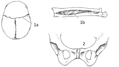
Classification of Articulations 1a Fibrous (Synarthrosis) 1b Fibrous (Amphiarthrosis) 2 Cartilaginous (Amphiarthrosis) 3 Synovial 3a Articulating Bones 3b Anterior Synovial Membrane 3c Synovial Fluid 3d Tendon 3e Bursa Sacs 3f Articulating Cartilage 3g Meniscus 3h Posterior Synovial Membrane 3i Sub-patellar Bursa Sac 3j Cruciate Ligament
Color each joint a different color for use as a reference when identifying on your skeleton |

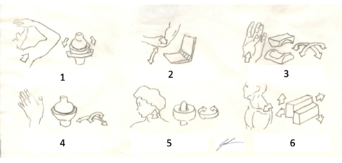
Types of Synovial Joints
|
1. Ball and Socket 2. Hinge 3. Saddle 4. Condylar (Ellipsoid) 5. Pivot 6. Planar (Gliding)
Color each synovial joint a different color for use as a reference when identifying on your skeleton |
Activity 2:
Lines of Pull and Movements:
Lines of pull of the description of movements that can be generated by a muscle contraction. These lines of pull are based on the origin and insertion of the muscle, the anatomical location of the muscle as it crosses the articulation that is being moved and the anatomical shape of the articulation. For most of the articulations of the body, these motions will occur in the coronal or sagittal planes, however there are special movements that occur in the transverse plane. These movements are generally referred to as being horizontal movements of the appendage.
These lines of pull will be described by defined antagonistic pairs (pairs of movements that perform the opposite of each other) of movements at the joint. Most of movements will result in a paired movement patterning between agonistic muscles (muscles that allow for motion in the direction that is desired) and antagonistic muscles (muscles that allow for motion in the opposite direction to the desired direction). The agonistic muscle also will involve muscles that are furthered described as being primary or accessory to the movement. Where the primary movers are those muscles that will produce the majority of the active force needed for the movement to occur and the accessory muscles will either provide a limited amount of force to the movement or will stabilize the articulation within the pattern of movement.
The movements include:
Flexion: movement of the extremity toward the torso/thorax
Extension: movement of the extremity away from the torso/thorax
Abduction: movement away from the mid-line of the body
Adduction: movement toward the mid-line of the body
Rotation: movement around the central axis of the body or extremity
Circumduction: movement of the arm through all three anatomical planes
Deviation: movement of the hand and wrist toward the radius or the ulna
Supination: rotating hand and wrist, or foot and ankle, so that palm of hand, or sole of foot, is facing anterior (as is holding a bowl of soup)
Pronation: rotating hand and wrist, or foot and ankle, so that palm of hand, or sole of foot, is facing posterior
Vertebral/Trunk Rotation: rotating of the vertebral column (or segments of the vertebral column) about the central long axis of the body
Inversion: rotation of the foot and ankle, medially and superiorly
Eversion: rotation of the foot and ankle laterally and inferiorly
Plantarflexion: movement of the toes and foot (and ankle) toward the ground (away from the leg)
Dorsiflexion: movement of the toes and foot (and ankle) toward the anterior leg (away from the ground)
Horizontal Adduction: movement of the appendage in an abducted position toward the mid-line of the body
Horizontal Abduction: movement of the appendage in an abducted position away the mid-line of the body
1. Using the following articulations, take turns within your group to mimic the movements that would be allowed at the articulation. As you perform the movement, have one person in your group indicate a muscle that would allow the movement to occur.
a. Shoulder (Glenohumeral)
b. Elbow (Humeroulnar)
c. Forearm (Proximal and Distal Radioulnar)
d. Wrist (Radiocarpal)
e. Head and Neck (Atlooccipital)
f. Head and Neck (Atloaxial)
g. Vertebral
h. Hip (Femoroacetabular)
i. Knee (Tibiofemoral)
j. Ankle (Tibiotalar)
Patterns of Movement and Kinematic Chain:
Kinematics is the description of the movements of the bones at the joints (articulations) that allow for locomotion (movement) to occur either within the limb or body segment or the body as a whole. There are two distinct features that we must remember, and both relate to what is called the kinematic chain. Kinematic chain is the linking of the articulations of the body that leads to the desired movement. In the kinematic chain you can either have an open chain or a closed chain. The open chain is where the distal (terminal) end of the chain is free to move and the proximal (initial) segment of the chain is fixed. While a closed chain will have both the distal (terminal) end and the proximal (initial) segment of the chain are fixed.
The motion that is allowed by the articulations interactions with the muscles follows the physical principles of levers. Based on where the muscles insert on the bone, three basic lever systems are developed all of which provides a level of mechanical advantage to the articulation allowing for movement to occur. In addition to the lever system, the arrangement of tendinous insertion and pennation of the muscle fascicles (fibers) within the muscle gaster summate to determine the overall mechanical advantage that each articulation provides for the kinematics of movement for the body.
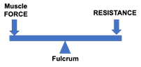
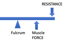
The first-class (type 1) lever system is the simplest of the lever system. In can be thought of as a teeter-tooter where the muscle pull is on the opposite side of the joint from the resistance placed on the joint. This type of lever is seen at the Atlooccipital (between CV1 and the Occipital bone of the cranium) articulation.
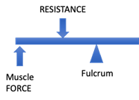
The second-class (type 2) lever system is more readily seen, but slightly more complex than, the type-1 lever for the human body. The system has the muscle pull is on the same side of the joint as the resistance placed on the joint but will have the resistance placed on the joint falls between the joint moving and muscle pull causing the movement, similar to the action of using a nutcracker. This type of lever is seen at the Tibiotalar and Talofibular (ankle) articulation.
The third-class (type 3) lever system is the most complex system used in the body. It typically seen in articulation that has a moveable pivot point for the fulcrum. This is necessary to provide the maximal mechanical advantage from the lever, as the muscle pull will be closer to the fulcrum than the resistance on the same side of the joint. This lever is similar to the action of using a shovel and is seen at the Humeroulnar (elbow) articulation.
2. As you perform the movements, as a group determine the type of lever system being used and if the movement is an open chain or a closed chain pattern of movement
a. Shoulder (Glenohumeral)
i. Lever system:
ii. Movement:
iii. Kinematic Chain:
b. Elbow (Humeroulnar)
i. Lever system:
ii. Movement:
iii. Kinematic Chain:
c. Forearm (Proximal and Distal Radioulnar)
i. Lever system:
ii. Movement:
iii. Kinematic Chain:
d. Wrist (Radiocarpal)
i. Lever system:
ii. Movement:
iii. Kinematic Chain:
e. Head and Neck (Atlooccipital)
i. Lever system:
ii. Movement:
iii. Kinematic Chain:
f. Head and Neck (Atloaxial)
i. Lever system:
ii. Movement:
iii. Kinematic Chain:
g. Vertebral
i. Lever system:
ii. Movement:
iii. Kinematic Chain:
h. Hip (Femoroacetabular)
i. Lever system:
ii. Movement:
iii. Kinematic Chain:
i. Knee (Tibiofemoral)
i. Lever system:
ii. Movement:
iii. Kinematic Chain:
j. Ankle (Tibiotalar)
i. Lever system:
ii. Movement:
iii. Kinematic Chain:
3. Have your instructor check your answers and then clean up your lab area.