
Lab 6 Musculoskeletal Anatomy Part 2: Skeletal Muscle
- Page ID
- 53332
Objectives:
At the end of this lab, you will be able to…
- Using appropriate anatomical terminology correctly identify muscles by using correct anatomical (Latin) name
- Using appropriate anatomical terminology correctly identify type of motion generated by the muscles of importance
- Differentiate between classifications of architecture of muscles by shape
- Describe the agonist/antagonist relationships between muscles surrounding joints of importance
- Correctly identify and mimic generalized movement patterns based on individual muscles or muscle groups
Pre-Lab Exercises:
After reading through the lab activities prior to lab, complete the following before you start your lab.
1. The naming rules for muscles include names based on: .
2. How many biceps muscles are found in the body?
3. The muscles on the dorsal side of the thorax will the trunk while the muscles on the ventral side of the abdominal cavity will the trunk.
4. Color the images for use as a reference for identifying the muscles and showing actions of the muscles.
Materials:
- Muscle Models
- Stickers
- Felt pens
Activity 1: Naming Principles, Origins and Insertions:
The methods to name muscles include: the fiber arrangement, the location, the size, the number of origins (heads), the shape, the action, the origin and insertions or the bone that the muscle overlays. In some cases, we will combine these methods for naming as some muscles may have one or more method of classification in common. Which is why we must be very careful to include the entirety of the name of the muscle, such as biceps brachii, and not just simply use biceps. As there are multiple Bicipital muscles in the body and without the complete terminology you may have someone thinking of the different biceps (femoral) from the biceps (brachii) that you are talking about.
When we discuss by fiber arrangement you will see muscle names such as: rectus (parallel angle to long axis of body), transverse (perpendicular angle to long axis of body), or oblique (oblique angle to long axis of body). With discussion by location, you will see muscle names such as: superficial (closer to surface), profundus (deeper or underlying musculature), side of the bone (anterior or posterior) and the associated bone that underlies the muscle. We can also use the muscles comparative size within a group of muscles there is an indication for comparing three muscles maximus, minimus and medius (largest, smallest, middle of the three being compared) versus groups of two muscles major and minor (largest and smallest of the three being compared) or by the length brevis and longus (shortest and longest of the two being compared). There will also be names based on the number of origins (heads) of the muscles such as triceps (three muscle origins/heads), biceps (two muscle origins/heads), and quadriceps (four muscle origins/heads). Then we can discuss the muscle name by the shape of the muscle such as deltoid (triangular, or like Greek letter Δ), trapezius (trapezoidal), rhomboideus (rhomboid), teres (tear-drop), serratus (saw-tooth), or quadratus (quadrilateral). There is also the naming method that provides the muscles origin and the muscles insertion into a single name such as the sternocleidomastoid. Lastly, we can use the action of the muscle to names of muscles such as tensors (tenses the connective tissues), flexors (flexes the joint or articulation), extensors (extends the joint or articulation), supinator (supinates the distal end of the extremities), and pronators (pronates the distal end of the extremities).
Procedures:
1. Review the naming principles by calling out a muscle based on one of the naming rules and pointing to that muscle
2. Check your group members for completeness of the coloring of muscles necessary for completing the identifications.
| ANTERIOR - POSTERIOR |
|
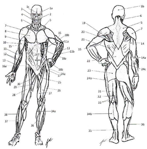
1a=Temporalis 1b=Epicranial Aponeurosa 1c=Frontalis 2=Orbicularis Oris 3=Orbicularis Oculi 4=Masseter 5=Trapezius 6=Sternocleidomastoid 7=Deltoid 8=Pectoralis Major 9=Serratus Anterior 10=Biceps Brachii 11=Brachioradialis 11a=Extensor Carpi Radialis Longus and Brevis 11b=Common Extensors 12=Common Flexors 13=Extensor Carpi Radialis 14=Triceps Brachii 14a=Triceps Tendon 15=Brachialis 16a=External Abdominal Oblique 16b=Internal Abdominal Oblique 17=Rectus Abdominis 18a=Internal Intercostal 18b=External Intercostal 19=Teres Minor 20=Teres Major 21=Latissimus Dorsi 22=Thoracolumbar Fascia 23=Gluteus Maximus 24a=Tensor Fascia Latte 24b=Iliotibial Band 24c=Gluteus Medius 25=Sartorius 26=Vastus Lateralis 27=Vastus Medialis 28=Rectus Femoris 29=Adductor Magnus 30=Adductor Longus 31=Biceps Femoris 32=Semitendinosus 33=Semimembranosus 34a=Medial Head Gastrocnemius 34b=Lateral Head Gastrocnemius 35=Calcaneal (Achilles) Tendon 36=Soleus (Deep to Tendon on posterior) 37=Tibialis Anterior 38=Peroneal Muscles Color each muscle a different color for use as a reference when identifying muscles. |
Muscles of the axilla and pectoral girdle are broken into key groups: pectoral muscles, deltoid muscles and the rotator cuff. Each muscle is involved with stabilization of the Scapula and movement of the Humerus during locomotion of the upper extremity of the body and movement of the thoracic cage during ventilation.
Pectoral Girdle Muscles
|
Muscle |
Origin |
Insertion |
Action |
|
Pectoralis Major |
Clavicle; Body of sternum; Costal |
Lateral lip of Intertubercular groove |
Horizontal Adduct, Medially Rotate Humerus @ shoulder |
|
Pectoralis Minor |
Costal 2-5 |
Coracoid process |
Protract Scapula Elevates Ribs 2-5 in breathing |
|
Subclavius |
Clavicle |
Costal 1 |
Depress clavicle |
|
Anterior Deltoid |
Clavicle; Acromion of Scapula |
Deltoid Tuberosity |
Flex, Abduct Humerus @ shoulder |
|
Middle Deltoid |
Clavicle; Acromion & Spine of scapula |
Deltoid Tuberosity |
Abduct Humerus @ shoulder |
|
Serratus Anterior |
Costal 1-7 |
Anterior surface of Medial Border of Scapula |
Stabilizes scapula during Upper Extremity movements |
|
Intercostal |
Internal: Upper Rib
External: Lower Rib |
Internal: Lower Rib
External: Upper Rib |
Internal: retracts ribs together External: protracts ribs away from each other |
Rotator Cuff & Accessory Muscles
|
Muscle |
Origin |
Insertion |
Action |
|
Posterior Deltoid |
Spine of Scapula |
Deltoid Tuberosity |
Extend, Abduct Humerus |
|
Trapezius |
Occipital; S.P. of T-vertebrae; Ligamentum Nuchae |
Lateral 1/3 of clavicle, Acromion & spine of scapula |
Elevation, Depression & Retract Scapula |
|
Latissimus Dorsi |
S.P. of T-vertebrae and L-Vertebrae; Iliac crest; lower 3 costal |
Floor Intertubercular groove (Humerus) |
Extend, Adduct, Medially Rotate Humerus @ shoulder |
|
Supraspinatus1 |
Supraspinous Fossa |
Greater Tubercle |
Elevate, Abduct, Medially Rotate Humerus @ shoulder |
|
Infraspinatus1 |
Infraspinous Fossa |
Greater Tubercle |
Medially Rotate Humerus @ shoulder |
|
Subscapularis1 |
Subscapular Fossa |
Lesser Tubercle |
Laterally Rotate Humerus @ shoulder |
|
Teres Minor1 |
Inferior lateral margin of Scapula |
Greater Tubercle |
Medially Rotate, Adduct Humerus @ shoulder |
|
Teres Major |
Dorsal surface of Inferior Angle of Scapula |
Medial lip of Intertubercular groove |
Adduct Humerus @ shoulder |
1Indicates the Rotor Cuff Muscles
Thorax
The muscles of the thorax are going to be involved with postural support and trunk movements that allow for an erect posture to be maintained. Along with being involved in the movement of the limbs that allow for locomotion to occur. These muscles are generally discussed in groups based on anatomical location (anterior and posterior) and then layering (superficial, intermediate, or deep). The superficial posterior muscles are involved with the movement of the limbs and scapula. The intermediate posterior muscles are involved with scapular movement and postural support. The deep posterior muscles are involved with the support and movement of the vertebral column and individual vertebrae via the transverse group muscles. The anterior group is typically identified as being the abdominal group that is involved with postural support and movement of the pelvis and trunk around each other.
Superficial & Intermediate Posterior Muscles
|
Muscle |
Origin |
Insertion |
Action |
|
Trapezius |
Occipital; S.P. of T-vertebrae; Ligamentum Nuchae |
Lateral 1/3 of clavicle, Acromion & spine of scapula |
Elevate, Depress & Retract Scapula |
|
Levator Scapulae |
T.P. of C-vertebrae 1-4 |
Superior Angle of Scapula |
Elevate & inferiorly rotate Scapula |
|
Latissimus Dorsi |
S.P. of T-vertebrae and L-Vertebrae; Iliac crest; lower 3 costal |
Floor Intertubercular groove (Humerus) |
Extend, Adduct, Medially Rotate Humerus Assist with Extending trunk |
|
Rhomboideus Major |
S.P. of T-vertebrae 2-5 |
Medial Border of Scapula (inferior to Minor) |
Elevate, Retract & inferiorly rotate scapula |
|
Rhomboideus Minor |
Ligamentum Nuchae; S.P. of C-vertebrae 7 & T-Vertebrae 1 |
Medial Border of Scapula & base of Spine |
Elevate, Retract & inferiorly rotate scapula |
|
Serratus Posterior Superior |
Ligamentum Nuchae; S.P. of C-vertebrae 4-7 & T-vertebrae 1-4 |
Costal 2-5 |
Assist w/ respiration |
|
Serratus Posterior Inferior |
S.P. of T-vertebrae 5-12 & L-vertebrae1-2 |
Costal 9-12
|
Assist w/ respiration |
Deep Posterior Muscles:
|
Muscle |
Origin |
Insertion |
Action |
|
Iliocostalis Lumborum |
Iliac Crest; Sacrum |
Costal 6-12 |
Bilaterally: Extend Vertebral Column Unilaterally: Ipsilateral Lateral flex |
|
Iliocostalis Thoracis |
Costal 6-12 (Medial to Lumborum) |
Costal 1-6 |
Bilaterally: Extend Vertebral Column Unilaterally: Ipsilateral Lateral flex |
|
Iliocostalis Cervicis |
Costal 1-6 (Medial to Thoracis) |
T.P. of C-vertebrae 4-6 |
Bilaterally: Extend Vertebral Column Unilaterally: Ipsilateral Lateral flex |
|
Longissimus Thoracis |
S.P. of L-vertebrae 4-5; Sacrum |
Costal 4-12 & associated T.P. of vertebrae |
Bilaterally: Extend Vertebral Column Unilaterally: Ipsilateral Lateral flex |
|
Longissimus Cervicis |
T.P. of T-vertebrae 1-6 |
T.P. of C-vertebrae 2-6 |
Bilaterally: Extend Vertebral Column Unilaterally: Ipsilateral Lateral flex |
|
Longissimus Capitis |
C-vertebrae 3-7 |
Mastoid Process |
Bilaterally: Extend Vertebral Column Unilaterally: Ipsilateral Lateral flex |
|
Splenius Capitis |
Ligamentum Nuchae; S.P. of C-vertebrae 7 & T-vertebrae 1-3 |
Occipital & Mastoid Processes |
Bilaterally: Extend Head & Neck Unilaterally: Ipsilateral Lateral flex and rotate |
|
Splenius Cervicis |
S.P. of T-vertebrae 3-6 |
T.P. of C-Vertebrae 1-4 |
Bilaterally: Extend Head & Neck Unilaterally: Ipsilateral Lateral flex and rotate |
|
Spinal Thoracis |
S.P. of T-vertebrae 11 to L-Vertebrae 2 |
S.P. of T-vertebrae 1-8 |
Bilaterally: Extend Vertebral Column Unilaterally: Ipsilateral Lateral flex |
|
Transverse Group |
|
|
|
|
Semispinalis: Spans 4-to-5 vertebral spaces; Allow for vertebral extension and contralateral rotation of vertebrae |
|||
|
Multifidus: Spans 3-to-4 vertebral spaces; Allow for vertebral extension contralateral vertebral rotation & lateral flexion of vertebrae |
|||
|
Rotatores: Spans 1-to-2 spaces; Allow for contralateral vertebral rotation |
|||
Abdominal Muscles:
|
Muscle |
Origin |
Insertion |
Action |
|
Rectus Abdominis |
Symphysis Pubis; Pubic Crest |
Xiphoid process; Costal Cartilage (5-7) |
Flex trunk Compress Abdominal Cavity |
|
Transverse Abdominis |
Costal Arch; Thoracolumbar Fascia; Iliac Crest; Inguinal ligament |
Xiphoid process; Linea Alba; Pubic Crest; Pectineal Line |
Flex trunk Compress Abdominal Cavity |
|
External Abdominal Oblique |
Costal 5-12; connects w/ Serratus Anterior & Latissimus Dorsi |
Iliac Crest; Pubic Crest; Pubic Symphysis; Linea Alba |
Bilaterally: Flex trunk Compress Abdominal Cavity Unilaterally: Ipsilateral Trunk rotation and lateral flex |
|
Internal Abdominal Oblique |
Thoracolumbar Fascia; Iliac Crest; Inguinal ligament |
Costal 7-12; Costal arch; Xiphoid process; Linea Alba; Pubic Crest; Pectineal Line |
Bilaterally: Flex trunk Compress Abdominal Cavity Unilaterally: Contralateral Trunk rotation and lateral flex |
|
Diaphragm |
Lower 3 costal bones; Xiphoid process |
Transverse Abdominis; Rectus Abdominus; Linea Alba |
Inspiration and Forced Exhalation during breathing |
|
Quadratus Lumborum |
Iliac Crest; Iliolumbar Ligament; T.P. of lower L-vertebrae |
Costal 12; T.P. of upper L-vertebrae |
Bilaterally: Assist Forced Exhalation, Extend Trunk Unilaterally: Ipsilateral trunk rotation & lateral flexion |
|
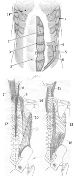
Muscles of the Thorax 1=Rectus Abdominis 2=Abdominal Sheath 3=Linea Alba 4=External Abdominal Oblique 5=Internal Abdominal Oblique 6=Transvers Abdominus 7=Splenius Capitis 8=Splenius Cervicis 9=Iliocostalis Cervicis 10=Iliocostalis Thoracis 11=Iliocostalis Lumborum 12=Splenius Thoracis 13=Longissimus Thoracis 14=Longissimus Cervicis 15=Longissimus Capitis 16= Quadratus Lumborum 17=Serratus Anterior 18- Pectoralis Major
Color each muscle a different color for use as a reference when identifying muscles. |
Procedures:
1. Obtain models, stickers, felt pens
2. Write the names of the trunk muscles that you are responsible for knowing on to the stickers
Rectus Abdominus, External Abdominal Oblique, Longissimus, Latissimus Dorsi, Pectoralis Major, Trapezius, Rhomboideus Major, Rhomboideus Minor, Deltoid
3. Select a “team leader” and a group member to be the “mannequin” for your group.
a. Using the colored images as reference have members of the group take turns labeling the muscles on your group member
b. As the muscle is labeled, ask the “mannequin” to demonstrate the movement that the muscle will produce.
4. Write the names of the key trunk muscles on the stickers
a. Pectoralis Minor, Supraspinatus, Infraspinatus, Teres Minor, Internal Abdominal Oblique, Serratus Anterior, Intercostals
5. Change “team leader” and using the images as reference, label the muscles on the torso models that your group has.
6. Have your instructor check your work and then move to the next activity.
Activity 3: Muscles that move the Upper Extremity:
Muscles of the brachium are broken into key groups the anterior group that is responsible for flexion of the brachium and antebrachium, and the posterior group that is responsible for extension of the brachium and antebrachium.
Anterior Brachium Muscles
|
Muscle |
Origin |
Insertion |
Action |
|
Coracobrachialis |
Coracoid Process |
Anterior mid-shaft of the Humerus |
Flex Humerus @ shoulder |
|
Bicep Brachii |
Long Head: Superior Glenoid Fossa Short Head: Coracoid Process |
Radius and Ulna via Bicipital Aponeurosis; Radial Tuberosity |
Flex, Supinates Forearm @ elbow Flex Humerus @ shoulder |
|
Brachialis |
Mid-shaft of Humerus |
Coronoid Process and Ulnar Tuberosity of the Ulna |
Flex Forearm @ elbow |
Posterior Brachium Muscles
|
Muscle |
Origin |
Insertion |
Action |
|
Triceps Brachii |
Long Head: Inferior Glenoid Fossa Medial Head: Medial Upper 1/3-Shaft of Humerus Lateral Head: Lateral to Radial Groove of Humerus |
Olecranon Process of Ulna |
All Heads: Extend Forearm @ elbow Long Head: Extend Humerus @ Shoulder |
|
Anconeus |
Lateral Epicondyle of Humerus |
Lateral surface of Olecranon Process of Ulna |
Extend forearm @ elbow |
Muscles of the antebrachium are comprised of two compartments that are either anterior (or common flexors) or posterior (or common extensors). Each compartment will be responsible for movement within the antebrachium and movement of the carpals and digits of the hand in antagonistic patterns that are necessary for dexterous movements.
Antebrachium Anterior/Medial (comes from Medical Epicondyle/Medial Condyle, a.k.a. Common Flexor Tendon) Muscles
|
Muscle |
Origin |
Insertion |
Action |
|
Pronator Teres |
Common Flexor Tendon (CFT) |
Lateral Mid-shaft of Radius |
Pronates Forearm |
|
Flexor Carpi Radialis |
(CFT) |
Base of 2nd Metacarpal |
Flex & Radial Deviate Wrist & Hand |
|
Flexor Carpi Ulnaris |
CFT; Medial Olecranon process |
Pisiform; hook of Hamate; Base of 5th Metacarpal |
Flex & Ulnar Deviate Wrist & Hand |
|
Palmaris Longis |
CFT |
Palmar Aponeurosis |
Tense Palmar Aponeurosis |
|
Flexor Digitorum Superficialis |
CFT; Coronoid Process; Proximal Radius |
Margins of Middle Phalanx of medial 4 digits |
Flex middle Phalange of Digits 2-5@ IP joints Assist w/ Flex wrist & hand |
|
Flexor Digitorum Profundis |
Anterior medial surface of Ulna |
Distal phalanx of medial 4 digits |
Flex distal Phalange of Digits 2-5 @ IP joints Assist w/ Flex wrist & hand |
|
Flexor Pollicis Longus |
Anterior surface of Radius & Interosseous membrane |
Distal phalanx of thumb |
Flex distal Phalange of Digit 1 @ IP joints Assist w/ Flex wrist & hand |
|
Pronator Quadratus |
Anterior Distal end of Ulna |
Anterior Distal end of Radius |
Pronates Forearm |
Antebrachium Posterior/Lateral (comes from Lateral Epicondyle/Condyle, a.k.a. Common Extensor Tendon) Muscles
|
Muscle |
Origin |
Insertion |
Action |
|
Brachioradialis |
Lateral Supracondylar Ridge (Humerus) |
Lateral Distal surface of Radius |
Flex forearm @ elbow |
|
Extensor Carpi Radialis Longus |
Lateral Supracondylar ridge (Humerus) |
Base of 2nd Metacarpal |
Flex forearm @ elbow Extend & Radial deviate wrist & hand |
|
Extensor Carpi Radialis Brevis |
Common Extensor Tendon (CET) |
Base of 3rd Metacarpal |
Extend & Radial deviate wrist & hand |
|
Extensor Digitorum |
CET |
Extensor Expansion |
Extend distal Phalange of digits 2-5 @ IP joints |
|
Extensor Digit Minimi |
CET |
Extensor Expansion of Digit 5 |
Extend distal phalange of digit 5 @ IP joints |
|
Extensor Carpi Ulnaris |
CET |
Base of 5th Metacarpal |
Extend and Ulnar deviate wrist & hand |
|
Supinator |
Lateral Epicondyle & Supinator Crest of Ulna |
Posterior & Lateral proximal 1/3 of Radius |
Supinate Forearm |
|
Abductor Pollicis Longus |
Posterior surface of middle Ulna/Radius/ Interosseous Membrane |
Base of 1st Metacarpal |
Abduct digit 1 @ MCP Radial deviate wrist & hand |
|
Extensor Pollicis Brevis |
Middle 1/3 of Posterior Radius/ Interosseous Membrane |
Base of Proximal Phalanx of Thumb |
Extend proximal Phalange of digit 1 |
|
Extensor Pollicis Longus |
Posterior Surface of Middle 1/3 of Ulna/ Interosseous Membrane |
Base of Distal Phalanx of Thumb |
Extend distal Phalange of digit 1 Assist w/ Extend wrist & hand |
|
Extensor Indices |
Posterior Surface of Ulna/ Interosseous Membrane |
Extensor Digitorum tendon to Index finger |
Extend distal Phalange of digit 2 Assist w/ Extend wrist & hand |
|
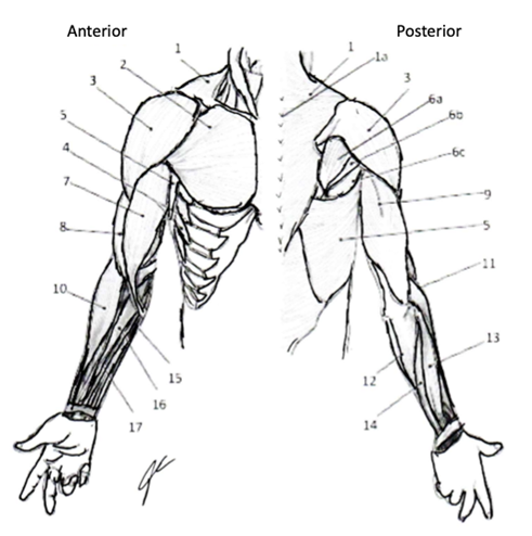
Muscles of Pectoral Girdle and Upper Extremity 1=Trapezius 1a=Ligamentum Nuchae 2=Pectoralis Major 3=Deltoid 4=Serratus Anterior 5=Latissimus Dorsi 6a=Infraspinatus 6b=Teres Minor 6c=Teres Major 7=Biceps Brachii 8=Brachialis 9=Triceps Brachii 10=Brachioradialis 11=Extensor Carpi Radialis Longus 12=Extensor Carpi Ulnaris 13=Extensor Digitorum 14=Extensor Digiti Minimi 15=Flexor Carpi Ulnaris 16=Flexor Digitorum Superficialis 17=Flexor Carpi Radialis
Color each a different color to use as reference for identifying muscles in lab |
Procedures:
1. Obtain models, stickers, felt pens
2. Write the names of the upper extremity muscles that you are responsible for knowing on to the stickers
Biceps Brachii, Triceps Brachii, Brachioradialis, Extensor Carpi Radialis Longus, Extensor Carpi Ulnaris, Extensor Digitorum, Flexor Carpi Radialis, Flexor Carpi Ulnaris, Flexor Digitorum Superficialis
3. Select a “team leader” and a group member to be the “mannequin” for your group.
a. Using the colored images as reference have members of the group take turns labeling the muscles on your group member
b. As the muscle is labeled, ask the “mannequin” to demonstrate the movement that the muscle will produce.
4. Have your instructor check your work and then move to the next activity.
Activity 4: Muscles that move the Lower Extremity:
Hip & Thigh:
The muscles of the hip and pelvis are involved with maintaining trunk postural support (erect posture) and movement of the lower extremity necessary for locomotion. The muscles are identified by the anatomical location (anterior/medial and posterior/lateral) and then by the actions (hip flexors, hip extensors, hip abductors, hip adductors, and rotators) that are necessary for moving the body through space.
Anterior/Medial Hip Muscles:
|
Muscle |
Origin |
Insertion |
Action |
|
Rectus Femoris |
Anterior Inferior Iliac Spine (AIIS) |
Tibial Tuberosity (via Patella Ligament) |
Flex thigh @ Hip Extend leg @ knee |
|
Sartorius |
Anterior Superior Iliac Spine (ASIS) |
Medial Plateau of Tibia |
Flex, Abduct, Externally rotate thigh @ hip Flex leg @ knee |
|
Pectineus |
Pectineal line of Superior Ramus of Pubis |
Pectineal line of Femur |
Adduct thigh @ Hip |
|
Iliopsoas |
Iliacus: Bowl of Pelvis Psoas: Body of 5 Lumbar Vertebrae |
Lesser Trochanter of Femur |
Standing: Flex trunk, Posterior tilt pelvis Sitting: Flex thigh @ hip |
|
Gracilis |
Inferior Ramus of Pubis |
Medial surface of Tibia (infracondylar) |
Adduct and Internally rotate thigh @ Hip Flex leg @ knee |
|
Adductor Longus |
Body of Pubis |
Linea Aspera of Femur |
Adduct thigh @ Hip |
|
Adductor Brevis |
Inferior Ramus of the Pubis |
Femur |
Adduct thigh @ Hip |
|
Adductor Magnus |
Ischiopubic Ramus; Ischial Tuberosity |
Linea Aspera; Adductor Tubercle |
Adduct, Flex and Extend thigh @ hip |
Posterior/Lateral Hip Muscles:
|
Muscle |
Origin |
Insertion |
Action |
|
Gluteus Maximus |
Ilium (inferior gluteal line); Sacrum; Sacrotuberous Ligament |
Gluteal tuberosity; Iliotibial Band (ITB) |
Extend, Externally rotates thigh @ Hip |
|
Piriformis |
Pelvic surface of Sacrum |
Greater Trochanter |
Abducts, Externally rotates thigh @ hip |
|
Gluteus Medius |
Ilium (superior gluteal line) |
Greater Trochanter |
Abducts, Internally rotates thigh @ hip Controls Lateral tilt of pelvis in gait |
|
Obturator Internus |
Obturator Membrane |
Greater Trochanter |
Externally rotates, Abducts thigh @ Hip |
|
Obturator Externus |
Obturator Membrane |
Trochanteric Fossa |
Externally rotates, Abducts thigh @ Hip |
|
Superior Gemellus |
Ischial Spine |
Greater Trochanter |
Externally rotates, Abducts thigh @ Hip |
|
Inferior Gemellus |
Ischial Tuberosity |
Greater Trochanter |
Externally rotates, Abducts thigh @ Hip |
|
Quadratus Femoris |
Ischial Tuberosity |
Intertrochanteric Crest |
Externally rotates thigh @ Hip Stabilize hip during LE movements |
|
Gluteus Minimus |
Ilium (between gluteal lines) |
Greater Trochanter |
Abducts, Internally rotates thigh @ Hip |
|
Tensor Fascia Lata (TFL) |
Iliac Crest |
Iliotibial Band (ITB), Lateral Epicondyle of Femur & Head of Fibula |
Abducts thigh @ hip Controls Lateral tilt of pelvis in gait |
|
Biceps Femoris |
Long Head: Ischial Tuberosity Short Head: Linea Aspera |
Fibular Head; Lateral Plateau of Tibia |
Both: flex leg @ Knee Long Head: extend thigh @ Hip |
|
Semimembranosus |
Ischial Tuberosity |
Medial Condyle of Tibia |
Extend thigh @ Hip Flex leg @ Knee |
|
Semitendinosus |
Ischial Tuberosity |
Medial Condyle of Tibia |
Extend thigh @ Hip Flex leg @ Knee |
Thigh
The muscles of the thigh are involved with propulsion of the body during gait and the postural support, at the tibiofemoral joint, necessary for standing. The muscles are grouped by location as being either anterior or posterior thigh muscles. The anterior group is typically identified as being comprised of the quadriceps (vastus muscles and rectus femoris) and adductors. The poster group is typically referred to as the hamstrings but will also include the adductor magnus and gastrocnemius muscles in some of its actions. Actions of flexion and extension of the lower extremity occur through a coordinated action of the anterior thigh and posterior leg muscles for extension, and posterior muscles of the thigh and anterior muscles of the leg for flexion, of the lower extremity.
Anterior Thigh:
|
Muscle |
Origin |
Insertion |
Action |
|
Vastus Medialis1 |
Medial surface of Femur |
Tibial Tuberosity (via Patella Ligament) |
Extend leg @ knee |
|
Vastus Lateralis1 |
Lateral surface of Femur |
Tibial Tuberosity (via Patella Ligament) |
Extend leg @ knee |
|
Vastus Intermedius1 |
Anterior surface of Femur |
Tibial Tuberosity (via Patella Ligament) |
Extend leg @ knee |
|
Rectus Femoris1 |
AIIS |
Tibial Tuberosity (via Patella Ligament) |
Flex thigh @ hip Extend leg @ knee |
|
Gracilis |
ASIS |
Medial Plateau of Tibia; Medial Condyle of Femur |
Adduct and Internally rotate thigh @ Hip Flex leg @ knee |
|
Sartorius |
ASIS |
Medial Plateau of Tibia; Medial Condyle of Femur |
Flex, Abduct, Externally rotate thigh @ hip Flex leg @ knee |
Posterior Thigh Muscles:
|
Muscle |
Origin |
Insertion |
Action |
|
Bicep Femoris2 |
Long Head: Ischial Tuberosity Short Head: Linea Aspera |
Fibular Head; Lateral Plateau of Tibia |
Both: flex leg @ Knee Long Head: extend thigh @ Hip |
|
Semimembranosus2 |
Ischial Tuberosity |
Medial Condyle of Tibia |
Extend thigh @ Hip Flex leg @ Knee |
|
Semitendinosus2 |
Ischial Tuberosity |
Medial Condyle of Tibia |
Extend thigh @ Hip Flex leg @ Knee |
|
Gastrocnemius |
Medial and Lateral Condyle of Femur |
Calcaneus (via Calcaneal Tendon) |
Assist Flex of leg @ Knee Plantarflex foot @ Ankle |
|
Adductor Magnus |
Ramus of the Pubis |
Linea Aspera; Medial Plateau of Tibia |
Adduct, Flex, Extend thigh @ Hip |
|
Popliteus |
Tibial Plateau |
Lateral Condyle of Femur |
Internally rotate femur @ Knee Assist Flex of leg @ Knee |
1 Indicates Quadriceps group; 2 Indicates Hamstring group
|
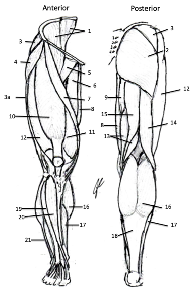
Muscles of the Hip and Lower Extremity 1=Iliopsoas 2=Gluteus Maximus 3=Gluteus Medius 3a=Iliotibial Band 4=Tensor Fascia Lata 5=Pectineus 6=Adductor Longus 7=Gracilis 8=Sartorius 9=Adductor Magnus 10=Rectus Femoris 11=Vastus Medialis 12=Vastus Lateralis 13=Semimembranosus 14=Biceps Femoris 15=Semitendinosus 16=Gastrocnemius 17= Soleus 18=Calcaneal (Achilles') Tendon 19=Extensor Digitorum Longus 20=Tibialis Anterior 21=Peroneus (Fibularis) Longus
Color each with a different color for use as reference for identify muscles. |
Leg:
The muscles of the leg are involved with movement of the tibia and fibula about the femur during postural support and locomotion. The actions are highly involved with propulsion of limb during gait and controlling limb during loading of limb upon landing. Muscles are broken into two groups the anterior group (the dorsiflexors), lateral (peroneal), and posterior (the plantarflexors). The dorsiflexors are involved with clearing the toes and stabilizing the foot during gait. The plantarflexors and are involved with propelling the limb forward in gait and assisting with the production of force necessary to stand and jump.
Anterior Muscles of the Leg:
|
Muscle |
Origin |
Insertion |
Action |
|
Tibialis Anterior |
Superior 2/3 lateral surface of Tibia |
Medial Cuneiform; Base 1st Metatarsal |
Dorsiflex and Invert foot @ Ankle & IT joints |
|
Extensor Digitorum Longus |
Superior 2/3 of Fibula |
Middle and Distal Phalanx of lateral 4 digits |
Extend distal Phalange of digits 2-5 @ IP joints Assist w/ Dorsiflex foot @ ankle |
|
Extensor Hallucis Longus |
Middle 1/3 of Fibula |
Base of distal Phalanx of Hallux |
Extend distal Phalange of digit 1 @ IP joint Assist w/ Dorsiflex foot @ ankle |
|
Peroneus Tertius |
Distal end of Fibula |
Base of 5th Metatarsal |
Dorsiflex and Evert foot @ Ankle & IT joints |
|
Extensor Hallucis & Digitorum Brevis |
Sinus Tarsi (Calcaneus Bone) |
Base of proximal Phalanx of Hallux & Long extensor tendons |
Extend middle Phalange of Digits 1-5 @ IP joint |
Lateral Muscles of the Leg:
|
Muscle |
Origin |
Insertion |
Action |
|
Peroneus Longus |
Superior 2/3 of Fibula |
Base of 1st Metatarsal; Medial Cuneiform |
Plantarflex and Evert foot @ Ankle & IT joints |
|
Peroneus Brevis |
Distal end of Fibula |
Base of 5th Metatarsal |
Plantarflex and Evert foot @ Ankle& IT joints |
|
Peroneus Tertius |
Distal end of Fibula |
Base of 5th Metatarsal |
Dorsiflex and Evert foot @ Ankle & IT joints |
Posterior Muscles of the Leg:
|
Muscle |
Origin |
Insertion |
Action |
|
Gastrocnemius |
Medial & Lateral Condyle of Femur |
Calcaneus (via Calcaneal Tendon) |
Assist Flex of leg @ Knee Plantarflex foot @ Ankle |
|
Soleus |
Posterior Surface of Tibia & Fibula |
Calcaneus (via Calcaneal Tendon) |
Plantarflex foot @ Ankle Control Dorsiflex foot @ Ankle during gait |
|
Plantaris |
Lateral Condyle of Femur |
Calcaneus (via Calcaneal Tendon) |
Plantarflex foot @ Ankle |
|
Flexor Digitorum Longus |
Tibia |
Distal Phalanx of lateral 4 digits |
Flex distal Phalange @ IP Assist w/ Plantarflex foot @ Ankle |
|
Flexor Hallucis Longus |
Fibula |
Distal Phalanx of Hallux |
Flex distal Phalange of digit 1 @ IP |
|
Tibialis Posterior |
Tibia; Fibula; Interosseous Membrane |
Navicular; Medial Cuneiform |
Plantarflex and Invert foot @ Ankle |
|
Popliteus |
Lateral Condyle of Femur |
Tibial Plateau |
Internally rotate femur @ Knee Assist Flex of leg @ Knee |
Procedures:
1. Obtain models, stickers, felt pens
2. Write the names of the lower-extremity muscles that you are responsible for knowing on to the stickers
Rectus Femoris, Vastus Lateralis, Vastus Medius, Tensor Fascia Lata, Biceps Femoris, Semitendinosus, Semimembranosus, Gastrocnemius
3. Select a “team leader” and a group member to be the “mannequin” for your group.
a. Using the colored images as reference have members of the group take turns labeling the muscles on your group member
b. As the muscle is labeled, ask the “mannequin” to demonstrate the movement that the muscle will produce.
4. Write the names of the key hip muscles on the stickers
Gluteus Maximus, Gluteus Medius, Gluteus Minimus, Piriformis
5. Change “team leader”.
a. Using the colored images as reference have members of the group take turns labeling the muscles on your torso model
b. As the muscle is labeled, demonstrate the movement that the muscle will produce.
6. Have your instructor check your work and then move to the next activity.
Activity 5: Muscles of the Face, Head and Neck:
Face and Neck:
The muscles of the face have a variety of roles for the human body. They are involved with the conveying of psychosocial cues (facial expressions) along with respiration, mastication and feeding behaviors. The extrinsic eye muscles are involved with movements of the eye to keep pupils centered on the visual field during tracking of object without head motions or movement of the remainder of the body. The muscles of the neck are layered in a superficial to deep fashion and are involved with movement of the head and cervical regions along with movement of the hyoid involved with vocalization.
Facial Muscles
|
Muscle |
Origin |
Insertion |
Action |
|
Epicranius |
Frontal Bone (Frontal Belly) Superior Nuchal Line (Occipital Belly) |
Epicranial Neurosa |
Raise Eyebrows, Wrinkle forehead
Hold Scalp posteriorly |
|
Nasalis |
Maxilla and alar cartilage of nose |
Dorsum of nose |
Flare nostrils |
|
Buccinator |
Alveolar Processes of Mandible & Maxilla |
Orbicularis Oris |
Press cheek against teeth, draws lips laterally Assist with chewing |
|
Depressor Anguli Oris |
Body of Mandible |
Skin @ inferior corner of mouth |
Opens mouth (pulls lips inferiorly/laterally) |
|
Depressor Labii Inferioris |
Body of Mandible, lateral to midline |
Skin @ inferior lip |
Depresses lower lip |
|
Levator Anguli Oris |
Lateral Maxilla |
Skin @ superior corner of mouth |
Elevates angle of mouth laterally/superiorly |
|
Levator Labii Superioris |
Zygomatic bone & Maxilla |
Skin & muscles of superior lip |
Elevates upper lips |
|
Mentalis |
Central Mandible |
Skin of chin |
Elevate & protrude lower lip |
|
Orbicularis Oris |
Maxilla & Mandible CT of other facial muscles |
Encircles mouth skin & muscles @ angles of mouth |
Closes & Protrude Lips Compress Lips to Teeth, forms mouth for speech |
|
Risorius |
Deep Fascia of Masseter |
Skin @ angle of mouth |
Pulls lips laterally |
|
Zygomaticus Major |
Zygomatic Bone |
Skin @ superolateral edge of mouth |
Moves lips for smiling |
|
Zygomaticus Minor |
Zygomatic Bone |
Skin of superior lip |
Raise lips to expose Maxillar Teeth |
|
Corrugator Supercilli |
Medial end of Superciliary Arch |
Skin superior to Supraorbital Margin & Superciliary Arch |
Draw eyebrow inferiorly Wrinkle forehead |
|
Levator Palpebrae Superioris |
Lesser Wing of Sphenoid |
Superior Tarsal Plate & skin of superior eyelid |
Opens eyes |
|
Orbicularis Oculi |
Medial Wall &/or Margin of Orbit |
Skin surrounding eyelids |
Closes eyes |
|
Platysma |
Fascia of Deltoid & Pectoralis Major & Acromion Process of Scapula |
Skin of Cheek & Mandible |
Depresses lower lip and mandible Tenses cervical fascia |
|
Temporalis |
Superior & Inferior Temporal Lines |
Coronoid Process of Mandible |
Elevates & retracts Mandible for mastication (chewing) |
|
Masseter |
Zygomatic Arch |
Coronoid Process, Lateral Surface & Angle of Mandible |
Elevates mandible Mastication (chewing) |
|
Medial Pterygoid |
Maxilla, Palatine & medial surface of lateral Pterygoid Plate |
Medial surface of Ramus of Mandible |
Elevates & Protracts Mandible Lateral Glide of Mandible |
|
Lateral Pterygoid |
Greater Wing of Sphenoid, lateral surface of lateral Pterygoid Plate |
Condylar Process of Mandible |
Protracts & Depress Mandible (open mouth) |
Extrinsic Muscles of the Eye
|
Muscle |
Origin |
Insertion |
Action |
|
Medial Rectus |
Common Tendinous Rind (CTR) |
Anteromedial Surface of Eye |
Adducts eye |
|
Lateral Rectus |
Common Tendinous Rind (CTR) |
Anterolateral Surface of Eye |
Abducts eye |
|
Inferior Rectus |
Common Tendinous Rind (CTR) |
Anteroinferior Surface of Eye |
Adducts eye, Medially rotate eye, moves eye inferiorly |
|
Superior Rectus |
Common Tendinous Rind (CTR) |
Anterosuperior Surface of Eye |
Adducts eye, Medially rotate eye, moves eye superiorly |
|
Inferior Oblique |
Anterior Orbital surface of Maxilla |
Posteroinferior lateral Surface of Eye |
Abducts eye, moves eye superiorly, Laterally rotates |
|
Superior Oblique |
Sphenoid Bone |
Posterosuperior lateral Surface of Eye |
Abducts eye, moves eye inferiorly, Medially rotates |
Muscles of the Neck
|
Muscle |
Origin |
Insertion |
Action |
|
Scalene |
T.P. of C-vert |
Superior Surface of 1st & 2nd costal |
Bilaterally: elevate ribs 1-2, assist with respiration Unilaterally: Ipsilateral lateral flexion & rotate head & neck |
|
Longissimus Capitis |
C-vertebrae 3-7 |
Mastoid Process |
Bilaterally: extend head & neck Unilaterally: Ipsilateral lateral flex & rotate head & neck |
|
Splenius Capitis |
Ligamentum Nuchae; S.P. of C-vert 7 & T-vert 1-3 |
Occipital & Mastoid Processes |
Bilaterally: extend head & neck Unilaterally: Contralateral rotation of head & neck |
|
Splenius Cervicis |
S.P. of T-vert 3-6 |
T.P. of C-Vert 1-4 |
Bilaterally: extend head & neck Unilaterally: Ipsilateral lateral flex and rotate head & neck |
|
Sternocleidomastoid (SCM) |
Manubrium and sternal end of clavicle |
Mastoid Process |
Bilaterally: flex head & neck, asst. with respiration Unilaterally: Contralateral flexion and rotation of head & neck |
|
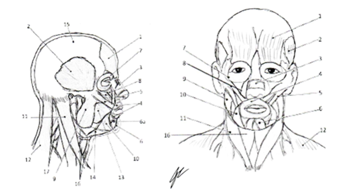
Muscles of the Face and Neck 1=Frontalis 2=Temporalis 3=Nasalis 4=Orbicularis Oris 5=Zygomaticus 6=Mentalis 6a=Depressor Labii Inferioris 7= Orbicularis Oculi 8=Levator Labii Superioris 9=Masseter 10= Triagnularis 11=Sternocleidomastoid 12=Trapezius 13=Buccinator 14=Digastricus 15=Epicranial Aponeurosa 16=Hyoid Muscles 17=Cervical Extensors Color each a different color for use as a reference for identification of muscles. |
Procedures:
1. Obtain models, stickers, felt pens
2. Write the names of the facial and neck muscles that you are responsible for knowing on to the stickers
a. Orbicularis Oris, Temporalis, Masseter, Sternocleidomastoid, Scalene, Trapezius [upper fibers]
3. Select a “team leader” and a group member to be the “mannequin” for your group.
a. Using the colored images as reference have members of the group take turns labeling the muscles on your group member
b. As the muscle is labeled, ask the “mannequin” to demonstrate the movement that the muscle will produce.
4. Have your instructor check your work and then clean-up your lab area.