1.4: Skin and Integument
- Page ID
- 53236
\( \newcommand{\vecs}[1]{\overset { \scriptstyle \rightharpoonup} {\mathbf{#1}} } \)
\( \newcommand{\vecd}[1]{\overset{-\!-\!\rightharpoonup}{\vphantom{a}\smash {#1}}} \)
\( \newcommand{\dsum}{\displaystyle\sum\limits} \)
\( \newcommand{\dint}{\displaystyle\int\limits} \)
\( \newcommand{\dlim}{\displaystyle\lim\limits} \)
\( \newcommand{\id}{\mathrm{id}}\) \( \newcommand{\Span}{\mathrm{span}}\)
( \newcommand{\kernel}{\mathrm{null}\,}\) \( \newcommand{\range}{\mathrm{range}\,}\)
\( \newcommand{\RealPart}{\mathrm{Re}}\) \( \newcommand{\ImaginaryPart}{\mathrm{Im}}\)
\( \newcommand{\Argument}{\mathrm{Arg}}\) \( \newcommand{\norm}[1]{\| #1 \|}\)
\( \newcommand{\inner}[2]{\langle #1, #2 \rangle}\)
\( \newcommand{\Span}{\mathrm{span}}\)
\( \newcommand{\id}{\mathrm{id}}\)
\( \newcommand{\Span}{\mathrm{span}}\)
\( \newcommand{\kernel}{\mathrm{null}\,}\)
\( \newcommand{\range}{\mathrm{range}\,}\)
\( \newcommand{\RealPart}{\mathrm{Re}}\)
\( \newcommand{\ImaginaryPart}{\mathrm{Im}}\)
\( \newcommand{\Argument}{\mathrm{Arg}}\)
\( \newcommand{\norm}[1]{\| #1 \|}\)
\( \newcommand{\inner}[2]{\langle #1, #2 \rangle}\)
\( \newcommand{\Span}{\mathrm{span}}\) \( \newcommand{\AA}{\unicode[.8,0]{x212B}}\)
\( \newcommand{\vectorA}[1]{\vec{#1}} % arrow\)
\( \newcommand{\vectorAt}[1]{\vec{\text{#1}}} % arrow\)
\( \newcommand{\vectorB}[1]{\overset { \scriptstyle \rightharpoonup} {\mathbf{#1}} } \)
\( \newcommand{\vectorC}[1]{\textbf{#1}} \)
\( \newcommand{\vectorD}[1]{\overrightarrow{#1}} \)
\( \newcommand{\vectorDt}[1]{\overrightarrow{\text{#1}}} \)
\( \newcommand{\vectE}[1]{\overset{-\!-\!\rightharpoonup}{\vphantom{a}\smash{\mathbf {#1}}}} \)
\( \newcommand{\vecs}[1]{\overset { \scriptstyle \rightharpoonup} {\mathbf{#1}} } \)
\(\newcommand{\longvect}{\overrightarrow}\)
\( \newcommand{\vecd}[1]{\overset{-\!-\!\rightharpoonup}{\vphantom{a}\smash {#1}}} \)
\(\newcommand{\avec}{\mathbf a}\) \(\newcommand{\bvec}{\mathbf b}\) \(\newcommand{\cvec}{\mathbf c}\) \(\newcommand{\dvec}{\mathbf d}\) \(\newcommand{\dtil}{\widetilde{\mathbf d}}\) \(\newcommand{\evec}{\mathbf e}\) \(\newcommand{\fvec}{\mathbf f}\) \(\newcommand{\nvec}{\mathbf n}\) \(\newcommand{\pvec}{\mathbf p}\) \(\newcommand{\qvec}{\mathbf q}\) \(\newcommand{\svec}{\mathbf s}\) \(\newcommand{\tvec}{\mathbf t}\) \(\newcommand{\uvec}{\mathbf u}\) \(\newcommand{\vvec}{\mathbf v}\) \(\newcommand{\wvec}{\mathbf w}\) \(\newcommand{\xvec}{\mathbf x}\) \(\newcommand{\yvec}{\mathbf y}\) \(\newcommand{\zvec}{\mathbf z}\) \(\newcommand{\rvec}{\mathbf r}\) \(\newcommand{\mvec}{\mathbf m}\) \(\newcommand{\zerovec}{\mathbf 0}\) \(\newcommand{\onevec}{\mathbf 1}\) \(\newcommand{\real}{\mathbb R}\) \(\newcommand{\twovec}[2]{\left[\begin{array}{r}#1 \\ #2 \end{array}\right]}\) \(\newcommand{\ctwovec}[2]{\left[\begin{array}{c}#1 \\ #2 \end{array}\right]}\) \(\newcommand{\threevec}[3]{\left[\begin{array}{r}#1 \\ #2 \\ #3 \end{array}\right]}\) \(\newcommand{\cthreevec}[3]{\left[\begin{array}{c}#1 \\ #2 \\ #3 \end{array}\right]}\) \(\newcommand{\fourvec}[4]{\left[\begin{array}{r}#1 \\ #2 \\ #3 \\ #4 \end{array}\right]}\) \(\newcommand{\cfourvec}[4]{\left[\begin{array}{c}#1 \\ #2 \\ #3 \\ #4 \end{array}\right]}\) \(\newcommand{\fivevec}[5]{\left[\begin{array}{r}#1 \\ #2 \\ #3 \\ #4 \\ #5 \\ \end{array}\right]}\) \(\newcommand{\cfivevec}[5]{\left[\begin{array}{c}#1 \\ #2 \\ #3 \\ #4 \\ #5 \\ \end{array}\right]}\) \(\newcommand{\mattwo}[4]{\left[\begin{array}{rr}#1 \amp #2 \\ #3 \amp #4 \\ \end{array}\right]}\) \(\newcommand{\laspan}[1]{\text{Span}\{#1\}}\) \(\newcommand{\bcal}{\cal B}\) \(\newcommand{\ccal}{\cal C}\) \(\newcommand{\scal}{\cal S}\) \(\newcommand{\wcal}{\cal W}\) \(\newcommand{\ecal}{\cal E}\) \(\newcommand{\coords}[2]{\left\{#1\right\}_{#2}}\) \(\newcommand{\gray}[1]{\color{gray}{#1}}\) \(\newcommand{\lgray}[1]{\color{lightgray}{#1}}\) \(\newcommand{\rank}{\operatorname{rank}}\) \(\newcommand{\row}{\text{Row}}\) \(\newcommand{\col}{\text{Col}}\) \(\renewcommand{\row}{\text{Row}}\) \(\newcommand{\nul}{\text{Nul}}\) \(\newcommand{\var}{\text{Var}}\) \(\newcommand{\corr}{\text{corr}}\) \(\newcommand{\len}[1]{\left|#1\right|}\) \(\newcommand{\bbar}{\overline{\bvec}}\) \(\newcommand{\bhat}{\widehat{\bvec}}\) \(\newcommand{\bperp}{\bvec^\perp}\) \(\newcommand{\xhat}{\widehat{\xvec}}\) \(\newcommand{\vhat}{\widehat{\vvec}}\) \(\newcommand{\uhat}{\widehat{\uvec}}\) \(\newcommand{\what}{\widehat{\wvec}}\) \(\newcommand{\Sighat}{\widehat{\Sigma}}\) \(\newcommand{\lt}{<}\) \(\newcommand{\gt}{>}\) \(\newcommand{\amp}{&}\) \(\definecolor{fillinmathshade}{gray}{0.9}\)Learning Objectives:
At the end of the lab, you will be able to…
- Correctly identify the type of cells comprising the integument by layer
- Differentiate the epidermis by strata
- Explain the role of the skin as it relates to the regulation of homeostasis of the body
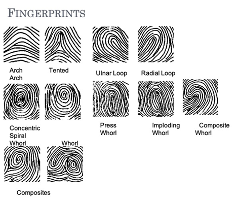
- Differentiate the types of fingerprint patterns and how to read the fingerprint
Pre-Lab Exercise:
After reading through the lab activities prior to lab, complete the following before you start your lab.
1. The layers of the epidermis from deepest to most superficial are .
2. True/False: Subcutaneous tissues are made of dense irregular connective tissues that tightly binds the skin to the underlying tissues of the body. .
3. When examining fingerprints, the print that loops toward the thumb would be referenced as a .
4. Color the images for use as a reference for identifying the models.
Materials
- Colored Pencils
- Model of Skin
- Stickers
- Felt-tip pen
- Inkpad
- Flashcard
- Microscope
- Slide of Skin
Activity 1
Layers of the Skin
The skin is comprised of three layers of tissues (epidermis, dermis, and subcutaneous tissue {hypodermis}). Each layer can function independently or in conjunction with each other to fulfill the generalized role of protection of the body. These layers of cells and tissues will combine with the accessory organs integrated into the integument to perform its function to help with maintaining homeostasis for the body.
The outer most of the layers (epidermis) is made of stratified squamal epithelial tissue that is layered into 5 distinct stratas. The stratification of specialized cells (keratinocytes) establishes an impermeable membrane around the entirety of the body. Deep to the epidermis is the dermis that is primarily made of dense irregular connective tissue. Within the dermis there will be all of the accessory organs that allow for thermoregulation (hair, sudoriferous glands, and the dermal arterioles and capillaries) and an oil secretion (sebaceous glands) that protects the body for dehydrating, along with the gathering sensory information from the external environment. The deepest layer is the hypodermis, where loose connective tissue (areolar connective tissue) will bind the skin to the underlying tissues and adipose tissue that will insulate the body and protect underlying from physical injury
Methods:
1. Obtain model of the skin, felt pen and stickers from your instructor
2. On the stickers, write the names of the layers of the skin and proceed to label the structural layers of the skin
Epidermis, Dermis, Hypodermis (Subcutaneous Adipose Tissue), Dermal Arteries and Veins, Hair, Hair Follicle, Arector Pili, Dermal Papilla
3. After labeling the layers of the skin, write the names of the structures of the skin responsible for protecting the body and obtaining sensory information from the external environment.
Ruffini Endings, Pacinian Corpuscles, Root Hair Plexus, Merkle’s Discs, Meissner’s Corpuscles
4. Take turns within your group labeling the structures of the skin responsible for receiving sensory information
5. Discuss in your group: Why are sensory receptors located at various depths within the dermis? Share your thoughts with your instructor.
6. Move the model to the side of your work area and move to activity 2
|
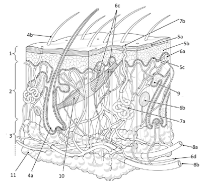
Layers and Structures in the skin: 1=Epidermis 2=Dermis 3=Hypodermis (Adipose/Subcutaneous) 4a= Hair Follicle 4b=Hair Proper 5a=Stratified Keratinocytes 5b=Stratum Basale 5c=Dermal Papilla/Ridges 6a= Meissner's Corpuscles 6b= Pacinian Corpuscles 6c=Merkle’s Discs, Thermoreceptors/Nociceptors 6d=Afferent Nerve 7a=Coiled Gland of Sudoriferous (Sweat) Gland 7b=Duct of Sudoriferous (Sweat) Gland 8a=Dermal Arterioles 8b=Dermal Venuoles 9=Sebaceous Gland 10=Erector Pili Muscle 11=Adipose Tissue Use you colored pencils to color each layer of the skin and the various structures using a different color |
|
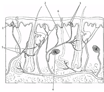
Layers and Structures in the skin: 1=Nociceptors 2=Meissner's Corpuscles 3=Merkle’s Discs 4=Pacinian Corpuscles 5=Root Hair Plexus 6=Ruffini Endings Use you colored pencils to color each of the various structures using a different color
|
Activity 2
The epidermis is comprised on Stratified Squamal Epithelium. The stratification is presented so that there are 5-distinct layers (strata) within the epidermis (Stratum Basale, Stratum Spinosum, Stratum Granulosum, Stratum Lucidium, and Stratum Corneum). The layering and density of cells within each layer can vary based on the area of the body (such as the plantar surface of the foot and palmar surface of the hand having a larger layer of spinosum).
1. Obtain microscope and slide of the skin
2. Label the image of the epidermis to show the appropriate strata
|
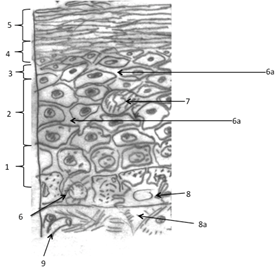
Stratum of the Epidermis: 1=Stratum Basale 2=Stratum Spinosum 3=Stratum Granulosum 4=Stratum Lucidum 5=Stratum Corneum 6=Melanocyte 6a=Pseudopodia of Melanocyte 7=Langerhans Cell 8=Merkle’s Discs 8a=Neuron of Merkle’s Discs 9=Capillary Use you colored pencils to color each layer of cells in a different color |
3. Using your microscope to exam the skin slide.
a. Focus to the superficial edge of the skin and magnify to highest power. Once at highest power (400x total magnification), draw and label the slide in the area provided.

Activity 3
Fingerprints and Footprints are found on palmar and plantar surfaces of the hand and feet at the distal ends of digits. These are specialized areas that have a high density of reticular connective tissues in the dermal papilla that generates ridging within the epidermis. These ridges function to increase the surface area to allow for greater contact between the skin and the surface that they are in contact with. The ridging develops into prototypical forms that are quasi-unique among individuals.
1. Obtain materials from your instructor
2. Select the partner to be printed first.
3. The person to be printed needs to clean the end of each finger with alcohol swabs
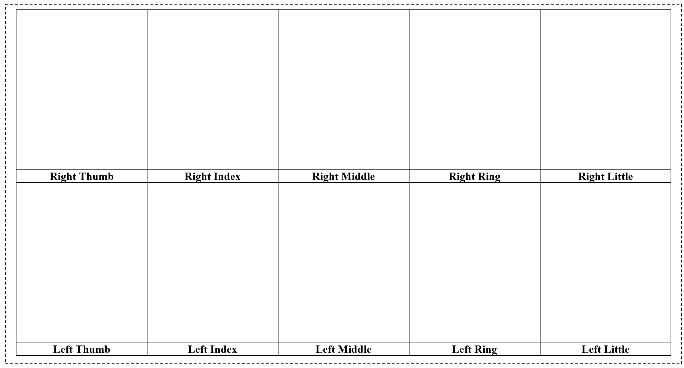
4. While the first person being printed is cleaning their digit, the partner that is going to do the fingerprinting needs to ready the flash card by labeling areas for each finger and open the ink pad
5. The person to do the fingerprinting needs to stand to the side of your partner and grasp their hand, folding all fingers but the thumb into a fist
a. Controlling the distal end of the Right thumb, place the thumb onto the ink pad, and then rolling in one smooth ulna-to-radius across the flash card where “Right Thumb” is indicated.
b. Fold the thumb into the fist and have the person being printed extend their index finger.
c. Repeat the procedure to print the index finger
d. Repeat for the remaining fingers and then switch hands
6. The person that just got printed should now wash the ink off.
7. Repeat the procedures for the second partner.
8. Compare your prints to the examples shown below.
Flash Card Design for Fingerprinting