
1.3: ORGANIZATION of the BODY CELLS and TISSUES
- Page ID
- 53231
\( \newcommand{\vecs}[1]{\overset { \scriptstyle \rightharpoonup} {\mathbf{#1}} } \)
\( \newcommand{\vecd}[1]{\overset{-\!-\!\rightharpoonup}{\vphantom{a}\smash {#1}}} \)
\( \newcommand{\dsum}{\displaystyle\sum\limits} \)
\( \newcommand{\dint}{\displaystyle\int\limits} \)
\( \newcommand{\dlim}{\displaystyle\lim\limits} \)
\( \newcommand{\id}{\mathrm{id}}\) \( \newcommand{\Span}{\mathrm{span}}\)
( \newcommand{\kernel}{\mathrm{null}\,}\) \( \newcommand{\range}{\mathrm{range}\,}\)
\( \newcommand{\RealPart}{\mathrm{Re}}\) \( \newcommand{\ImaginaryPart}{\mathrm{Im}}\)
\( \newcommand{\Argument}{\mathrm{Arg}}\) \( \newcommand{\norm}[1]{\| #1 \|}\)
\( \newcommand{\inner}[2]{\langle #1, #2 \rangle}\)
\( \newcommand{\Span}{\mathrm{span}}\)
\( \newcommand{\id}{\mathrm{id}}\)
\( \newcommand{\Span}{\mathrm{span}}\)
\( \newcommand{\kernel}{\mathrm{null}\,}\)
\( \newcommand{\range}{\mathrm{range}\,}\)
\( \newcommand{\RealPart}{\mathrm{Re}}\)
\( \newcommand{\ImaginaryPart}{\mathrm{Im}}\)
\( \newcommand{\Argument}{\mathrm{Arg}}\)
\( \newcommand{\norm}[1]{\| #1 \|}\)
\( \newcommand{\inner}[2]{\langle #1, #2 \rangle}\)
\( \newcommand{\Span}{\mathrm{span}}\) \( \newcommand{\AA}{\unicode[.8,0]{x212B}}\)
\( \newcommand{\vectorA}[1]{\vec{#1}} % arrow\)
\( \newcommand{\vectorAt}[1]{\vec{\text{#1}}} % arrow\)
\( \newcommand{\vectorB}[1]{\overset { \scriptstyle \rightharpoonup} {\mathbf{#1}} } \)
\( \newcommand{\vectorC}[1]{\textbf{#1}} \)
\( \newcommand{\vectorD}[1]{\overrightarrow{#1}} \)
\( \newcommand{\vectorDt}[1]{\overrightarrow{\text{#1}}} \)
\( \newcommand{\vectE}[1]{\overset{-\!-\!\rightharpoonup}{\vphantom{a}\smash{\mathbf {#1}}}} \)
\( \newcommand{\vecs}[1]{\overset { \scriptstyle \rightharpoonup} {\mathbf{#1}} } \)
\(\newcommand{\longvect}{\overrightarrow}\)
\( \newcommand{\vecd}[1]{\overset{-\!-\!\rightharpoonup}{\vphantom{a}\smash {#1}}} \)
\(\newcommand{\avec}{\mathbf a}\) \(\newcommand{\bvec}{\mathbf b}\) \(\newcommand{\cvec}{\mathbf c}\) \(\newcommand{\dvec}{\mathbf d}\) \(\newcommand{\dtil}{\widetilde{\mathbf d}}\) \(\newcommand{\evec}{\mathbf e}\) \(\newcommand{\fvec}{\mathbf f}\) \(\newcommand{\nvec}{\mathbf n}\) \(\newcommand{\pvec}{\mathbf p}\) \(\newcommand{\qvec}{\mathbf q}\) \(\newcommand{\svec}{\mathbf s}\) \(\newcommand{\tvec}{\mathbf t}\) \(\newcommand{\uvec}{\mathbf u}\) \(\newcommand{\vvec}{\mathbf v}\) \(\newcommand{\wvec}{\mathbf w}\) \(\newcommand{\xvec}{\mathbf x}\) \(\newcommand{\yvec}{\mathbf y}\) \(\newcommand{\zvec}{\mathbf z}\) \(\newcommand{\rvec}{\mathbf r}\) \(\newcommand{\mvec}{\mathbf m}\) \(\newcommand{\zerovec}{\mathbf 0}\) \(\newcommand{\onevec}{\mathbf 1}\) \(\newcommand{\real}{\mathbb R}\) \(\newcommand{\twovec}[2]{\left[\begin{array}{r}#1 \\ #2 \end{array}\right]}\) \(\newcommand{\ctwovec}[2]{\left[\begin{array}{c}#1 \\ #2 \end{array}\right]}\) \(\newcommand{\threevec}[3]{\left[\begin{array}{r}#1 \\ #2 \\ #3 \end{array}\right]}\) \(\newcommand{\cthreevec}[3]{\left[\begin{array}{c}#1 \\ #2 \\ #3 \end{array}\right]}\) \(\newcommand{\fourvec}[4]{\left[\begin{array}{r}#1 \\ #2 \\ #3 \\ #4 \end{array}\right]}\) \(\newcommand{\cfourvec}[4]{\left[\begin{array}{c}#1 \\ #2 \\ #3 \\ #4 \end{array}\right]}\) \(\newcommand{\fivevec}[5]{\left[\begin{array}{r}#1 \\ #2 \\ #3 \\ #4 \\ #5 \\ \end{array}\right]}\) \(\newcommand{\cfivevec}[5]{\left[\begin{array}{c}#1 \\ #2 \\ #3 \\ #4 \\ #5 \\ \end{array}\right]}\) \(\newcommand{\mattwo}[4]{\left[\begin{array}{rr}#1 \amp #2 \\ #3 \amp #4 \\ \end{array}\right]}\) \(\newcommand{\laspan}[1]{\text{Span}\{#1\}}\) \(\newcommand{\bcal}{\cal B}\) \(\newcommand{\ccal}{\cal C}\) \(\newcommand{\scal}{\cal S}\) \(\newcommand{\wcal}{\cal W}\) \(\newcommand{\ecal}{\cal E}\) \(\newcommand{\coords}[2]{\left\{#1\right\}_{#2}}\) \(\newcommand{\gray}[1]{\color{gray}{#1}}\) \(\newcommand{\lgray}[1]{\color{lightgray}{#1}}\) \(\newcommand{\rank}{\operatorname{rank}}\) \(\newcommand{\row}{\text{Row}}\) \(\newcommand{\col}{\text{Col}}\) \(\renewcommand{\row}{\text{Row}}\) \(\newcommand{\nul}{\text{Nul}}\) \(\newcommand{\var}{\text{Var}}\) \(\newcommand{\corr}{\text{corr}}\) \(\newcommand{\len}[1]{\left|#1\right|}\) \(\newcommand{\bbar}{\overline{\bvec}}\) \(\newcommand{\bhat}{\widehat{\bvec}}\) \(\newcommand{\bperp}{\bvec^\perp}\) \(\newcommand{\xhat}{\widehat{\xvec}}\) \(\newcommand{\vhat}{\widehat{\vvec}}\) \(\newcommand{\uhat}{\widehat{\uvec}}\) \(\newcommand{\what}{\widehat{\wvec}}\) \(\newcommand{\Sighat}{\widehat{\Sigma}}\) \(\newcommand{\lt}{<}\) \(\newcommand{\gt}{>}\) \(\newcommand{\amp}{&}\) \(\definecolor{fillinmathshade}{gray}{0.9}\)Learning Objectives
Students will be able to…
1. Differentiate between the various organs and tissues of the body
2. Correctly troubleshoot issues with microscope
3. Be able to focus and change magnifications of view on the microscope
4. Differentiate between the cytology of the various types of tissues
5. Identify and explain the functions of the various organelles of the cells of the body
Pre-Lab Exercise:
After reading through the lab activities prior to lab, complete the following before you start your lab.
1. There are types of tissues.
2. True/False: Tissues are the building blocks of the human body. .
3. When using a microscope, you only use coarse adjustment at a magnification of .
4. Color the images for use as a reference for identifying the models and dissected specimens
Materials:
• Stickers
• Felt Tip Pen
• Cell Model
• Microscopes
• Slides: Lung and Bronchiole, Kidney, Skin, Urinary Bladder, Ileum, Fibrocartilage, Elastic Cartilage, Bone, Ligament, Areolar Connective Tissue, Reticular Connective Tissue, Skeletal Muscle Cardiac Muscle, Nerve Smear
Organization of the Body
We must think of the body as being built in layers of ascending complexity beginning with the atom and ending with entire organism. Each level of complexity is developed through an increase in the various components that are interacting within that level. This begins with the atom and the subatomic components (electrons, neutrons, protons) followed by the interaction of atoms with other atoms forming molecules that will interact with other molecules forming the macromolecules. We tend to think about in these macromolecules as being carbohydrates, lipids, and proteins, but also include molecules like adenosine triphosphate (ATP) and nucleic acids. From these macromolecules we have interactions that eventually from the organelles and cells that will interact with each other leading to the formation of the tissues. Tissues are conglomerations of cells that share a similar function for the body that will work and interact with each other. Developing into regulated (or control) organs, a conglomeration of tissues with a shared function within the homeostasis of the body. These organs eventually coordinate their independent functions into the systems that comprise the body that we typically think about when discussing human anatomy and physiology.
There are four distinct types of tissues. The first type of tissue that we should be aware of is the epithelial tissue. The epithelial cells are found throughout the body and are typically found as a tissue that lines the body. In their function as a barrier tissue, these cells will be attached to these tissues by a layer of connective tissue layer described as the basal membrane. The next type of tissue is the connective tissue. Connective tissue is comprised of cells that produce different types of protein fibers that are exuded from cells that develop a matrix of protein and fluids that connect different tissues of the body into a network of tissues that provides functional units of the organ systems of the body. There is a vast array of connective tissue structures and functions throughout the body. Each type of connective tissue has different cells that provide the materials for the matrix and the matrix of the connective tissue will differ to match the desired function of the connective tissue type. The third type of tissue is muscle tissue. Muscle tissue is a special type of cell that unlike the cell types already described are deemed to be an “excitable” tissue. Meaning that they function by generating electrical currents within the tissue to perform the function of the tissue. In this way, all muscle tissue, regardless of the distinct type will exhibit the following qualities: irritability, extensibility, elasticity, and contractility. Each one of these qualities provides the foundation for the difference in physiology of the muscle tissue. The final type of tissue is nervous tissue. Nerve tissue is made of specialized excitable cells termed neurons and support cells termed glia. Neurons are specialized columnar epithelial cells that function to transmit electrical signals between cells and tissues. While glial cells are hoist of various types of cells that support the function and “health” of the neurons.
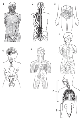
When we examine the body, it is generally done based on a systems approach. These systems include the musculoskeletal (skeletal muscle and skeletal) systems, the nervous system, the cardiorespiratory (cardiovascular and respiratory) systems, the immune system, the excretory system, the digestive system, the neuroendocrine system, the reproductive system and the integument system. While each system will have an independent function, they function in a coordinate manner so as to ensure that the body is able to remain in a stable state and respond effectively to any stimulus that might disrupt this stability.
|
Systems of the Body 1= Musculoskeletal 2= Nervous 3= Digestive 4= Neuroendocrine 5= Cardiovascular 6= Lymphatic/Immune 7=Respiratory 8= Urinary Use you colored pencils to color each plane in a different color |
|
Organ System |
Function |
Organ System |
Function |
|
Integument |
Functions to serve as barrier between the body and the external environment. Provides as a means for sensory information as to the conditions of the external environment and a means to thermoregulate to maintain a homeostatic core body temperature |
Respiratory |
Functions to exchange volatile chemicals (gasses) between the body and the external environment. Provides a means to regulate the chemistry of the plasma via gas exchange at the alveoli. |
|
Musculoskeletal |
Functions to serve as the mechanical lever systems for movements of the body and rigid structures to provide stable morphology of the body. Involved with metabolic and immune regulation, ion balance, and thermogenesis to maintain a homeostatic internal environment |
Immune |
Functions to serve as a means to protect the body from foreign or toxic materials. Provides responses to eliminate infectious agents, inflame areas following injury to “splint” and ensure limited damage following the injury and the repair of the tissues |
|
Nervous: |
Functions to serve as a means to transmit information from various tissues of the body to other parts of the body via specific cells (neurons). Provides the ability to instantaneously regulate homoeostasis via reflex loops and through specific central structures establish memories to provide anticipation for reflex loops and coordinate functions between tissues. |
Gastrointestinal |
Functions as an open tube through the body to ingest and digest materials necessary to tissue repair and energetic balance. Provides a sequestered area to mechanically and chemically digest and then absorb nutrients without over expression of immune response to foreign materials |
|
Neuroendocrine |
Functions to produce and release chemical signals to regulate the metabolic functions of tissues. Provides a means to signal tissues of the metabolic stress being encountered by different regions of the body and then regulate, and control, the metabolism of cells and tissues to ensure that homeostasis is maintained. |
Excretory |
Functions to eliminate metabolic waste products and toxins from the body. Provides a means to regulate the chemistry of the body to ensure homeostatic balance of ions, water, and chemicals within the blood and tissues of the body. |
|
Cardiovascular |
Functions to serve as transportation medium of chemicals and specific cells throughout the body. Heart functions as hydraulic pump moving blood through the body via tubule structures (vessels). Provides as a means for conveying chemical information as to the conditions of the internal environment, transportation of metabolites, and a means to thermoregulate to maintain a homeostatic core body temperature through heat exchanges. |
Reproductive |
Functions to form gametes and regulate maturation of the body to allow for sexual reproduction and for females the system is involved with pregnancy and care of the infant. Highly integrated within the neuroendocrine system. Provides a means to allow for offspring to be formed and cared for during infancy and early childhood. |
Activity 1: Identifying Cellular Organelles
Cell and Organelles
Cells are the building blocks of the body. Each cell itself is made of various components that perform functions that all for the cell to maintain its own homeostasis.
|
Organelle |
Associated Function |
|
Cell Membrane |
Phospholipid bilayer that surrounds outer surface of the cell forming “protective” envelope |
|
Integral Membrane Proteins |
Proteins embedded into cell membrane that either attach internally to membrane anchored vesicle/vacuole, or externally to function as “marker” of cell or adhesion point to join with other cells |
|
Transmembrane Protein |
Proteins that span the membrane that allow for materials to move between the external and internal cellular environments |
|
Nucleus |
Organelle that is responsible for “housing” genetic materials (DNA, mRNA, tRNA, rRNA) of the cell |
|
Nucleolus |
Subregion within the nucleus that contains ribosome proteins prior to movement to cytosol or endoplasmic reticulum |
|
Endoplasmic Reticulum |
Phospholipid membrane that anchors to the outer membrane of the nucleus and runs throughout the cell, identified as being “smooth” or “rough” depending on presence of ribosomes. Responsible for production of lipids and proteins (from embedded ribosomes). |
|
Ribosome |
Responsible for translation and protein synthesis, comprised of 2 components (protein component and nucleic acid component, rRNA) |
|
Lysosomes |
Vesicle responsible for sequestering digestive enzymes for use by the cell on materials following pino-, or phago-cytosis |
|
Peroxisomes |
Vesicle housing peroxides and digestive enzymes responsible for cellular digestion of materials following pinocytosis, or phagocytosis. Involved with fatty-acid oxidation |
|
Vacuoles |
Vesicle responsible for “storage” of cellular materials and compounds |
|
Mitochondria |
Organelle responsible for aerobic phosphorylation of ADPàATP |
|
Golgi Apparatus |
Stacked membrane organelle connected with endoplasmic reticulum that “coats” materials for secretion, or release, from the cell |
|
Cilia |
Membrane extensions comprised of cytoskeletal protein core and cell membrane “envelope” responsible for motility, and locomotion, of the cell (or in colony movement of materials) |
|
Microvilli |
Membrane extensions comprised of rigid cytoskeleton that increases the total surface of the cell membrane |
|
Flagella |
Elongated membrane extension comprised of cytoskeletal protein core and cell membrane “envelope” responsible for locomotion of the cell |
|
Cytoskeleton-Proteins |
Functional proteins responsible for maintenance of cell shape in response to external/internal environmental cues |
|
Centrioles |
Protein based organelle only functional during mitosis that is responsible for segregation of chromosomes between daughter cells |
|
Cytosol |
Internal environment of cell comprised of electrolytes, proteins and non-organelle materials |
|
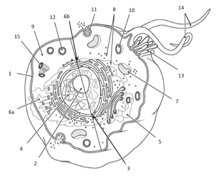
Organelles and Structures of the Cell 1= Cell Membrane 2= Nucleolus 3= Nuclear Envelope/Nuclear Membrane 4= Nucleus 5= Golgi Apparatus/Golgi Bodies 6a= Smooth Endoplasmic Reticulum 6b= Rough Endoplasmic Reticulum 7= Mitochondria 8= Ribosomes 9= Cytoplasm 10=Lysosome 11= Clathrin Pit/Endocytosis or Exocytosis 12= Peroxisome 13= Cilia 14= Flagella 15= Centriole Use you colored pencils to color each part of the cell in a different color for reference when identifying.
|
1. Obtain cell model, stickers and felt pens from your instructor
2. Write the organelle names on the stickers
3. Taking turns within your lab group,
a. Each member will label one organelle of the cell on the model.
b. Check that they have labeled the organelle correctly and if correct move the next member. If it is not correct, a member in your group should correct the label.
c. Proceed to the next member in the group and continue until all labels have been used.
d. Once you have labeled the entire model, have your instructor check your work
4. Clean-up your labels from the cell model
Activity 2: Tissues and Histology (study of the tissues by using a microscope)
Using the Microscope
The microscope is a key tool for examining the cells and tissues of the body. In order to successfully examine tissues, you must be able to use the microscope correctly. Before we start off with studying tissues and cells it is important to able to use the microscope correctly is having the knowledge of magnification of the tissue through the microscope and what to do if something goes wrong.
Principles of Magnification
Microscopes have 3 magnifications: Scanning, Low and High. Each objective will have written the magnification. In addition to this, the ocular lens (eyepiece) has a magnification. The total magnification is the ocular x objective
Total Magnification
|
Objective Lens |
Ocular Lens |
Total Magnification |
|
4x |
10x |
40x |
|
10x |
10x |
100x |
|
40x |
10x |
400x |
Focusing Specimens
1. Always start with the scanning objective. Odds are, you will be able to see something on this setting. Use the Coarse Knob to focus, image may be small at this magnification, but you won't be able to find it on the higher powers without this first step. Do not use stage clips, try moving the slide around until you find something.
2. Once you've focused on Scanning, switch to Low Power. Use the Coarse Knob to refocus. Again, if you haven't focused on this level, you will not be able to move to the next level.
3. Now switch to High Power. (If you have a thick slide, or a slide without a cover, do NOT use the high- power objective). At this point, ONLY use the Fine Adjustment Knob to focus specimens.
4. If the specimen is too light or too dark, try adjusting the diaphragm.
5. If you see a line in your viewing field, try twisting the eyepiece, the line should move, that is the pointer, and is useful for pointing out things to your lab partner or teacher. Drawing Specimens
1. Use pencil - you can erase and shade areas
2. All drawings should include clear and proper labels (and be large enough to view details). Drawings should be labeled with the specimen name and magnification.
3. Labels should be written on the outside of the circle. The circle indicates the viewing field as seen through the eyepiece, specimens should be drawn to scale (if your specimen takes up the whole viewing field, make sure your drawing reflects that).
Troubleshooting
Occasionally you may have trouble with working your microscope. Here are some common problems and solutions.
1. If the Image is too dark!
Make sure your light is on
Adjust the diaphragm
2. There's a spot in my viewing field, even when I move the slide the spot stay in the same place!
Your lens is dirty. Use lens paper, and only lens paper to carefully clean the objective and ocular lens. The ocular lens can be removed to clean the inside.
3. I can't see anything under high power!
Remember the steps, if you can't focus under scanning and then low power, you won't be able to focus anything under high power.
4. Only half of my viewing field is lit, it looks like there's a half-moon in there!
You probably don't have your objective fully clicked into place.
Part A: Epithelial Tissue
The description of epithelium is based on the shape of the cell or based on the number of layers of cells contained in the tissue. The identification that is based on layers of epithelium is given by the number of layers of cells between the basal membrane of the tissue and the outer most cells. It is generally divided into either being a single layer of cells or appearing to have more than a single layer of cells. In which we discuss epithelial cells as: simple (only 1 cell layer of epithelium), stratified (more than 1 cell layer of epithelium), or pseudostratified (appears to have multiple layers of cells, but all cells make contact with the basal membrane). The layer identification is then combined with the general shape of the epithelial cell to give the complete identification. The shapes are described as: squamous (flat elongated cells associated with lining of tissues and organs, primary cell of the epidermis), cuboidal (cube shaped cells that are associated with absorbing materials but may also be involved with secretory functions of glands), or columnar (column shaped cells that are associated with secreting and absorbing materials from the extracellular spaces).
Additionally, epithelial cells can be discussed based on cellular structures. Ciliated epithelium is typically columnar (or cuboidal) that use the cilia and microvilli to establish a “brush boarder” within the tissue. These brush boarders are used to either increase total surface area for interaction between substances with the epithelial cells or assist with the movement of materials along surface of tissue. There are also some specialized columnar cells. One such cell are the secretory cells that are identified as “goblet” and the prototypical cell used for most tissues involved with secretions within lumens of the body. Within the types of epithelium are specialized secretory cells (cells that secrete materials into the extracellular fluids. These are “glandular tissues” are described using 3 general classifications. There are merocrine glands, which release only secretions from the cell into ducts or onto tissues. There are apocrine glands that release small parts of the cell that are “squeezed off” from the cell into the ducts for secretion. Lastly there are holocrine glands that release entire cells into the ducts for secretion. The other type of secretory glandular epithelial is the serosa epithelial. These epithelial cells secrete a mucous coating that allows for a decrease the friction between two interacting surfaces of tissues within the body.
Procedures:
1. Examine the slides of epithelial tissues under scanning and high magnification.
2. For each power, one person in your group will draw what is seen in the ocular of the microscope and the other person will draw the image at the other magnification.
3. Simple Squamous (Lung and Bronchiole Slide)


4. Simple Columnar (Ileum Slide)

5. Simple Cuboid (Kidney Slide)


6. Pseudostratified Ciliated Columnar (Trachea Slide)


7. Transitional Epithelial (Urinary Bladder Slide)


8. Stratified Squamous Epithelial (Keratinized) (Skin)


9. Compare your slides and diagrams within your group.
Part B: Connective Tissue
We generally use three distinct classes of tissues to describe the array of cells and tissues that comprise the connective tissues. There is true connective tissue, which form a protein matrix that connects tissues to each other that are classified as being either dense or loose. There is supportive: forms solid matrixes that form the rigid or semi-rigid structures of the body. Then there is the liquid: protein matrix is dissolved within an aqueous solution, used for transportation connection between tissues and includes tissues such as blood and lymph.
Procedures:
1. Examine the slides of connective tissues under scanning and high magnification.
2. For each power, one person in your group will draw what is seen in the ocular of the microscope and the other person will draw the image at the other magnification.
3. Dense Regular Connective Tissue (Ligament Slide)


4. Dense Irregular Connective Tissue (Skin Slide)

5. Loose Areolar Connective Tissue
6. Reticular Connective Tissue
7. Elastic Cartilage
8. Hyaline Cartilage (Trachea Slide)
9. Fibrocartilage
10. Bone
11. Compare your slides and diagrams within your group.
Part C: Muscle Tissue
There are three (3) different types of muscle cells that recognized in the human body. This recognition is noted by the presence of (striated) or lack of organized intracellular structures (smooth) referred to transverse tubules (T-tubules). Within this typing of cells striated muscle tissues are additionally given names based on where in the body they are located (skeletal or cardiac) in the body, figure 15. Histologically striated (skeletal) muscle is a poly-nucleated (having more than 1 nucleus) cell with elongated striated muscle that attach to the skeletal structures via tendons and with nervous system stimulation allow for movement to occur. While smooth muscle is amorphic (no regular shape) muscle tissue with no visible striations that form a ring of muscle tissue surrounding lumens and organs of the body. Within the smooth muscle, the contractile proteins are arranged in the spiral to the long axis of the cell as opposed to cylinders that parallel the long axis seen in the skeletal and cardiac muscle. Lastly the cardiac muscle is a “Y-shaped” striated muscle that forms a network of overlapping muscle tissue connected with intercalated disks to all for coordination of muscle contraction.
Procedures:
1. Examine the slides of muscle tissues under scanning and high magnification.
2. For each power, one person in your group will draw what is seen in the ocular of the microscope and the other person will draw the image at the other magnification.
3. Skeletal Muscle


4. Cardiac Muscle


5. Compare your slides and diagrams within your group.
Part D: Nervous
Neurons are classified by shape and by function relative to that shape and number of axon projections. With the identification being unipolar (one axonal projection), bipolar (two axonal projections), or multipolar (multiple axonal projections). The key glial cells are the myelinating cells (Schwann and oligodendrocyte) that support and insult the axon of the neuron, and the astrocytes, microglia and oligoglia that support the health of the neuron via metabolic activities or functioning as immune-like cells.
Procedures:
1. Examine the slides of nervous tissues under scanning and high magnification.
2. For each power, one person in your group will draw what is seen in the ocular of the microscope and the other person will draw the image at the other magnification.
3. Nerve Smear


4. Compare your slides and diagrams within your group.