
26.2: Exercise
- Page ID
- 105943

\( \newcommand{\vecs}[1]{\overset { \scriptstyle \rightharpoonup} {\mathbf{#1}} } \)
\( \newcommand{\vecd}[1]{\overset{-\!-\!\rightharpoonup}{\vphantom{a}\smash {#1}}} \)
\( \newcommand{\dsum}{\displaystyle\sum\limits} \)
\( \newcommand{\dint}{\displaystyle\int\limits} \)
\( \newcommand{\dlim}{\displaystyle\lim\limits} \)
\( \newcommand{\id}{\mathrm{id}}\) \( \newcommand{\Span}{\mathrm{span}}\)
( \newcommand{\kernel}{\mathrm{null}\,}\) \( \newcommand{\range}{\mathrm{range}\,}\)
\( \newcommand{\RealPart}{\mathrm{Re}}\) \( \newcommand{\ImaginaryPart}{\mathrm{Im}}\)
\( \newcommand{\Argument}{\mathrm{Arg}}\) \( \newcommand{\norm}[1]{\| #1 \|}\)
\( \newcommand{\inner}[2]{\langle #1, #2 \rangle}\)
\( \newcommand{\Span}{\mathrm{span}}\)
\( \newcommand{\id}{\mathrm{id}}\)
\( \newcommand{\Span}{\mathrm{span}}\)
\( \newcommand{\kernel}{\mathrm{null}\,}\)
\( \newcommand{\range}{\mathrm{range}\,}\)
\( \newcommand{\RealPart}{\mathrm{Re}}\)
\( \newcommand{\ImaginaryPart}{\mathrm{Im}}\)
\( \newcommand{\Argument}{\mathrm{Arg}}\)
\( \newcommand{\norm}[1]{\| #1 \|}\)
\( \newcommand{\inner}[2]{\langle #1, #2 \rangle}\)
\( \newcommand{\Span}{\mathrm{span}}\) \( \newcommand{\AA}{\unicode[.8,0]{x212B}}\)
\( \newcommand{\vectorA}[1]{\vec{#1}} % arrow\)
\( \newcommand{\vectorAt}[1]{\vec{\text{#1}}} % arrow\)
\( \newcommand{\vectorB}[1]{\overset { \scriptstyle \rightharpoonup} {\mathbf{#1}} } \)
\( \newcommand{\vectorC}[1]{\textbf{#1}} \)
\( \newcommand{\vectorD}[1]{\overrightarrow{#1}} \)
\( \newcommand{\vectorDt}[1]{\overrightarrow{\text{#1}}} \)
\( \newcommand{\vectE}[1]{\overset{-\!-\!\rightharpoonup}{\vphantom{a}\smash{\mathbf {#1}}}} \)
\( \newcommand{\vecs}[1]{\overset { \scriptstyle \rightharpoonup} {\mathbf{#1}} } \)
\(\newcommand{\longvect}{\overrightarrow}\)
\( \newcommand{\vecd}[1]{\overset{-\!-\!\rightharpoonup}{\vphantom{a}\smash {#1}}} \)
\(\newcommand{\avec}{\mathbf a}\) \(\newcommand{\bvec}{\mathbf b}\) \(\newcommand{\cvec}{\mathbf c}\) \(\newcommand{\dvec}{\mathbf d}\) \(\newcommand{\dtil}{\widetilde{\mathbf d}}\) \(\newcommand{\evec}{\mathbf e}\) \(\newcommand{\fvec}{\mathbf f}\) \(\newcommand{\nvec}{\mathbf n}\) \(\newcommand{\pvec}{\mathbf p}\) \(\newcommand{\qvec}{\mathbf q}\) \(\newcommand{\svec}{\mathbf s}\) \(\newcommand{\tvec}{\mathbf t}\) \(\newcommand{\uvec}{\mathbf u}\) \(\newcommand{\vvec}{\mathbf v}\) \(\newcommand{\wvec}{\mathbf w}\) \(\newcommand{\xvec}{\mathbf x}\) \(\newcommand{\yvec}{\mathbf y}\) \(\newcommand{\zvec}{\mathbf z}\) \(\newcommand{\rvec}{\mathbf r}\) \(\newcommand{\mvec}{\mathbf m}\) \(\newcommand{\zerovec}{\mathbf 0}\) \(\newcommand{\onevec}{\mathbf 1}\) \(\newcommand{\real}{\mathbb R}\) \(\newcommand{\twovec}[2]{\left[\begin{array}{r}#1 \\ #2 \end{array}\right]}\) \(\newcommand{\ctwovec}[2]{\left[\begin{array}{c}#1 \\ #2 \end{array}\right]}\) \(\newcommand{\threevec}[3]{\left[\begin{array}{r}#1 \\ #2 \\ #3 \end{array}\right]}\) \(\newcommand{\cthreevec}[3]{\left[\begin{array}{c}#1 \\ #2 \\ #3 \end{array}\right]}\) \(\newcommand{\fourvec}[4]{\left[\begin{array}{r}#1 \\ #2 \\ #3 \\ #4 \end{array}\right]}\) \(\newcommand{\cfourvec}[4]{\left[\begin{array}{c}#1 \\ #2 \\ #3 \\ #4 \end{array}\right]}\) \(\newcommand{\fivevec}[5]{\left[\begin{array}{r}#1 \\ #2 \\ #3 \\ #4 \\ #5 \\ \end{array}\right]}\) \(\newcommand{\cfivevec}[5]{\left[\begin{array}{c}#1 \\ #2 \\ #3 \\ #4 \\ #5 \\ \end{array}\right]}\) \(\newcommand{\mattwo}[4]{\left[\begin{array}{rr}#1 \amp #2 \\ #3 \amp #4 \\ \end{array}\right]}\) \(\newcommand{\laspan}[1]{\text{Span}\{#1\}}\) \(\newcommand{\bcal}{\cal B}\) \(\newcommand{\ccal}{\cal C}\) \(\newcommand{\scal}{\cal S}\) \(\newcommand{\wcal}{\cal W}\) \(\newcommand{\ecal}{\cal E}\) \(\newcommand{\coords}[2]{\left\{#1\right\}_{#2}}\) \(\newcommand{\gray}[1]{\color{gray}{#1}}\) \(\newcommand{\lgray}[1]{\color{lightgray}{#1}}\) \(\newcommand{\rank}{\operatorname{rank}}\) \(\newcommand{\row}{\text{Row}}\) \(\newcommand{\col}{\text{Col}}\) \(\renewcommand{\row}{\text{Row}}\) \(\newcommand{\nul}{\text{Nul}}\) \(\newcommand{\var}{\text{Var}}\) \(\newcommand{\corr}{\text{corr}}\) \(\newcommand{\len}[1]{\left|#1\right|}\) \(\newcommand{\bbar}{\overline{\bvec}}\) \(\newcommand{\bhat}{\widehat{\bvec}}\) \(\newcommand{\bperp}{\bvec^\perp}\) \(\newcommand{\xhat}{\widehat{\xvec}}\) \(\newcommand{\vhat}{\widehat{\vvec}}\) \(\newcommand{\uhat}{\widehat{\uvec}}\) \(\newcommand{\what}{\widehat{\wvec}}\) \(\newcommand{\Sighat}{\widehat{\Sigma}}\) \(\newcommand{\lt}{<}\) \(\newcommand{\gt}{>}\) \(\newcommand{\amp}{&}\) \(\definecolor{fillinmathshade}{gray}{0.9}\)Sponges (Phylum Porifera)
Sponges have the most simplified body plan of animals, with no true tissues (cell-level organization), no symmetry, and structural components called spicules. The spicules can be made of calcium carbonate or silica.
Procedure
View the preserved specimens and slides on display.
Questions
- The preserved sponge specimens will be on display, but may differ from the ones directly mentioned in the lab handout. Please make observations on the available specimens and fill in the chart below.
Name of Specimen Physical Description Sponge Structures Visible (osculum, other spores, spicules) - What type of symmetry is displayed in the sponge specimens?
- View the Grantia slides. There will not be slides available of the spicules but view the pictures in the lab materials.
- What is the function of spicules?
- Do sponges contain true tissues?
- Can you find any collar cells in the slide?
- What is the function of the collar cells?
- Can you find any epidermal cells in the sponge slide?
- What is the function of the epidermal cells?
Cnidarians (Phylum Cnidaria)
Cnidarians have radial symmetry. The body parts of a radially symmetrical animal are arranged around a central axis so that each part extends from the center. The animal can be cut along the axis in more than one plane to produce identical halves. Animals that exhibit radial symmetry tend to be sessile (immobile). Radial symmetry allows them to reach out in all directions.
Cnidarians have two tissue layers. The outer layer is the epidermis. It is formed from ectoderm. The inner layer, the gastrodermis, secretes digestive juices into the inner space called the gastrovascular cavity. The gastrodermis is formed from endoderm.
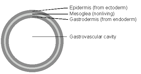
Figure \(\PageIndex{1}\): Cnidarian tissue layers. Cnidarians do not have mesoderm and therefore do not have organs.
A nonliving gelatinous material called mesoglea separates the two tissue layers. A nerve net is located between the epidermis and mesoglea. The body contains long structures called tentacles that can be moved to capture prey. The tentacles contain stinging cells called cnidocytes and within each one is a capsule called a nematocyst, which discharges to either trap or sting the prey. Contractile (muscle-like) fibers are found in both the epidermis and the gastrodermis. Their movements are not complex because they do not have a brain.
Cnidarians have a hydrostatic skeleton. The contractile fibers act against the fluid-filled gastrovascular cavity. The movements are like a balloon; the animal can be short and thick or long and thin. Cnidarians have a saclike gut and extracellular digestion.
Two body forms are found among the Cnidarians, a polyp and a medusa. A polyp is attached and has the tentacles and mouth directed upward. A medusa is free-floating and has the mouth and tentacles on the ventral surface. It resembles an upside-down polyp. Some species have both a polyp and a medusa in their life cycle, others have one or the other form dominant.
Procedure
Hydra
- Use a dropper to place a live Hydra on a slide. Examine the Hydra using a dissection microscope.
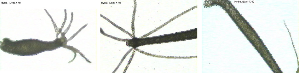
Figure \(\PageIndex{2}\): Three images of live Genus Hydra 40x magnification.
- Hydra reproduce both sexually and asexually by budding. Try to find a live Hydra with buds. If you cannot find a live Hydra budding, look for budding in a prepared slide of Hydra.
- Add a drop of vinegar to the slide containing Hydra. Describe what happened to the cnidocytes.

Figure \(\PageIndex{3}\): Hydra cnidocytes discharge their nematocysts when exposed to a 5% vinegar solution.
- Examine microscope slides of hydra l.s. and hydra c.s. Look for the presence of two tissue layers. Identify stinging cells (Cnidocytes) in a slide of the whole animal.

Figure \(\PageIndex{4}\): Hydra l.s. 100x, c.s. 100x, and c.s. 200x showing the two tissue layers.

Figure \(\PageIndex{5}\): Portion of a Hydra tentacle showing cnidocytes

Figure \(\PageIndex{6}\): Left: Hydra l.s. X 40. Right: Hydra l.s. with ingested food X 40


View other preserved specimens and slides on display (anthozoans, scyphozoans).
Questions
- Does the hydra illustrate the polyp or the medusa stage?
- How many germ layers does the hydra contain?
- What type of symmetry is seen in the hydra?
- Can you find the hydra tentacles? How many tentacles does your hydra specimen contain?
- Name the stinging cells present on the tentacles that are unique to cnidarians.
- Explain the movement of the hydra if live specimens are available. If they are not, review the video on hydra movement.
- If there are live hydra specimens do not add the vinegar as indicated on the lab website. Vinegar causes them to expel the cnidocytes. Review the nematocysts video, which shows a jellyfish discharging the nematocyst cells.
- View the preserved cnidarian specimens will be on display. They may differ from the ones directly mentioned in the lab website but there should be some medusa cnidarians as well as corals and sea anemones. Please make observations on the available specimens. And fill in the chart below.
Name of Specimen Physical Description Polyp or Medusa Stage?
Lophotrochozoa
Flatworms (Phylum Platyhelminthes)
Procedure
View specimens and slides on display.
Questions
- Observe the live planaria, if present, under the dissecting scope. If there are no live specimens, review the planarian video.
- What type of symmetry does the planaria display?
- Does the planaria exhibit cephalization?
- Can you locate the planaria eyespots? What do the eyespots sense?
- View the large planaria model. Make sure you can identify the pharynx, the eyespots, and the flame cells.
- Does the planaria have a complete or incomplete digestive system?
- What is the function of the flame cell?
- Are planaria hermaphrodites?
- Scientists say planaria have ladder like organs. Why?
View the preserved liver fluke specimens. Liver flukes are an example of a parasitic flatworm.
Questions
- Where does the adult liver fluke live?
- When the liver fluke egg hatches, what organism does it infect first?
- Can humans become infected?
View the preserved tapeworm and the slides of the tapeworm scolex (head) and proglottids (reproductive bodies). This is another example of a parasitic flatworm.
Questions
- What structures are located on the scolex to help the tapeworm attach to the host?
- Are tapeworms hermaphrodites?
- Name two livestock that can be infected by tapeworms.
- If a human is infected, where does the tapeworm live?
Molluscs (Phylum Mollusca)
Molluscs include bivalves (clams, scallops), gastropods (snails, slugs), cephalopods (octopus, squid), and polyplacophorans (chitons). Molluscs have a body divided into the head, visceral mass, mantle, and muscular foot. In many members of this group the mantle layer secretes a calcium carbonate shell for protection. Many molluscs have a radula, which is a toothed tongue-like structure they can use to scrape food particles off a surface.
Procedure
View the preserved specimens and slides on display.
Please make observations on the available specimens and fill in the chart below.
| Name of Specimen | Physical Description | Gastropod, chiton, bi-valve or cephalopod |
|---|---|---|
Questions
- What is the function of the foot?
- What is the function of the gills?
- What does the mantle secrete for the clam?
- What is contained within the visceral mass?
Segmented Worms (Phylum Annelida)
Procedure
View the preserved specimens and slides on display.
Questions
- What type of symmetry does the earthworm display?
- Does the earthworm exhibit cephalization?
- Does the earthworm exhibit segmentation?
The preserved annelida specimens will be on display, but may differ from the ones directly mentioned in the lab handout. Please make observations on the available specimens and fill in the table below.
| Name of Specimen | Physical Description | Leech, Earthworm, or Marine Worm |
|---|---|---|
Ecdysozoa
Roundworms (Phylum Nematoda)
Procedure
View the preserved specimens and slides on display.
- View the specimens and preserved slides of Ascaris
- What type of symmetry is seen in the roundworm?
- Does it exhibit cephalization?
- Ascaris is a parasite that swims constantly in human intestines. What structure protects the nematode from being digested?
- View the Trichinella slide. Trichinella is also a parasite. It can infect humans as well as other mammals like pigs, bears, and rodents. If untreated, it can lead to death.
- What mammal tissue does this roundworm infect?
- Draw a picture of the Trichinella as viewed under the microscope.
- If there are live vinegar eel specimens available, view them. If there are no live specimens available, review the vinegar eels video.
- Describe the movement of the vinegar eels.
- Do they have a complete or incomplete digestive system?
Arthropods (Phylum Arthropoda)
Arthopods include the following
- Chelicerates
- Arachnids—(Spiders, Scorpions, Harvestmen, Ticks and Mites)
- Horseshoe Crabs
- Myriapods
- Millipedes
- Centipedes
- Pancrustaceans
- Crustaceans
- Decopods
- Isopods
- Krill
- Copepods
- Barnacle
- Insects
- Crustaceans
Arthropods have an exoskeleton that cannot enlarge as the animal grows. As a result, it must be shed from time to time to allow growth.
The young of some arthropods look like the adults. The change from young to adult that these species undergo is called incomplete metamorphosis.
In many species, the egg hatches to produce a larva (pl. larvae) that does not look like the adult. At some point in the maturation process, the larva will produce a pupa (pl. pupae). In this phase its tissues will become reorganized into the adult form. This type of development is called complete metamorphosis.
Procedure
View preserved specimens and slides on display.
Questions
- View the preserved arthropod specimens available. There will be at least one example of each lineage group discussed on the website but not all of the specimens may be available. Use the table below to organize your observations.
Name of Specimen Lineage Exoskeleton? Jointed Appendages? Specialized Segments - What type of symmetry is displayed in the arthropods?
- Do the arthropods exhibit cephalization?
Crayfish Dissection
Procedure
Obtain a crayfish and identify the structures shown in the photograph below. You should familiarize yourself with the names of the external structures shown in the photograph below.

Figure \(\PageIndex{7}\): Crayfish external anatomy.
The photographs below show the ventral surface of male and female crayfish. Click on the photographs to view them. Identify the sex of your crayfish. Find somebody in class that has the opposite sex and view the ventral surface of their crayfish. Notice that crayfish have five pairs of swimmerets. In males, the anterior two pairs are large and less flexible than those of females. They are used to transfer sperm to the female.
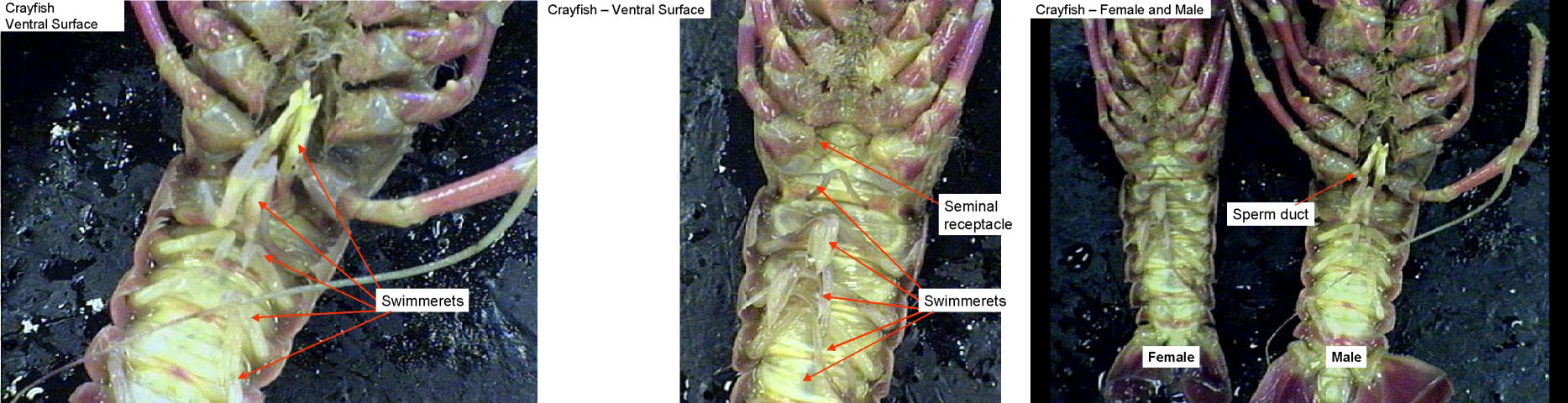
Figure \(\PageIndex{8}\): ventral surface showing swimmerettes, and ventral surface of male and female crayfish.
Find the seminal receptacles located between the third and fourth walking legs. These structures function to receive sperm from the male during mating.
Observe the sperm duct in males located at the base of the fifth walking leg.
Begin your dissection by cutting along the midline of the carapace from the posterior edge to an area just behind the two eyes (see the photographs below). Next, cut laterally just behind the eye until you reach the ventral edge. Carefully remove the piece of carapace that you have cut to expose the gills underneath. As you peel this piece of carapace away, it may be necessary to reach underneath with the point of a scissors or needle to brush away and detach any tissue from the interior of the animal that is attached to the carapace.

Figure \(\PageIndex{9}\): Crayfish dissection; carapace removal.
Your crayfish should look like the first photograph below. Next, remove the carapace on the other side.
Using a needle, carefully separate the first row of gills and notice that there is another row underneath. Remove one of the legs and observe how the gills are attached to the walking legs.
The first two photographs below show the gills on the left side of a crayfish. The third photograph is a crayfish with the entire carapace removed. The first row of gills on each side has been moved aside to expose a row underneath it.

Figure \(\PageIndex{10}\):Crayfish with carapace removed.

Figure \(\PageIndex{11}\): Crayfish leg showing gill attachment
Brush all of the gills to the side and then carefully cut the membrane (epidermis) that covers the internal organs.
The heart is a small diamond-shaped structure located below where the posterior edge of the carapace was. It may be difficult to see the blood vessels attached to the heart. Openings (ostia) in the side of the heart should be visible. These allow blood to enter the circulatory system.
Large white digestive glands can be seen on each side of the stomach. They produce digestive enzymes.

Figure \(\PageIndex{12}\): Crayfish internal anatomy, showing stomach, digestive glands, gills, heart, and ostium.
Remove the digestive glands and then carefully remove the stomach. Notice how the stomach is attached to the mouth. Cut open the stomach and observe the tooth-like structures at the anterior end for grinding food. This is called the gastric mill.
The green glands are positioned ventrally near the anterior end of the body cavity. They are spherical and will appear to be embedded within the surface. Their function is excretion. Find the openings for the green glands on the outer surface near the base of each antenna.
Find the brain near the anterior end and the two ventral nerve cords attached to the brain.

Figure \(\PageIndex{13}\): Left: Crayfish ventral surface. Middle: Crayfish viewed from above. The stomach has been removed revealing the nerve cords and green glands underneath. Right: Crayfish viewed from above looking into the head region. The stomach has been removed. The brain can be seen.
Questions
- Make sure you can identify the following external structures: antenna, chiliped, cephalothorax, abdomen, and walking legs
- Do you have a male or female crayfish?
- How many swimmerets does your crayfish have?
- How many rows of gills does the crayfish have?
- Where do the gills attach?
- Can you find the stomach and the digestive glands?
- What does the stomach attach to directly?
- Try to locate the green glands. What is the function of this structure?
Review Questions
Answer the review questions below. The phyla we viewed today were the porifera, the cnidaria, the nematoda and the arthropoda.
- Which phyla exhibited bilateral symmetry?
- Which phyla had no true tissues?
- Which phyla contained parasitic organisms?
- Which phyla were coelomates?
- Which phyla exhibited cephaliziation?
- Which phyla that you viewed today contained specialized appendages?
- Which phyla exhibited radial symmetry?
- Which phyla were pseudocoelomates?
- Which phyla had a complete digestive system?
- Which phyla were multicellular?
- Which phyla were asymmetrical?
- Which phyla were acoelomates?
Deuterostomia
Echinoderms (Phylum Echinodermata)
Echinoderms are coelomate, and deuterostomes.
Echinoderms include sea stars (starfishes), sea urchins, sand dollars, sea cucumbers, and sea lilies. There are 6,000 species of echinoderms; they are all marine.
Although echinoderm adults have radial symmetry, they evolved from ancestors that were bilaterally symmetrical. They have free-swimming, bilateral larvae that metamorphose (change as they mature) into adults with radial symmetry.
The adult body usually has five-part organization.
They possess an internal skeleton (endoskeleton) composed of calcium carbonate plates just beneath the surface of the skin. The plates often bear spines that protrude through the skin.
Echinoderms have numerous tube feet underneath each arm. The tube feet are connected to a system of pipes referred to as the water vascular system. Water enters the system by a sieve plate on the aboral surface. Each tube foot has a fleshy bulb or ampulla attached so that the entire structure looks like an medicine dropper or pipette. When muscles surrounding the ampulla contract, fluid inside the bulb moves down into the tube foot, extending it.
Large digestive glands produce enzymes necessary for digestion. Sexes are separate and gametes are shed into the water. The gonads are large due to the necessity of releasing large numbers of gametes into the marine environment. Coelomic fluid circulates substances and carries amoeboid cells that clean up particulate wastes. Gas exchange is done with numerous tiny gills that extend from the surface of the skin.
The nervous system consists of a central nerve ring with nerve branches extending into the arms. They do not have a brain.
Identify the blastopore. What structure in the adult does this give rise to? Notice that the embryo shows bilateral symmetry yet the adult is radial. The ancestral echinoderms were bilateral and bilateral symmetry is maintained in the larval stage.

Figure \(\PageIndex{14}\): Echinoderm development (various stages)
Procedure
View the preserved echinoderm specimens will be on display. Please make observations on the available specimens and fill in the chart below.
| Name of Specimen | Physical Description |
Chordates (Phylum Chordata)
Phylum Chordata includes two groups of invertebrate chordates (the cephalochordates and the urochordates) and the subphylum Vertebrata (vertebrates). We will be dissecting several vertebrates soon.
The diagram below shows evolutionary relationships among the major clades of chordates but avoids classification into subphylum and classes. For example, the close relationship between Myxini and the rest of the vertebrates can be seen even though there is uncertainty in the classification of Myxini.

Figure \(\PageIndex{15}\): Evolutionary relationships among the major clades of chordates.
Chordates exhibit bilateral symmetry.
Chordates have the following characteristics at some point in their life history:
- a dorsal, hollow nerve cord.
- a dorsal supporting rod called a notochord. This is replaced by a vertebral column in vertebrates.
- pharyngeal clefts (pouches). These develop into openings to the exterior (gill slits) in some chordates. Gill slits functioned as a mechanism for filter-feeding in primitive vertebrates. The gills of fish function in gas exchange.
- a postanal tail. In most other kinds of animals, the digestive tract extends the entire length of the animal.
Procedure
View specimens and slides of the three major groups of chordates on display
Questions
- View the lancelet slide. The lancelet is an example of a cephalochordate. It contains all four chordate characteristics. List the four chordate characteristics below.
- View the lancelet model. Make sure you can identify all four chordate characteristics on the model.
- Chooses several vertebrates on display and fill in the chart below. Can you identify all four chordate characteristics on each specimen? Why or why not?
| Name of Specimen | Physical Description |