Biology LibreTexts

15.4: Protein Synthesis

  • Page ID
    30734
  • \( \newcommand{\vecs}[1]{\overset { \scriptstyle \rightharpoonup} {\mathbf{#1}} } \)

    \( \newcommand{\vecd}[1]{\overset{-\!-\!\rightharpoonup}{\vphantom{a}\smash {#1}}} \)

    \( \newcommand{\id}{\mathrm{id}}\) \( \newcommand{\Span}{\mathrm{span}}\)

    ( \newcommand{\kernel}{\mathrm{null}\,}\) \( \newcommand{\range}{\mathrm{range}\,}\)

    \( \newcommand{\RealPart}{\mathrm{Re}}\) \( \newcommand{\ImaginaryPart}{\mathrm{Im}}\)

    \( \newcommand{\Argument}{\mathrm{Arg}}\) \( \newcommand{\norm}[1]{\| #1 \|}\)

    \( \newcommand{\inner}[2]{\langle #1, #2 \rangle}\)

    \( \newcommand{\Span}{\mathrm{span}}\)

    \( \newcommand{\id}{\mathrm{id}}\)

    \( \newcommand{\Span}{\mathrm{span}}\)

    \( \newcommand{\kernel}{\mathrm{null}\,}\)

    \( \newcommand{\range}{\mathrm{range}\,}\)

    \( \newcommand{\RealPart}{\mathrm{Re}}\)

    \( \newcommand{\ImaginaryPart}{\mathrm{Im}}\)

    \( \newcommand{\Argument}{\mathrm{Arg}}\)

    \( \newcommand{\norm}[1]{\| #1 \|}\)

    \( \newcommand{\inner}[2]{\langle #1, #2 \rangle}\)

    \( \newcommand{\Span}{\mathrm{span}}\) \( \newcommand{\AA}{\unicode[.8,0]{x212B}}\)

    \( \newcommand{\vectorA}[1]{\vec{#1}}      % arrow\)

    \( \newcommand{\vectorAt}[1]{\vec{\text{#1}}}      % arrow\)

    \( \newcommand{\vectorB}[1]{\overset { \scriptstyle \rightharpoonup} {\mathbf{#1}} } \)

    \( \newcommand{\vectorC}[1]{\textbf{#1}} \)

    \( \newcommand{\vectorD}[1]{\overrightarrow{#1}} \)

    \( \newcommand{\vectorDt}[1]{\overrightarrow{\text{#1}}} \)

    \( \newcommand{\vectE}[1]{\overset{-\!-\!\rightharpoonup}{\vphantom{a}\smash{\mathbf {#1}}}} \)

    \( \newcommand{\vecs}[1]{\overset { \scriptstyle \rightharpoonup} {\mathbf{#1}} } \)

    \( \newcommand{\vecd}[1]{\overset{-\!-\!\rightharpoonup}{\vphantom{a}\smash {#1}}} \)

    \(\newcommand{\avec}{\mathbf a}\) \(\newcommand{\bvec}{\mathbf b}\) \(\newcommand{\cvec}{\mathbf c}\) \(\newcommand{\dvec}{\mathbf d}\) \(\newcommand{\dtil}{\widetilde{\mathbf d}}\) \(\newcommand{\evec}{\mathbf e}\) \(\newcommand{\fvec}{\mathbf f}\) \(\newcommand{\nvec}{\mathbf n}\) \(\newcommand{\pvec}{\mathbf p}\) \(\newcommand{\qvec}{\mathbf q}\) \(\newcommand{\svec}{\mathbf s}\) \(\newcommand{\tvec}{\mathbf t}\) \(\newcommand{\uvec}{\mathbf u}\) \(\newcommand{\vvec}{\mathbf v}\) \(\newcommand{\wvec}{\mathbf w}\) \(\newcommand{\xvec}{\mathbf x}\) \(\newcommand{\yvec}{\mathbf y}\) \(\newcommand{\zvec}{\mathbf z}\) \(\newcommand{\rvec}{\mathbf r}\) \(\newcommand{\mvec}{\mathbf m}\) \(\newcommand{\zerovec}{\mathbf 0}\) \(\newcommand{\onevec}{\mathbf 1}\) \(\newcommand{\real}{\mathbb R}\) \(\newcommand{\twovec}[2]{\left[\begin{array}{r}#1 \\ #2 \end{array}\right]}\) \(\newcommand{\ctwovec}[2]{\left[\begin{array}{c}#1 \\ #2 \end{array}\right]}\) \(\newcommand{\threevec}[3]{\left[\begin{array}{r}#1 \\ #2 \\ #3 \end{array}\right]}\) \(\newcommand{\cthreevec}[3]{\left[\begin{array}{c}#1 \\ #2 \\ #3 \end{array}\right]}\) \(\newcommand{\fourvec}[4]{\left[\begin{array}{r}#1 \\ #2 \\ #3 \\ #4 \end{array}\right]}\) \(\newcommand{\cfourvec}[4]{\left[\begin{array}{c}#1 \\ #2 \\ #3 \\ #4 \end{array}\right]}\) \(\newcommand{\fivevec}[5]{\left[\begin{array}{r}#1 \\ #2 \\ #3 \\ #4 \\ #5 \\ \end{array}\right]}\) \(\newcommand{\cfivevec}[5]{\left[\begin{array}{c}#1 \\ #2 \\ #3 \\ #4 \\ #5 \\ \end{array}\right]}\) \(\newcommand{\mattwo}[4]{\left[\begin{array}{rr}#1 \amp #2 \\ #3 \amp #4 \\ \end{array}\right]}\) \(\newcommand{\laspan}[1]{\text{Span}\{#1\}}\) \(\newcommand{\bcal}{\cal B}\) \(\newcommand{\ccal}{\cal C}\) \(\newcommand{\scal}{\cal S}\) \(\newcommand{\wcal}{\cal W}\) \(\newcommand{\ecal}{\cal E}\) \(\newcommand{\coords}[2]{\left\{#1\right\}_{#2}}\) \(\newcommand{\gray}[1]{\color{gray}{#1}}\) \(\newcommand{\lgray}[1]{\color{lightgray}{#1}}\) \(\newcommand{\rank}{\operatorname{rank}}\) \(\newcommand{\row}{\text{Row}}\) \(\newcommand{\col}{\text{Col}}\) \(\renewcommand{\row}{\text{Row}}\) \(\newcommand{\nul}{\text{Nul}}\) \(\newcommand{\var}{\text{Var}}\) \(\newcommand{\corr}{\text{corr}}\) \(\newcommand{\len}[1]{\left|#1\right|}\) \(\newcommand{\bbar}{\overline{\bvec}}\) \(\newcommand{\bhat}{\widehat{\bvec}}\) \(\newcommand{\bperp}{\bvec^\perp}\) \(\newcommand{\xhat}{\widehat{\xvec}}\) \(\newcommand{\vhat}{\widehat{\vvec}}\) \(\newcommand{\uhat}{\widehat{\uvec}}\) \(\newcommand{\what}{\widehat{\wvec}}\) \(\newcommand{\Sighat}{\widehat{\Sigma}}\) \(\newcommand{\lt}{<}\) \(\newcommand{\gt}{>}\) \(\newcommand{\amp}{&}\) \(\definecolor{fillinmathshade}{gray}{0.9}\)
    The Central Dogma of Biology

    Your DNA, or deoxyribonucleic acid, contains the genes that determine who you are. How can this organic molecule control your characteristics? DNA contains instructions for all the proteins your body makes. Proteins, in turn, determine the structure and function of all your cells. What determines a protein’s structure? It begins with the sequence of amino acids that make up the protein. Instructions for making proteins with the correct sequence of amino acids are encoded in DNA.

    How proteins are made
    Figure \(\PageIndex{1}\): Transcription and translation (Protein synthesis) in a cell.

    DNA is found in chromosomes. In eukaryotic cells, chromosomes always remain in the nucleus, but proteins are made at ribosomes in the cytoplasm or on the rough endoplasmic reticulum (RER). How do the instructions in DNA get to the site of protein synthesis outside the nucleus? Another type of nucleic acid is responsible. This nucleic acid is RNA or ribonucleic acid. RNA is a small molecule that can squeeze through pores in the nuclear membrane. It carries the information from DNA in the nucleus to a ribosome in the cytoplasm and then helps assemble the protein. In short:

    DNA → RNA → Protein

    Discovering this sequence of events was a major milestone in molecular biology. It is called the central dogma of biology. The two processes involved in the central dogma are transcription and translation.

    Transcription translation; mRNA to protein
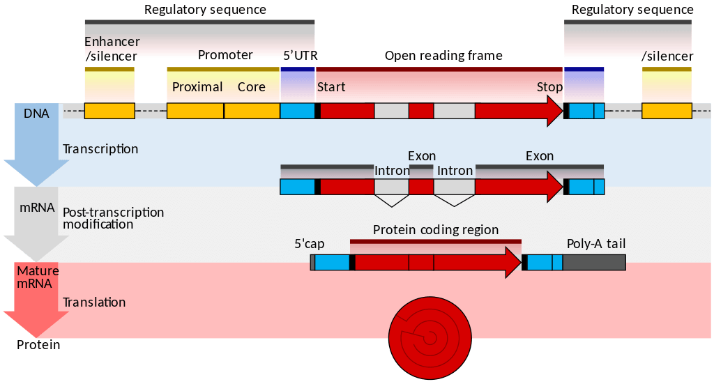
    Figure \(\PageIndex{2}\): An overview of transcription and translation. The top panel shows a gene. A gene is composed of the open reading frame (aka coding sequence) that is flanked by regulatory sequences. At the beginning of the gene, the regulatory sequence contains a promoter where RNA polymerase attaches and starts transcription. At the end of the open reading frame, the regulatory sequence contains a terminator (not shown.)The middle panel shows a pre mRNA which is modified by excising introns and keeping exons. This is called post transcription modification. A mature mRNA contains a 5' cap and poly-A tail. The bottom panel shows a synthesis of protein via translation.

    Transcription

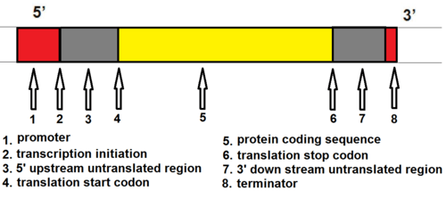
    Transcription is the first part of the central dogma of molecular biology: DNA → RNA. It is the transfer of genetic instructions in DNA to mRNA. Transcription happens in the nucleus of the cell. During transcription, a strand of mRNA is made that is complementary to a strand of DNA called a gene. A gene can easily be identified from the DNA sequence. A gene contains the basic three regions, promoter, coding sequence (reading frame), and terminator. There are more parts of a gene which are illustrated in Figure \(\PageIndex{3}\).

    gene regions
    Figure \(\PageIndex{3}\): The major components of a gene. 1. promoter, 2. transcription initiation, 3. 5' upstream untranslated region, 4. translation start codon site, 5. protein-coding sequence, 6. translation stop codon region, 7. 3' downstream untranslated region, and 8. terminator.

    Steps of Transcription

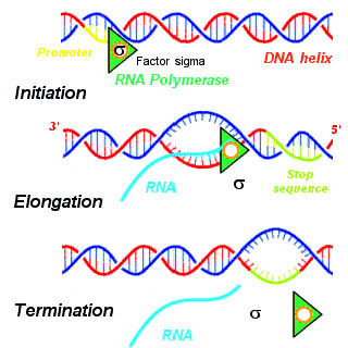
    Transcription takes place in three steps, called initiation, elongation, and termination. The steps are illustrated in Figure \(\PageIndex{4}\).

    1. Initiation is the beginning of transcription. It occurs when the enzyme RNA polymerase binds to a region of a gene called the promoter. This signals the DNA to unwind so the enzyme can “read” the bases in one of the DNA strands. The enzyme is ready to make a strand of mRNA with a complementary sequence of bases. The promoter is not part of the resulting mRNA
    2. Elongation is the addition of nucleotides to the mRNA strand.
    3. Termination is the ending of transcription. As RNA polymerase transcribes the terminator, it detaches from DNA. The mRNA strand is complete after this step.
      Transcription steps, initiation, elongation, and termination
      Figure \(\PageIndex{4}\): Transcription occurs in the three steps - initiation, elongation, and termination

    Processing mRNA

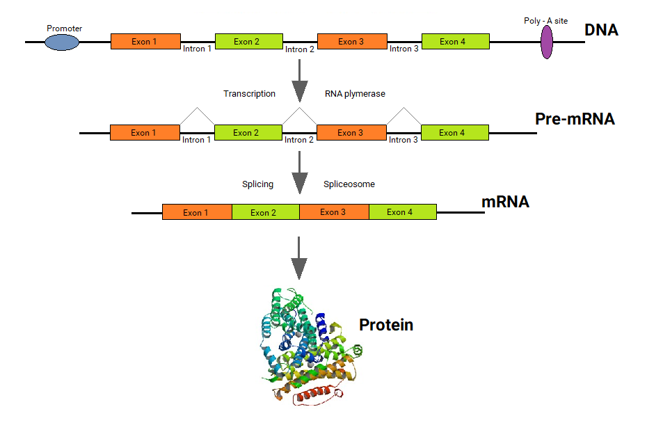
    In eukaryotes, the new mRNA is not yet ready for translation. At this stage, it is called pre-mRNA, and it must go through more processing before it leaves the nucleus as mature mRNA. The processing may include the addition of a 5' cap, splicing, editing, and 3' polyadenylation (poly-A) tail. These processes modify the mRNA in various ways. Such modifications allow a single gene to be used to make more than one protein. See Figure \(\PageIndex{5}\) as you read below:

    • 5' cap protects mRNA in the cytoplasm and helps in the attachment of mRNA with the ribosome for translation.
    • Splicing removes introns from the protein-coding sequence of mRNA. Introns are regions that do not code for the protein. The remaining mRNA consists only of regions called exons that do code for the protein.
    • Editing changes some of the nucleotides in mRNA. For example, a human protein called APOB, which helps transport lipids in the blood, has two different forms because of editing. One form is smaller than the other because editing adds an earlier stop signal in mRNA.
    • Polyadenylation adds a “tail” to the mRNA. The tail consists of a string of As (adenine bases). It signals the end of mRNA. It is also involved in exporting mRNA from the nucleus, and it protects mRNA from enzymes that might break it down.
    transcript splicing process
    Figure \(\PageIndex{5}\): Splicing removes introns from mRNA. Mature mRNA is translated to protein.

    Translation

    The translation is the second part of the central dogma of molecular biology: RNA --> Protein. It is the process in which the genetic code in mRNA is read to make a protein. The translation is illustrated in Figure \(\PageIndex{6}\). After mRNA leaves the nucleus, it moves to a ribosome, which consists of rRNA and proteins. Translation happens on the ribosomes floating in the cytosol, or on the ribosomes attached to the rough endoplasmic reticulum. The ribosome reads the sequence of codons in mRNA, and molecules of tRNA bring amino acids to the ribosome in the correct sequence.

    To understand the role of tRNA, you need to know more about its structure. Each tRNA molecule has an anticodon for the amino acid it carries. An anticodon is complementary to the codon for an amino acid. For example, the amino acid lysine has the codon AAG, so the anticodon is UUC. Therefore, lysine would be carried by a tRNA molecule with the anticodon UUC. Wherever the codon AAG appears in mRNA, a UUC anticodon of tRNA temporarily binds. While bound to mRNA, tRNA gives up its amino acid. With the help of rRNA, bonds form between the amino acids as they are brought one by one to the ribosome, creating a polypeptide chain. The chain of amino acids keeps growing until a stop codon is reached.

    Ribosomes, which are just made out of rRNA (ribosomal RNA) and protein, have been classified as ribozymes because the rRNA has enzymatic activity. The rRNA is important for the peptidyl transferase activity that bonds amino acids. Ribosomes have two subunits of rRNA and protein. The large subunit has three active sites called E, P, and A sites. These sites are important in the catalytic activity of ribosomes.

    Just as with mRNA synthesis, protein synthesis can be divided into three phases: initiation, elongation, and termination. In addition to the mRNA template, many other molecules contribute to the process of translation, such as ribosomes, tRNAs, and various enzymatic factors

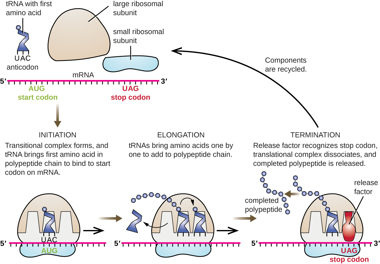
    Translation Initiation: The small subunit binds to a site upstream (on the 5' side) of the start of the mRNA. It proceeds to scan the mRNA in the 5'-->3' direction until it encounters the START codon (AUG). The large subunit attaches and the initiator tRNA, which carries methionine (Met), binds to the P site on the ribosome.

    Translation Elongation: The ribosome shifts one codon at a time, catalyzing each process that occurs in the three sites. With each step, a charged tRNA enters the complex, the polypeptide becomes one amino acid longer, and an uncharged tRNA departs. The energy for each bond between amino acids is derived from GTP, a molecule similar to ATP. Briefly, the ribosomes interact with other RNA molecules to make chains of amino acids called polypeptide chains, due to the peptide bond that forms between individual amino acids. Inside the ribosome, three sites participate in the translation process, the A, P, and E sites. Amazingly, the E. coli translation apparatus takes only 0.05 seconds to add each amino acid, meaning that a 200-amino acid polypeptide could be translated in just 10 seconds.

    Translation Termination: Termination of translation occurs when a stop codon (UAA, UAG, or UGA) is encountered (see Figure \(\PageIndex{7}\). When the ribosome encounters the stop codon, the growing polypeptide is released with the help of various releasing factors and the ribosome subunits dissociate and leave the mRNA. After many ribosomes have completed translation, the mRNA is degraded so the nucleotides can be reused in another transcription reaction.

    Translation Initiation & Elongation
    Figure \(\PageIndex{6}\): At the initiation phase, the initiator tRNA carrying methionine with its anticodon encounters the AUG start codon at the P-site of the ribosome. During elongation, the ribosome translocates in the 5' to 3' direction of the mRNA, at which point the amino acids of tRNA in P-site and amino acid of tRNA in the A-site of the large subunit bond to each other via a peptide bond. This repeated movement of the ribosome creates a long amino acid chain based on the codons in the mRNA. As the ribosome translocates, the tRNA leaves the ribosome through the E-site, while new tRNA enters the A-site in order to continue elongating the amino acid chain.
    Translation steps
    Figure \(\PageIndex{7}\). Summary of protein translation. During initiation, both ribosomal subunits, mRNA, and initiator tRNA come together. The initiator anticodon of initiator tRNA hydrogen bond with the start codon. During elongation, tRNA brings amino acids one by one to add to the polypeptide chain. During termination, the release factor recognizes stop codon, all components dissociate, and the completed polypeptide is released.

    What Happens Next?

    After a polypeptide chain is synthesized, it may undergo additional processes. For example, it may assume a folded tertiary shape due to interactions among its amino acids. It may also bind with other polypeptides or with different types of molecules, such as lipids or carbohydrates. Many proteins travel to the Golgi apparatus within the cytoplasm to be modified for the specific job they will do.

    Summary of Central Dogma

    Transcription translation
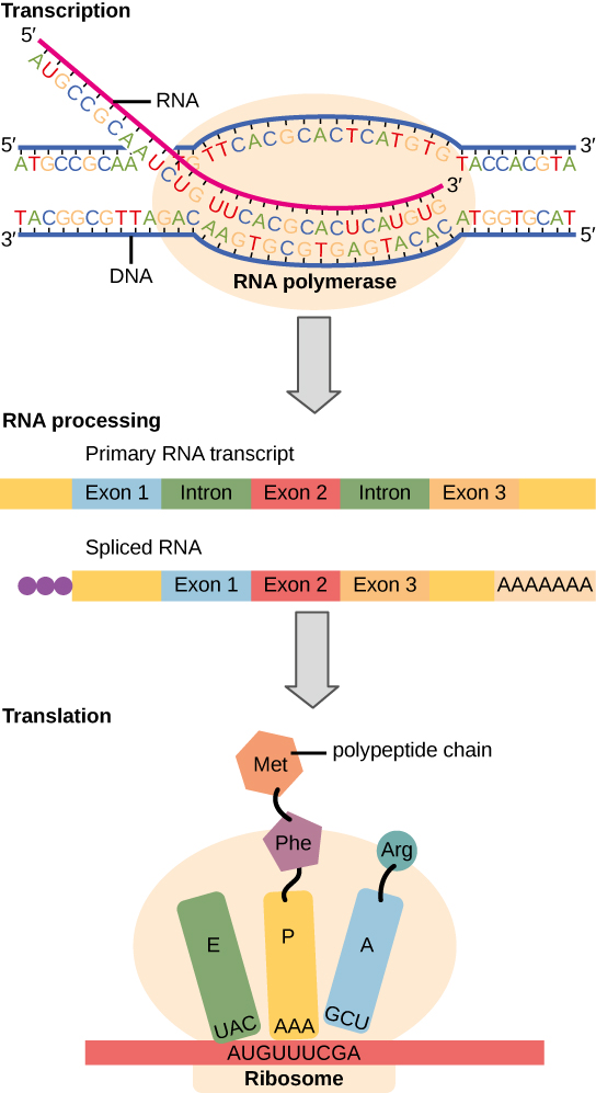
    Figure \(\PageIndex{8}\): The central dogma: Instructions on DNA are transcribed onto messenger RNA. Ribosomes are able to read the genetic information inscribed on a strand of messenger RNA and use this information to string amino acids together into a protein.

    Review

    1. Relate protein synthesis and its two major phases to the central dogma of molecular biology.
    2. Identify the steps of transcription, and summarize what happens during each step.
    3. Explain how mRNA is processed before it leaves the nucleus.
    4. Describe what happens during the translation phase of protein synthesis.
    5. What additional processes may a polypeptide chain undergo after it is synthesized?
    6. Where does transcription take place in eukaryotes?
    7. Where does translation take place?
    8. Which type of RNA (mRNA, rRNA, or tRNA) best fits each of the statements below? Choose only one type for each.
      1. Contains the codons
      2. Contains the anticodons
      3. Makes up the ribosome, along with proteins
    9. If the DNA has a triplet code of CAG in one strand (the strand used as a template for transcription),
      1. What is the complementary sequence on the other DNA strand?
      2. What is the complementary sequence in the mRNA? What is this sequence called?
      3. @hat is the resulting sequence in the tRNA? What is this sequence called? What do you notice about this sequence compared to the original DNA triplet on the template strand?
    10. The promoter is a region located in the:
      1. DNA
      2. mRNA
      3. tRNA
      4. Both A and B
    11. True or False. Introns in mRNA bind to tRNA at the ribosome.
    12. True or False. tRNAs can be thought of as the link between amino acids and codons in the mRNA.

    Explore More

    Messenger RNA molecules are "spliced" in order to create the mRNA involved in protein synthesis. Learn the process here:

    Attributions

    1. How proteins are made by Nicolle Rager, National Science Foundation, public domain via Wikimedia Commons
    2. Gene structure eukaryote by Thomas Shafee, licensed CC BY 4.0 via Wikimedia Commons
    3. Components of a gene by Mandeep Grewal, CC BY 4.0
    4. Transcription by Calibuon, released into the public domain via Wikimedia Commons
    5. Transcript and splicing by Ganeshmanohar, CC BY-SA 4.0 via Wikimedia Commons
    6. Initiation and elongation by Jordan Nguyen, CC BY-SA 4.0 via Wikimedia Commons
    7. Protein synthesis by OpenStax, CC BY 4.0
    8. Gene regulation by OpenStax, CC BY 4.0
    9. Text adapted from Human Biology by CK-12 licensed CC BY-NC 3.0

    This page titled 15.4: Protein Synthesis is shared under a CK-12 license and was authored, remixed, and/or curated by Suzanne Wakim & Mandeep Grewal via source content that was edited to the style and standards of the LibreTexts platform.

    CK-12 Foundation
    LICENSED UNDER
    CK-12 Foundation is licensed under CK-12 Curriculum Materials License