
8.3: Lymphatic System
- Page ID
- 30669

\( \newcommand{\vecs}[1]{\overset { \scriptstyle \rightharpoonup} {\mathbf{#1}} } \)
\( \newcommand{\vecd}[1]{\overset{-\!-\!\rightharpoonup}{\vphantom{a}\smash {#1}}} \)
\( \newcommand{\dsum}{\displaystyle\sum\limits} \)
\( \newcommand{\dint}{\displaystyle\int\limits} \)
\( \newcommand{\dlim}{\displaystyle\lim\limits} \)
\( \newcommand{\id}{\mathrm{id}}\) \( \newcommand{\Span}{\mathrm{span}}\)
( \newcommand{\kernel}{\mathrm{null}\,}\) \( \newcommand{\range}{\mathrm{range}\,}\)
\( \newcommand{\RealPart}{\mathrm{Re}}\) \( \newcommand{\ImaginaryPart}{\mathrm{Im}}\)
\( \newcommand{\Argument}{\mathrm{Arg}}\) \( \newcommand{\norm}[1]{\| #1 \|}\)
\( \newcommand{\inner}[2]{\langle #1, #2 \rangle}\)
\( \newcommand{\Span}{\mathrm{span}}\)
\( \newcommand{\id}{\mathrm{id}}\)
\( \newcommand{\Span}{\mathrm{span}}\)
\( \newcommand{\kernel}{\mathrm{null}\,}\)
\( \newcommand{\range}{\mathrm{range}\,}\)
\( \newcommand{\RealPart}{\mathrm{Re}}\)
\( \newcommand{\ImaginaryPart}{\mathrm{Im}}\)
\( \newcommand{\Argument}{\mathrm{Arg}}\)
\( \newcommand{\norm}[1]{\| #1 \|}\)
\( \newcommand{\inner}[2]{\langle #1, #2 \rangle}\)
\( \newcommand{\Span}{\mathrm{span}}\) \( \newcommand{\AA}{\unicode[.8,0]{x212B}}\)
\( \newcommand{\vectorA}[1]{\vec{#1}} % arrow\)
\( \newcommand{\vectorAt}[1]{\vec{\text{#1}}} % arrow\)
\( \newcommand{\vectorB}[1]{\overset { \scriptstyle \rightharpoonup} {\mathbf{#1}} } \)
\( \newcommand{\vectorC}[1]{\textbf{#1}} \)
\( \newcommand{\vectorD}[1]{\overrightarrow{#1}} \)
\( \newcommand{\vectorDt}[1]{\overrightarrow{\text{#1}}} \)
\( \newcommand{\vectE}[1]{\overset{-\!-\!\rightharpoonup}{\vphantom{a}\smash{\mathbf {#1}}}} \)
\( \newcommand{\vecs}[1]{\overset { \scriptstyle \rightharpoonup} {\mathbf{#1}} } \)
\(\newcommand{\longvect}{\overrightarrow}\)
\( \newcommand{\vecd}[1]{\overset{-\!-\!\rightharpoonup}{\vphantom{a}\smash {#1}}} \)
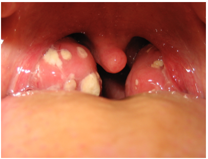
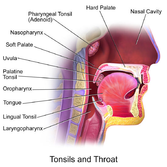
\(\newcommand{\avec}{\mathbf a}\) \(\newcommand{\bvec}{\mathbf b}\) \(\newcommand{\cvec}{\mathbf c}\) \(\newcommand{\dvec}{\mathbf d}\) \(\newcommand{\dtil}{\widetilde{\mathbf d}}\) \(\newcommand{\evec}{\mathbf e}\) \(\newcommand{\fvec}{\mathbf f}\) \(\newcommand{\nvec}{\mathbf n}\) \(\newcommand{\pvec}{\mathbf p}\) \(\newcommand{\qvec}{\mathbf q}\) \(\newcommand{\svec}{\mathbf s}\) \(\newcommand{\tvec}{\mathbf t}\) \(\newcommand{\uvec}{\mathbf u}\) \(\newcommand{\vvec}{\mathbf v}\) \(\newcommand{\wvec}{\mathbf w}\) \(\newcommand{\xvec}{\mathbf x}\) \(\newcommand{\yvec}{\mathbf y}\) \(\newcommand{\zvec}{\mathbf z}\) \(\newcommand{\rvec}{\mathbf r}\) \(\newcommand{\mvec}{\mathbf m}\) \(\newcommand{\zerovec}{\mathbf 0}\) \(\newcommand{\onevec}{\mathbf 1}\) \(\newcommand{\real}{\mathbb R}\) \(\newcommand{\twovec}[2]{\left[\begin{array}{r}#1 \\ #2 \end{array}\right]}\) \(\newcommand{\ctwovec}[2]{\left[\begin{array}{c}#1 \\ #2 \end{array}\right]}\) \(\newcommand{\threevec}[3]{\left[\begin{array}{r}#1 \\ #2 \\ #3 \end{array}\right]}\) \(\newcommand{\cthreevec}[3]{\left[\begin{array}{c}#1 \\ #2 \\ #3 \end{array}\right]}\) \(\newcommand{\fourvec}[4]{\left[\begin{array}{r}#1 \\ #2 \\ #3 \\ #4 \end{array}\right]}\) \(\newcommand{\cfourvec}[4]{\left[\begin{array}{c}#1 \\ #2 \\ #3 \\ #4 \end{array}\right]}\) \(\newcommand{\fivevec}[5]{\left[\begin{array}{r}#1 \\ #2 \\ #3 \\ #4 \\ #5 \\ \end{array}\right]}\) \(\newcommand{\cfivevec}[5]{\left[\begin{array}{c}#1 \\ #2 \\ #3 \\ #4 \\ #5 \\ \end{array}\right]}\) \(\newcommand{\mattwo}[4]{\left[\begin{array}{rr}#1 \amp #2 \\ #3 \amp #4 \\ \end{array}\right]}\) \(\newcommand{\laspan}[1]{\text{Span}\{#1\}}\) \(\newcommand{\bcal}{\cal B}\) \(\newcommand{\ccal}{\cal C}\) \(\newcommand{\scal}{\cal S}\) \(\newcommand{\wcal}{\cal W}\) \(\newcommand{\ecal}{\cal E}\) \(\newcommand{\coords}[2]{\left\{#1\right\}_{#2}}\) \(\newcommand{\gray}[1]{\color{gray}{#1}}\) \(\newcommand{\lgray}[1]{\color{lightgray}{#1}}\) \(\newcommand{\rank}{\operatorname{rank}}\) \(\newcommand{\row}{\text{Row}}\) \(\newcommand{\col}{\text{Col}}\) \(\renewcommand{\row}{\text{Row}}\) \(\newcommand{\nul}{\text{Nul}}\) \(\newcommand{\var}{\text{Var}}\) \(\newcommand{\corr}{\text{corr}}\) \(\newcommand{\len}[1]{\left|#1\right|}\) \(\newcommand{\bbar}{\overline{\bvec}}\) \(\newcommand{\bhat}{\widehat{\bvec}}\) \(\newcommand{\bperp}{\bvec^\perp}\) \(\newcommand{\xhat}{\widehat{\xvec}}\) \(\newcommand{\vhat}{\widehat{\vvec}}\) \(\newcommand{\uhat}{\widehat{\uvec}}\) \(\newcommand{\what}{\widehat{\wvec}}\) \(\newcommand{\Sighat}{\widehat{\Sigma}}\) \(\newcommand{\lt}{<}\) \(\newcommand{\gt}{>}\) \(\newcommand{\amp}{&}\) \(\definecolor{fillinmathshade}{gray}{0.9}\)The white patches on either side of the throat in this picture are signs of tonsillitis. The tonsils are small structures in the throat that are very common sites of infection. The white spots on the tonsils pictured here are evidence of infection. The patches consist of large amounts of dead bacteria, cellular debris, and white blood cells; in a word, pus. Children with recurrent tonsillitis may have their tonsils removed surgically to eliminate this type of infection. The tonsils are organs of the lymphatic system.
What Is the Lymphatic System?
The lymphatic system is a collection of organs involved in the production, maturation, and harboring of white blood cells called lymphocytes. It also includes a network of vessels that transport or filter the fluid known as lymph in which lymphocytes circulate. Figure \(\PageIndex{2}\)shows major lymphatic vessels and other structures that make up the lymphatic system. Besides the tonsils, organs of the lymphatic system include the thymus, the spleen, and hundreds of lymph nodes that are distributed along the lymphatic vessels.
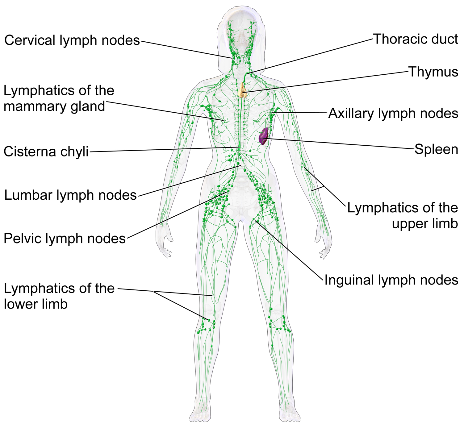 Figure \(\PageIndex{2}\): The lymphatic system includes the thymus, spleen, lymph vessels, and nodes.
The lymphatic vessels form a transportation network similar in many respects to the blood vessels of the cardiovascular system. However, unlike the cardiovascular system, the lymphatic system is not a closed system. Instead, lymphatic vessels carry lymph in a single direction, always toward the upper chest, where the lymph empties from lymphatic vessels into blood vessels.
Cardiovascular Function of the Lymphatic System
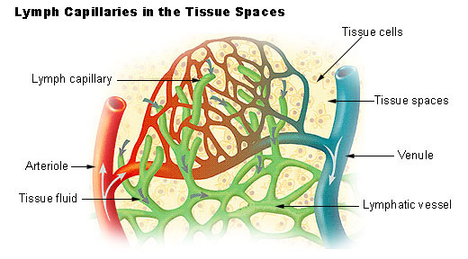
The return of lymph to the bloodstream is one of the major functions of the lymphatic system. When blood travels through capillaries of the cardiovascular system, it is under pressure, which forces some of the components of blood (such as water, oxygen, and nutrients) through the walls of the capillaries and into the tissue spaces between cells, forming tissue fluid, also called interstitial fluid (Figure \(\PageIndex{3}\)). Interstitial fluid bathes and nourishes cells and also absorbs their waste products. Much of the water from the interstitial fluid is reabsorbed into the capillary blood by osmosis. Most of the remaining fluid is absorbed by tiny lymphatic vessels called lymph capillaries. Once interstitial fluid enters the lymphatic vessels, it is called lymph. Lymph is very similar in composition to blood plasma. Besides water, lymph may contain proteins, waste products, cellular debris, and pathogens. It also contains numerous white blood cells, especially the subset of white blood cells known as lymphocytes. In fact, lymphocytes are the main cellular components of lymph.
The lymph that enters lymph capillaries in tissues is transported through the lymphatic vessel network to two large lymphatic ducts in the upper chest. From there, the lymph flows into two major veins (called subclavian veins) of the cardiovascular system. Unlike blood, lymph is not pumped through its network of vessels. Instead, lymph moves through lymphatic vessels via a combination of contractions of the vessels themselves and forces applied to the vessels externally by skeletal muscles. Lymphatic vessels also contain numerous valves that keep lymph flowing in just one direction, thereby preventing backflow.
Digestive Function of the Lymphatic System
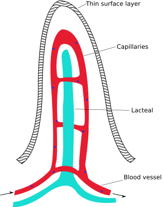
Lymphatic vessels called lacteals (Figure \(\PageIndex{4}\)) are present in the lining of the gastrointestinal tract, mainly in the small intestine. Each tiny villus in the lining of the small intestine has an internal bed of capillaries and lacteals. The capillaries absorb most nutrients from the digestion of food into the blood. The lacteals absorb mainly fatty acids from lipid digestion into the lymph, forming a fatty-acid-enriched fluid called chyle. Vessels of the lymphatic network then transport chyle from the small intestine to the main lymphatic ducts in the chest from which it drains into the blood circulation. The nutrients in chyle then circulate in the blood to the liver, where they are processed along with the other nutrients that reach the liver directly via the bloodstream.
Immune Function of the Lymphatic System
The primary function of the lymphatic system is host defense as part of the immune system. This function of the lymphatic system is centered on the production, maturation, and circulation of lymphocytes. Lymphocytes are leukocytes that are involved in the adaptive immune system. They are responsible for the recognition of, and tailored defense against, specific pathogens or tumor cells. Lymphocytes may also create a lasting memory of pathogens so they can be attacked quickly and strongly if they ever invade the body again. In this way, lymphocytes bring about long-lasting immunity to specific pathogens.
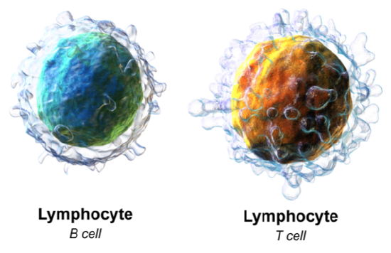
There are two major types of lymphocytes, called B cells and T cells, which are illustrated in Figure \(\PageIndex{5}\). Both B cells and T cells are involved in the adaptive immune response, but they play different roles. You can learn more about their immune functions by reading the concept Adaptive Immune System.
Production and Maturation of Lymphocytes
Like all other types of blood cells, including red blood cells as well as leukocytes, both B cells and T cells are produced from stem cells in the red marrow inside bones. After lymphocytes first form, they must go through a complicated maturation process before they are ready to search for pathogens. In this maturation process, they “learn” to distinguish self from non-self. Only those lymphocytes that successfully complete this maturation process go on to actually fight infections by pathogens.
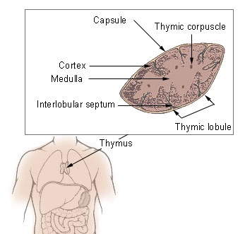
B cells mature in the bone marrow, which is why they are called B cells. After they mature and leave the bone marrow, they travel first to the circulatory system and then enter the lymphatic system to search for pathogens. T cells, on the other hand, mature in the thymus, which is why they are called T cells. The thymus is illustrated in Figure \(\PageIndex{6}\). It is a small lymphatic organ in the chest that consists of an outer cortex and inner medulla, all surrounded by a fibrous capsule. After maturing in the thymus, T cells enter the rest of the lymphatic system to join B cells in the hunt for pathogens. The bone marrow and thymus are called primary lymphoid organs because of their role in the production and/or maturation of lymphocytes.
Lymphocytes in Secondary Lymphoid Organs
The tonsils, spleen, and lymph nodes are referred to as secondary lymphoid organs. These organs do not produce or mature lymphocytes. Instead, they filter lymph and store lymphocytes. It is in these secondary lymphoid organs that pathogens (or their antigens) activate lymphocytes and initiate adaptive immune responses. Activation leads to the cloning of pathogen-specific lymphocytes, which then circulate between the lymphatic system and the blood, searching for and destroying their specific pathogens by producing antibodies against them.
Tonsils
There are actually four pairs of human tonsils. Three of the four are shown in Figure \(\PageIndex{7}\). The fourth pair, called tubal tonsils, is located at the back of the nasopharynx. The palatine tonsils are the tonsils that are visible on either side of the throat. All four pairs of tonsils encircle a part of the anatomy where the respiratory and gastrointestinal tracts intersect and where pathogens have ready access to the body. This ring of tonsils is called Waldeyer's ring.
Spleen
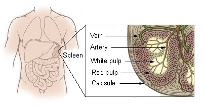
The spleen (Figure \(\PageIndex{8}\)) is the largest of the secondary lymphoid organs and is centrally located in the body. Besides harboring lymphocytes and filtering lymph, the spleen also filters blood. Most dead or aged red blood cells are removed from the blood in the red pulp of the spleen. Lymph is filtered in the white pulp of the spleen. In the fetus, the spleen has the additional function of producing red blood cells. This function is taken over by bone marrow after birth.
Lymph Nodes
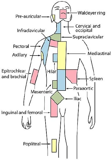
Each lymph node is a small but organized collection of lymphoid tissue (see green circular structures in Figure \(\PageIndex{1}\)) that contains many lymphocytes. Lymph nodes are located at intervals along the lymphatic vessels, and lymph passes through them on their way back to the blood. There are at least 500 lymph nodes in the human body. Many of them are clustered at the base of the limbs and in the neck. Figure \(\PageIndex{9}\) shows the major lymph node concentrations. The figure includes the spleen and the region named Waldeyer’s ring, consisting of the tonsils.
Lymph nodes near the surface of the body are obvious signs of immune system activity when they become enlarged and sometimes tender to the touch. Because it is easy to see and feel swollen lymph nodes, an individual can monitor his or her own health. It is important to be able to know the myths and realities of swollen lymph nodes.
Myth: You should see a doctor immediately whenever you have swollen lymph nodes.
Reality: Lymph nodes are constantly filtering lymph so it is expected that they will change in size with varying amounts of debris or pathogens that may be present. A minor, unnoticed infection may cause swollen lymph nodes that may last for a few weeks. Generally, lymph nodes that return to their normal size within three weeks are not a cause for concern.
Myth: Swollen lymph nodes mean you have a bacterial infection.
Reality: Although infection is the most common cause of swollen lymph nodes, not all infections are caused by bacteria. For example, mononucleosis commonly causes swollen lymph nodes, and it is caused by viruses. There are also other causes of swollen lymph nodes besides infections, such as cancer and certain medications.
Myth: A swollen lymph node means you have cancer.
Reality: Cancer is far less likely to be the cause of a swollen lymph node than is an infection.
Myth: Cancer in a lymph node always originates somewhere else. There is no cancer of the lymph nodes.
Reality: Cancers do commonly spread from their site of origin to nearby lymph nodes and then to other organs, but cancer may also originate in the lymph nodes. This type of cancer is called lymphoma.
Review
- What is the lymphatic system?
- Describe the composition of lymph.
- Outline the cardiovascular function of the lymphatic system.
- Describe the role of the lymphatic system in the absorption of nutrients from the digestive system.
- Summarize the function of the lymphatic system in host defense.
- Name primary lymphatic organs and their functions.
- What are the secondary lymphatic organs? State their functions in the adaptive immune system.
- How is interstitial fluid related to lymph?
- B and T cells are types of:
- Leukocytes
- Lymphocytes
- White blood cells
- All of the above
- For each of the following statements, indicate whether it applies to B cells, T cells, or both.
- These cells are born in the red bone marrow.
- These cells are part of the adaptive immune system.
- These cells mature in the bone marrow.
- Explain the difference between lymphocyte maturation and lymphocyte activation.
- True or False. The spleen produces lymphocytes.
- True or False. Tonsils are glands that produce lymph.
Explore More
Attributions
- Tonsillitis by Michaelbladon, Public Domain; via Wikimedia Commons
- Lymphatic System By Blausen.com staff (2014). "Medical gallery of Blausen Medical 2014". WikiJournal of Medicine 1 (2). DOI:10.15347/wjm/2014.010. ISSN 2002-4436. via Wikimedia Commons, licensed CC BY 3.0, via Wikimedia Commons
- Lymph Capillaries by US Government, Public Domain via Wikimedia Commons
- Intestinal Villus by Snow93, public domain via Wikimedia Commons
- White Blood Cells, CC BY 3.0, Blausen.com staff (2014). "Medical gallery of Blausen Medical 2014". WikiJournal of Medicine 1 (2). DOI:10.15347/wjm/2014.010. ISSN 2002-4436, licensed CC BY 3.0, via Wikimedia Commons
- Thymus by US Government, Public Domain via Wikimedia Commons
- Tonsils CC BY by Blausen.com staff (2014). "Medical gallery of Blausen Medical 2014". WikiJournal of Medicine 1 (2). DOI:10.15347/wjm/2014.010. ISSN 2002-4436. licensed CC BY 3.0, via Wikimedia Commons
- Spleen adapted from Illu Spleen by US government, public domain, via Wikimedia Commons
- Lymph Nodes by Fred the Oyster, public domain via Wikimedia Commons
- Text adapted from Human Biology by CK-12 licensed CC BY-NC 3.0