Biology LibreTexts

7.5: Blood

  • Page ID
    30662
  • \( \newcommand{\vecs}[1]{\overset { \scriptstyle \rightharpoonup} {\mathbf{#1}} } \)

    \( \newcommand{\vecd}[1]{\overset{-\!-\!\rightharpoonup}{\vphantom{a}\smash {#1}}} \)

    \( \newcommand{\id}{\mathrm{id}}\) \( \newcommand{\Span}{\mathrm{span}}\)

    ( \newcommand{\kernel}{\mathrm{null}\,}\) \( \newcommand{\range}{\mathrm{range}\,}\)

    \( \newcommand{\RealPart}{\mathrm{Re}}\) \( \newcommand{\ImaginaryPart}{\mathrm{Im}}\)

    \( \newcommand{\Argument}{\mathrm{Arg}}\) \( \newcommand{\norm}[1]{\| #1 \|}\)

    \( \newcommand{\inner}[2]{\langle #1, #2 \rangle}\)

    \( \newcommand{\Span}{\mathrm{span}}\)

    \( \newcommand{\id}{\mathrm{id}}\)

    \( \newcommand{\Span}{\mathrm{span}}\)

    \( \newcommand{\kernel}{\mathrm{null}\,}\)

    \( \newcommand{\range}{\mathrm{range}\,}\)

    \( \newcommand{\RealPart}{\mathrm{Re}}\)

    \( \newcommand{\ImaginaryPart}{\mathrm{Im}}\)

    \( \newcommand{\Argument}{\mathrm{Arg}}\)

    \( \newcommand{\norm}[1]{\| #1 \|}\)

    \( \newcommand{\inner}[2]{\langle #1, #2 \rangle}\)

    \( \newcommand{\Span}{\mathrm{span}}\) \( \newcommand{\AA}{\unicode[.8,0]{x212B}}\)

    \( \newcommand{\vectorA}[1]{\vec{#1}}      % arrow\)

    \( \newcommand{\vectorAt}[1]{\vec{\text{#1}}}      % arrow\)

    \( \newcommand{\vectorB}[1]{\overset { \scriptstyle \rightharpoonup} {\mathbf{#1}} } \)

    \( \newcommand{\vectorC}[1]{\textbf{#1}} \)

    \( \newcommand{\vectorD}[1]{\overrightarrow{#1}} \)

    \( \newcommand{\vectorDt}[1]{\overrightarrow{\text{#1}}} \)

    \( \newcommand{\vectE}[1]{\overset{-\!-\!\rightharpoonup}{\vphantom{a}\smash{\mathbf {#1}}}} \)

    \( \newcommand{\vecs}[1]{\overset { \scriptstyle \rightharpoonup} {\mathbf{#1}} } \)

    \( \newcommand{\vecd}[1]{\overset{-\!-\!\rightharpoonup}{\vphantom{a}\smash {#1}}} \)

    \(\newcommand{\avec}{\mathbf a}\) \(\newcommand{\bvec}{\mathbf b}\) \(\newcommand{\cvec}{\mathbf c}\) \(\newcommand{\dvec}{\mathbf d}\) \(\newcommand{\dtil}{\widetilde{\mathbf d}}\) \(\newcommand{\evec}{\mathbf e}\) \(\newcommand{\fvec}{\mathbf f}\) \(\newcommand{\nvec}{\mathbf n}\) \(\newcommand{\pvec}{\mathbf p}\) \(\newcommand{\qvec}{\mathbf q}\) \(\newcommand{\svec}{\mathbf s}\) \(\newcommand{\tvec}{\mathbf t}\) \(\newcommand{\uvec}{\mathbf u}\) \(\newcommand{\vvec}{\mathbf v}\) \(\newcommand{\wvec}{\mathbf w}\) \(\newcommand{\xvec}{\mathbf x}\) \(\newcommand{\yvec}{\mathbf y}\) \(\newcommand{\zvec}{\mathbf z}\) \(\newcommand{\rvec}{\mathbf r}\) \(\newcommand{\mvec}{\mathbf m}\) \(\newcommand{\zerovec}{\mathbf 0}\) \(\newcommand{\onevec}{\mathbf 1}\) \(\newcommand{\real}{\mathbb R}\) \(\newcommand{\twovec}[2]{\left[\begin{array}{r}#1 \\ #2 \end{array}\right]}\) \(\newcommand{\ctwovec}[2]{\left[\begin{array}{c}#1 \\ #2 \end{array}\right]}\) \(\newcommand{\threevec}[3]{\left[\begin{array}{r}#1 \\ #2 \\ #3 \end{array}\right]}\) \(\newcommand{\cthreevec}[3]{\left[\begin{array}{c}#1 \\ #2 \\ #3 \end{array}\right]}\) \(\newcommand{\fourvec}[4]{\left[\begin{array}{r}#1 \\ #2 \\ #3 \\ #4 \end{array}\right]}\) \(\newcommand{\cfourvec}[4]{\left[\begin{array}{c}#1 \\ #2 \\ #3 \\ #4 \end{array}\right]}\) \(\newcommand{\fivevec}[5]{\left[\begin{array}{r}#1 \\ #2 \\ #3 \\ #4 \\ #5 \\ \end{array}\right]}\) \(\newcommand{\cfivevec}[5]{\left[\begin{array}{c}#1 \\ #2 \\ #3 \\ #4 \\ #5 \\ \end{array}\right]}\) \(\newcommand{\mattwo}[4]{\left[\begin{array}{rr}#1 \amp #2 \\ #3 \amp #4 \\ \end{array}\right]}\) \(\newcommand{\laspan}[1]{\text{Span}\{#1\}}\) \(\newcommand{\bcal}{\cal B}\) \(\newcommand{\ccal}{\cal C}\) \(\newcommand{\scal}{\cal S}\) \(\newcommand{\wcal}{\cal W}\) \(\newcommand{\ecal}{\cal E}\) \(\newcommand{\coords}[2]{\left\{#1\right\}_{#2}}\) \(\newcommand{\gray}[1]{\color{gray}{#1}}\) \(\newcommand{\lgray}[1]{\color{lightgray}{#1}}\) \(\newcommand{\rank}{\operatorname{rank}}\) \(\newcommand{\row}{\text{Row}}\) \(\newcommand{\col}{\text{Col}}\) \(\renewcommand{\row}{\text{Row}}\) \(\newcommand{\nul}{\text{Nul}}\) \(\newcommand{\var}{\text{Var}}\) \(\newcommand{\corr}{\text{corr}}\) \(\newcommand{\len}[1]{\left|#1\right|}\) \(\newcommand{\bbar}{\overline{\bvec}}\) \(\newcommand{\bhat}{\widehat{\bvec}}\) \(\newcommand{\bperp}{\bvec^\perp}\) \(\newcommand{\xhat}{\widehat{\xvec}}\) \(\newcommand{\vhat}{\widehat{\vvec}}\) \(\newcommand{\uhat}{\widehat{\uvec}}\) \(\newcommand{\what}{\widehat{\wvec}}\) \(\newcommand{\Sighat}{\widehat{\Sigma}}\) \(\newcommand{\lt}{<}\) \(\newcommand{\gt}{>}\) \(\newcommand{\amp}{&}\) \(\definecolor{fillinmathshade}{gray}{0.9}\)
    Vampires

    From Bram Stoker’s famous novel about Count Dracula to the campy TV series Buffy the Vampire Slayer, fantasies featuring vampires, like the one in Figure \(\PageIndex{1}\), have been popular for decades. In fact, vampires are found in centuries-old myths from many cultures. In such myths, vampires are generally described as creatures that drink blood, preferably of the human variety, for sustenance. Dracula, for example, is based on Eastern European folklore about a human who attains immortality (and eternal damnation) by drinking the blood of others.

    Dracula
    Figure \(\PageIndex{1}\): Dracula

    Functions of Blood

    Blood performs many important functions in the body. Major functions of blood include:

    • supplying tissues with oxygen, which is needed by all cells for aerobic cellular respiration.
    • supplying cells with nutrients, including glucose, amino acids, and fatty acids.
    • removing metabolic wastes from cells, including carbon dioxide, urea, and lactic acid.
    • helping to defend the body from pathogens and other foreign substances.
    • forming clots to seal broken blood vessels and stop bleeding.
    • transporting hormones and other messenger molecules.
    • regulating the pH of the body, which must be kept within a narrow range (7.35 to 7.45).
    • helping to regulate body temperature (through vasoconstriction and vasodilation).

    What Is Blood?

    Blood is a fluid connective tissue that circulates throughout the body through blood vessels of the cardiovascular system. What makes blood so special that it features in widespread myths? Although blood accounts for less than 10 percent of human body weight, it is quite literally the elixir of life. As blood travels through the vessels of the cardiovascular system, it delivers vital substances such as nutrients and oxygen to all of the cells and carries away their metabolic wastes. It is no exaggeration to say that without blood, cells could not survive. Indeed, without the oxygen carried in the blood, cells of the brain start to die within just a matter of minutes.

    Blood centrifugation scheme
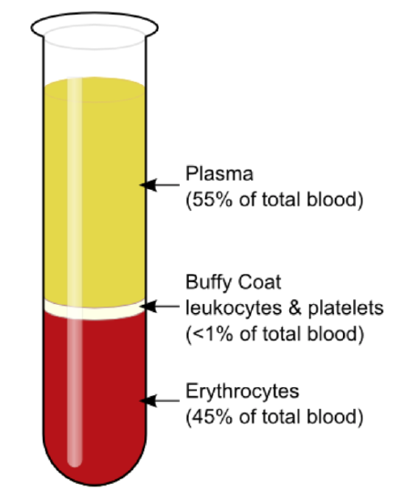
    Figure \(\PageIndex{2}\): If blood is centrifuged (spun at high speed), it separates into its major components based on density, as shown here: plasma, leukocytes (white blood cells) and platelets, and erythrocytes (red blood cells). Plasma makes up about 55% of the blood. Leukocytes and platelets make up less than 1%. Erythrocytes make up about 45%.

    The average adult body contains between 4.7 and 5.7 liters (5-6 quarts) of blood. More than half of that amount is fluid. Most of the rest of that amount consists of cells. The relative amounts of the various components in the blood are illustrated in Figure \(\PageIndex{2}\). The components are also described in the text.

    Blood Plasma

    Plasma is the liquid component of human blood and makes up about 55 percent of blood by volume. It is about 92 percent water and contains many dissolved substances. Most of these substances are proteins, but plasma also contains trace amounts of glucose, mineral ions, hormones, carbon dioxide, and other substances.

    Formed Elements

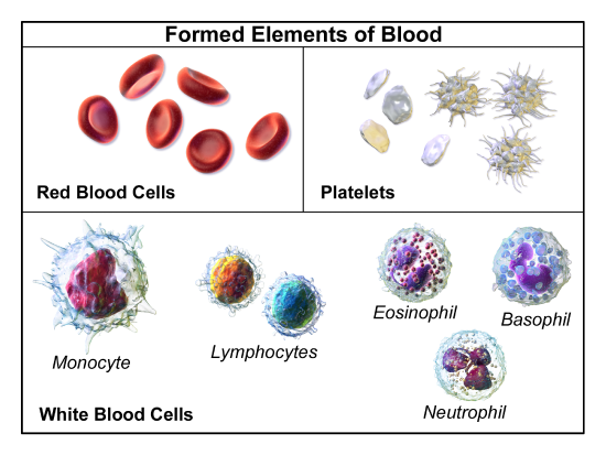
    The formed elements in the blood include red blood cells, white blood cells, and platelets. These different types of elements are pictured in Figure \(\PageIndex{3}\) and described in the sections that follow.

    Formed Elements
    Figure \(\PageIndex{3}\): Highly magnified blood cells in this image include donut-shaped red blood cells, rough-surfaced white blood cells, and small disc-shaped platelets.

    Red Blood Cells

    The most numerous cells in the blood are red blood cells, also called erythrocytes. One microliter of blood contains between 4.2 and 6.1 million red blood cells, and red blood cells make up about a quarter of all the cells in the human body. The cytoplasm of a mature red blood cell is almost completely filled with hemoglobin, the iron-containing protein that binds with oxygen and gives the cell its red color. Mature red blood cells lack a cell nucleus and most organelles in order to provide maximum space for hemoglobin. They are little more than sacks of hemoglobin.

    Red blood cells also carry proteins called antigens that determine blood type. Blood type is a genetic characteristic. The best known human blood type systems are the ABO and Rhesus systems. These are described in the next section.

    White Blood Cells (WBC)

    White blood cells are cells in the blood that defend the body against invading microorganisms and other threats. There are far fewer white blood cells (also called leukocytes) than red blood cells in the blood. There are normally only about 1,000 to 11,000 white blood cells per microliter of blood. Unlike red blood cells, white blood cells have a nucleus. White blood cells are part of the body’s immune system. They destroy and remove old or abnormal cells and cellular debris, as well as attack pathogens and foreign substances. There are two categories of WBCs, granulocytes (contain visible granules in the cytoplasm) and agranulocytes (do not contain granules). The Granulocytes include neutrophils, eosinophils, and basophils. The agranulocytes include lymphocytes and monocytes. The five types differ in their specific immune functions. The relative percent and functions of WBCs are summarized in Table \(\PageIndex{1}\):

    Table \(\PageIndex{1}\): Major Types of White Blood Cells
    Type of Leukocyte Percent of All Leukocytes Main Function(s)
    Neutrophil 62 Phagocytize (engulf and destroy) bacteria and fungi in the blood
    Eosinophil 2 Attack and kill large parasites; carry out allergic responses
    Basophil <1 Release histamines in inflammatory responses
    Lymphocyte 30 Attack and destroy virus-infected and tumor cells; create lasting immunity to specific pathogens
    Monocyte 5 Phagocytize pathogens and debris in tissues

    Platelets

    Platelets, also called thrombocytes, are actually cell fragments. Like red blood cells, they lack a nucleus and are more numerous than white blood cells. There are about 150,000 to 400,000 platelets per microliter of blood.

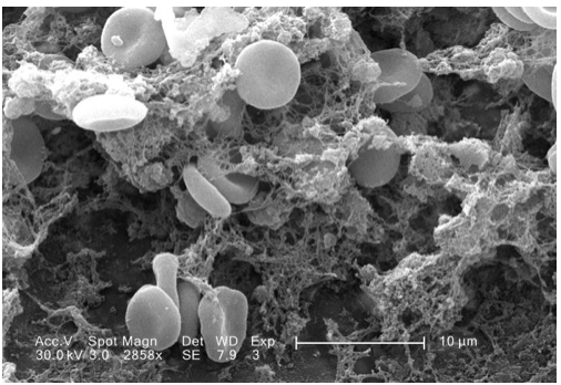
    The main function of platelets is blood clotting or coagulation. This is the process by which blood changes from a liquid to a gel, forming a plug in a damaged blood vessel. If blood clotting is successful, it results in hemostasis, which is the cessation of blood loss from the damaged vessel. A blood clot consists of both platelets and proteins, especially the protein fibrin. You can see a scanning electron microscope micrograph of a blood clot in Figure \(\PageIndex{4}\).

    Blood clot in scanning electron microscopy
    Figure \(\PageIndex{4}\): Red blood cells become trapped in a coagulating clot so they cannot escape through a break in a blood vessel.

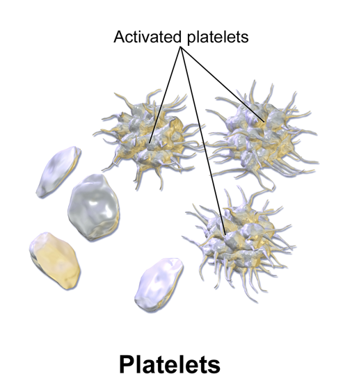
    Coagulation begins almost instantly after an injury to the endothelium of a blood vessel occurs. Platelets become activated and change their shape from spherical to star-shaped, as shown in Figure \(\PageIndex{5}\). This helps them aggregate with one another at the site of injury to start forming a plug in the vessel wall. Activated platelets also release substances into the blood that activate additional platelets and start a sequence of reactions leading to fibrin formation. Strands of fibrin crisscross the platelet plug and strengthen it, much as rebar strengthens concrete.

    activated Platelets
    Figure \(\PageIndex{5}\): The shape of platelets after they are activated helps them to stick together and form a plug for a damaged blood vessel.

    Formation and Degradation of Blood Cells

    Blood is considered to be a connective tissue because blood cells form inside bones. All three types of blood cells are made in the red marrow of bones in a process called hematopoiesis. The formation of blood cells occurs by the proliferation of stem cells in the marrow. These stem cells are self-renewing: when they divide, some of the daughter cells remain stem cells, so the pool of stem cells is not used up. Other daughter cells follow various pathways to differentiate into a variety of types of blood cells. Once the cells have differentiated, they cannot divide to form copies of themselves.

    Eventually, blood cells die and must be replaced through the formation of new blood cells from proliferating stem cells. After blood cells die, the dead cells are phagocytized (engulfed and destroyed) by white blood cells and removed from the circulation. This most often takes place in the spleen and liver.

    Blood Disorders

    Many human disorders primarily affect the blood. They include cancers, genetic disorders, poisoning by toxins, infections, and nutritional deficiencies.

    • Leukemia is a group of cancers of the blood-forming tissues in the bone marrow. It is the most common type of cancer in children, although most cases occur in adults. Leukemia is generally characterized by large numbers of abnormal white blood cells. Symptoms may include excessive bleeding and bruising, fatigue, fever, and an increased risk of infections. Leukemia is thought to be caused by a combination of genetic and environmental factors.
    • Hemophilia refers to any of several genetic disorders that cause dysfunction in the blood clotting process. People with hemophilia are prone to potentially uncontrollable bleeding even with otherwise inconsequential injuries. They also commonly suffer bleeding into the spaces between joints, which can cause crippling.
    • Carbon monoxide poisoning occurs when inhaled carbon monoxide (for example, in fumes from a faulty home furnace) binds irreversibly to the hemoglobin in red blood cells. As a result, oxygen cannot bind to the red cells for transport throughout the body, and this can quickly lead to suffocation. Carbon monoxide is extremely dangerous because it is colorless and odorless so it cannot be detected in the air by human senses.
    • HIV is a virus that infects certain types of white blood cells and interferes with the body’s ability to defend itself from pathogens and other causes of illness. HIV infection may eventually lead to AIDS or acquired immunodeficiency syndrome. AIDS is characterized by rare infections and cancers that people with a healthy immune system almost never acquire.
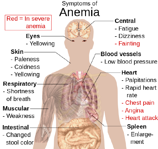
    Symptoms of anemia
    Figure \(\PageIndex{6}\): Anemia has wide-ranging effects on the human body because oxygen is essential for the normal functioning of cells in every organ system. The most severe symptoms include fainting, chest pain, angina, and heart attack.
    • Anemia is a disorder in which the blood has an inadequate volume of red blood cells. This reduces the amount of oxygen that the blood can carry and may cause weakness and fatigue. These and other signs and symptoms of anemia are shown in the figure below. Anemia has many possible causes, including excessive bleeding, inherited disorders such as sickle cell hemoglobin, or nutritional deficiencies (iron, folate, or B12). Severe anemia may require transfusions of donated blood.

    Feature: Myth vs. Reality


    Donating blood saves lives. In fact, with each blood donation, as many as three lives may be saved. The feeling that comes from knowing you have saved lives is well worth the short amount of time it takes to make a blood donation. Nonetheless, only a minority of potential donors actually donate blood. There are many myths about blood donation that contribute to the small percentage of donors. Knowing the facts may reaffirm your decision to donate if you are already a donor. If you aren’t a donor already, getting the facts may help you decide to become one.

    Myth: Your blood might become contaminated with an infection during the donation.

    Reality: There is no risk of contamination because only single-use, disposable catheters, tubing, and other equipment are used to collect blood for a donation.

    Myth: You are too old (or too young) to donate blood.

    Reality: There is no upper age limit on donating blood as long as you are healthy. The lower age limit is 16 years.

    Myth: You can’t donate blood if you have high blood pressure.

    Reality: As long as your blood pressure is below 180/100 at the time of donation, you can give blood. Even if you take blood pressure medication to keep your blood pressure below this level, you can donate.

    Myth: You can’t give blood if you have high cholesterol.

    Reality: Having high cholesterol does not affect your ability to donate blood. Taking cholesterol-lowering medication also does not disqualify you.

    Myth: You can’t donate blood if you have had a flu shot.

    Reality: Having a flu shot has no effect on your ability to donate blood. You can even donate on the same day that you receive a flu shot.

    Myth: You can’t donate blood if you take medication.

    Reality: As long as you are healthy, in most cases taking medication does not preclude you from donating blood.

    Myth: Your blood isn’t needed if it’s a common blood type.

    Reality: All types of blood are in constant demand.

    Review

    1. What is blood? Why is blood considered to be a connective tissue?
    2. Identify four physiological roles of blood in the body.
    3. Describe plasma and its components.
    4. Identify red blood cells and their major function.
    5. What are white blood cells? Which body system besides the cardiovascular system includes white blood cells?
    6. Explain how platelets cause coagulation.
    7. Summarize the formation and degradation of blood cells.
    8. Identify three disorders of the blood.
    9. For each of the descriptions below, choose the blood cell that best fits the description. Blood cells: red blood cells; white blood cells; platelets
      1. Has a nucleus
      2. Responsible for blood clotting
      3. Carbon monoxide binds to a protein in these cells
    10. What is another name for erythrocytes? What is another name for leukocytes?
    11. True or False. Plasma refers to the cytoplasm within blood cells.
    12. True or False. Platelets are cell fragments.

    Explore More

    https://bio.libretexts.org/link?16827#Explore_More

    In this TED talk, inventor Joe Landolina talks about his potentially life-saving invention: a medical gel that can instantly stop bleeding.

    Attributions

    1. Dracula by Clifton Chu; public domain via Pixy.org
    2. Blood Centrifugation by KnuteKnudsen licensed CC BY 3.0 via Wikimedia Commons
    3. Formed elements by Blausen.com staff (2014). "Medical gallery of Blausen Medical 2014". WikiJournal of Medicine 1 (2). DOI:10.15347/wjm/2014.010. ISSN 2002-4436. licensed CC BY 3.0 via Wikimedia Commons
    4. Blood clot in scanning electron microscopy by Janice Carr; CDC; public domain via Wikimedia Commons
    5. Platelets by Blausen.com staff (2014). "Medical gallery of Blausen Medical 2014". WikiJournal of Medicine 1 (2). DOI:10.15347/wjm/2014.010. ISSN 2002-4436.licensed CC BY 3.0 via Wikimedia Commons
    6. Symptoms of anemia by Mikael Häggström; Public domain via Wikimedia Commons
    7. Text adapted from Human Biology by CK-12 licensed CC BY-NC 3.0

    This page titled 7.5: Blood is shared under a CK-12 license and was authored, remixed, and/or curated by Suzanne Wakim & Mandeep Grewal via source content that was edited to the style and standards of the LibreTexts platform.

    CK-12 Foundation
    LICENSED UNDER
    CK-12 Foundation is licensed under CK-12 Curriculum Materials License