
3.3: Variation in Cells
- Page ID
- 30622

\( \newcommand{\vecs}[1]{\overset { \scriptstyle \rightharpoonup} {\mathbf{#1}} } \)
\( \newcommand{\vecd}[1]{\overset{-\!-\!\rightharpoonup}{\vphantom{a}\smash {#1}}} \)
\( \newcommand{\id}{\mathrm{id}}\) \( \newcommand{\Span}{\mathrm{span}}\)
( \newcommand{\kernel}{\mathrm{null}\,}\) \( \newcommand{\range}{\mathrm{range}\,}\)
\( \newcommand{\RealPart}{\mathrm{Re}}\) \( \newcommand{\ImaginaryPart}{\mathrm{Im}}\)
\( \newcommand{\Argument}{\mathrm{Arg}}\) \( \newcommand{\norm}[1]{\| #1 \|}\)
\( \newcommand{\inner}[2]{\langle #1, #2 \rangle}\)
\( \newcommand{\Span}{\mathrm{span}}\)
\( \newcommand{\id}{\mathrm{id}}\)
\( \newcommand{\Span}{\mathrm{span}}\)
\( \newcommand{\kernel}{\mathrm{null}\,}\)
\( \newcommand{\range}{\mathrm{range}\,}\)
\( \newcommand{\RealPart}{\mathrm{Re}}\)
\( \newcommand{\ImaginaryPart}{\mathrm{Im}}\)
\( \newcommand{\Argument}{\mathrm{Arg}}\)
\( \newcommand{\norm}[1]{\| #1 \|}\)
\( \newcommand{\inner}[2]{\langle #1, #2 \rangle}\)
\( \newcommand{\Span}{\mathrm{span}}\) \( \newcommand{\AA}{\unicode[.8,0]{x212B}}\)
\( \newcommand{\vectorA}[1]{\vec{#1}} % arrow\)
\( \newcommand{\vectorAt}[1]{\vec{\text{#1}}} % arrow\)
\( \newcommand{\vectorB}[1]{\overset { \scriptstyle \rightharpoonup} {\mathbf{#1}} } \)
\( \newcommand{\vectorC}[1]{\textbf{#1}} \)
\( \newcommand{\vectorD}[1]{\overrightarrow{#1}} \)
\( \newcommand{\vectorDt}[1]{\overrightarrow{\text{#1}}} \)
\( \newcommand{\vectE}[1]{\overset{-\!-\!\rightharpoonup}{\vphantom{a}\smash{\mathbf {#1}}}} \)
\( \newcommand{\vecs}[1]{\overset { \scriptstyle \rightharpoonup} {\mathbf{#1}} } \)
\( \newcommand{\vecd}[1]{\overset{-\!-\!\rightharpoonup}{\vphantom{a}\smash {#1}}} \)
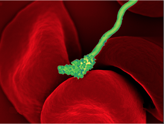
\(\newcommand{\avec}{\mathbf a}\) \(\newcommand{\bvec}{\mathbf b}\) \(\newcommand{\cvec}{\mathbf c}\) \(\newcommand{\dvec}{\mathbf d}\) \(\newcommand{\dtil}{\widetilde{\mathbf d}}\) \(\newcommand{\evec}{\mathbf e}\) \(\newcommand{\fvec}{\mathbf f}\) \(\newcommand{\nvec}{\mathbf n}\) \(\newcommand{\pvec}{\mathbf p}\) \(\newcommand{\qvec}{\mathbf q}\) \(\newcommand{\svec}{\mathbf s}\) \(\newcommand{\tvec}{\mathbf t}\) \(\newcommand{\uvec}{\mathbf u}\) \(\newcommand{\vvec}{\mathbf v}\) \(\newcommand{\wvec}{\mathbf w}\) \(\newcommand{\xvec}{\mathbf x}\) \(\newcommand{\yvec}{\mathbf y}\) \(\newcommand{\zvec}{\mathbf z}\) \(\newcommand{\rvec}{\mathbf r}\) \(\newcommand{\mvec}{\mathbf m}\) \(\newcommand{\zerovec}{\mathbf 0}\) \(\newcommand{\onevec}{\mathbf 1}\) \(\newcommand{\real}{\mathbb R}\) \(\newcommand{\twovec}[2]{\left[\begin{array}{r}#1 \\ #2 \end{array}\right]}\) \(\newcommand{\ctwovec}[2]{\left[\begin{array}{c}#1 \\ #2 \end{array}\right]}\) \(\newcommand{\threevec}[3]{\left[\begin{array}{r}#1 \\ #2 \\ #3 \end{array}\right]}\) \(\newcommand{\cthreevec}[3]{\left[\begin{array}{c}#1 \\ #2 \\ #3 \end{array}\right]}\) \(\newcommand{\fourvec}[4]{\left[\begin{array}{r}#1 \\ #2 \\ #3 \\ #4 \end{array}\right]}\) \(\newcommand{\cfourvec}[4]{\left[\begin{array}{c}#1 \\ #2 \\ #3 \\ #4 \end{array}\right]}\) \(\newcommand{\fivevec}[5]{\left[\begin{array}{r}#1 \\ #2 \\ #3 \\ #4 \\ #5 \\ \end{array}\right]}\) \(\newcommand{\cfivevec}[5]{\left[\begin{array}{c}#1 \\ #2 \\ #3 \\ #4 \\ #5 \\ \end{array}\right]}\) \(\newcommand{\mattwo}[4]{\left[\begin{array}{rr}#1 \amp #2 \\ #3 \amp #4 \\ \end{array}\right]}\) \(\newcommand{\laspan}[1]{\text{Span}\{#1\}}\) \(\newcommand{\bcal}{\cal B}\) \(\newcommand{\ccal}{\cal C}\) \(\newcommand{\scal}{\cal S}\) \(\newcommand{\wcal}{\cal W}\) \(\newcommand{\ecal}{\cal E}\) \(\newcommand{\coords}[2]{\left\{#1\right\}_{#2}}\) \(\newcommand{\gray}[1]{\color{gray}{#1}}\) \(\newcommand{\lgray}[1]{\color{lightgray}{#1}}\) \(\newcommand{\rank}{\operatorname{rank}}\) \(\newcommand{\row}{\text{Row}}\) \(\newcommand{\col}{\text{Col}}\) \(\renewcommand{\row}{\text{Row}}\) \(\newcommand{\nul}{\text{Nul}}\) \(\newcommand{\var}{\text{Var}}\) \(\newcommand{\corr}{\text{corr}}\) \(\newcommand{\len}[1]{\left|#1\right|}\) \(\newcommand{\bbar}{\overline{\bvec}}\) \(\newcommand{\bhat}{\widehat{\bvec}}\) \(\newcommand{\bperp}{\bvec^\perp}\) \(\newcommand{\xhat}{\widehat{\xvec}}\) \(\newcommand{\vhat}{\widehat{\vvec}}\) \(\newcommand{\uhat}{\widehat{\uvec}}\) \(\newcommand{\what}{\widehat{\wvec}}\) \(\newcommand{\Sighat}{\widehat{\Sigma}}\) \(\newcommand{\lt}{<}\) \(\newcommand{\gt}{>}\) \(\newcommand{\amp}{&}\) \(\definecolor{fillinmathshade}{gray}{0.9}\)Figure \(\PageIndex{1}\) shows a bacterial cell (colored green) attacking human red blood cells. The bacterium causes a disease called relapsing fever. The bacterial and human cells look very different in size and shape. Although all living cells have certain things in common — such as a plasma membrane and cytoplasm — different types of cells, even within the same organism, may have their own unique structures and functions. Cells with different functions generally have different shapes that suit them for their particular job. Cells vary not only in shape but also in size, as this example shows. In most organisms, however, even the largest cells are no bigger than the period at the end of this sentence. Why are cells so small?
Explaining Cell Size
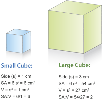
| Characteristic | Small Cube | Large Cube |
|---|---|---|
| sides (S) | \( 1 cm \) | \( 3 cm \) |
| Surface Area (SA) | \(6 S^2 = 6 \times 1^2 = 6 cm^2 \) | \(6 S^2 = 6 \times 3^2 = 54 cm^2 \) |
| Volume (V) | \(S^3 = 1^3 = 1 cm^3\) | \(S^3 = 3^3 = 27 cm^3\) |
| SA:V | \(SA/V = 6/1 = 6\) | \(SA/V = 54/27 = 2\) |
Most organisms, even very large ones, have microscopic cells. Why don't cells get bigger instead of remaining tiny and multiplying? What limits cell size?
The answers to these questions are clear once you know how a cell functions. To carry out life processes, a cell must be able to quickly pass substances into and out of the cell. For example, it must be able to pass nutrients and oxygen into the cell and waste products out of the cell. Anything that enters or leaves a cell must cross its outer surface. It is this need to pass substances across the surface that limits how large a cell can be.
Look at the two cubes in Figure \(\PageIndex{2}\). As this figure and table show, a larger cube has less surface area relative to its volume than a smaller cube. This relationship also applies to cells; a larger cell has less surface area relative to its volume than a smaller cell. A cell with a larger volume also needs more nutrients and oxygen and produces more wastes. Because all of these substances must pass through the surface of the cell, a cell with a large volume will not have enough surface area to allow it to meet its needs. The larger the cell is, the smaller its ratio of surface area to volume, and the harder it will be for the cell to get rid of its wastes and take in necessary substances. This is what limits the size of the cell.
Cell Form and Function
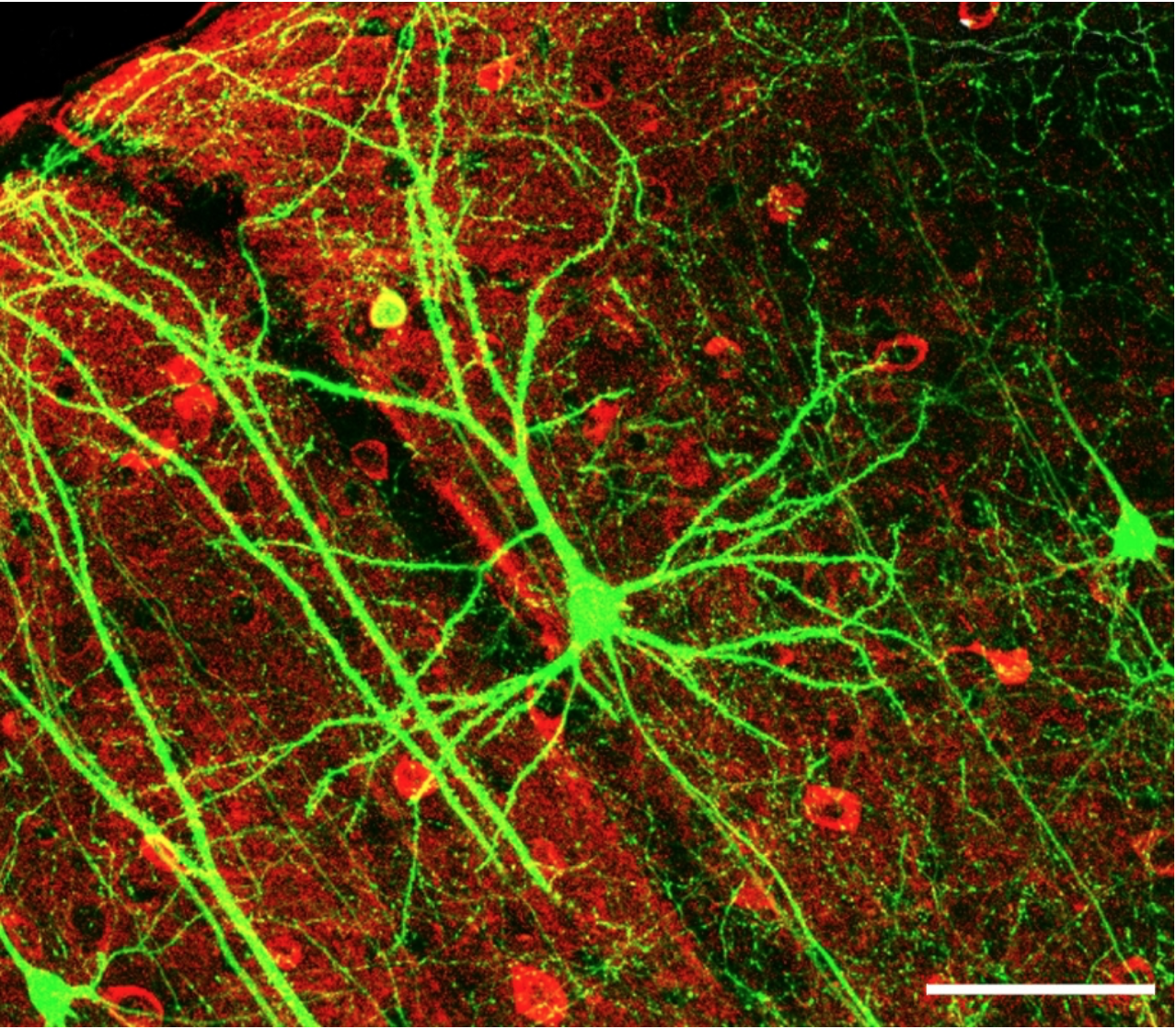
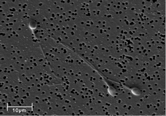
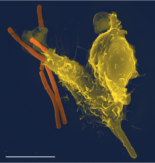
Cells with different functions often have different shapes. The cells in Figure \(\PageIndex{3}\) - Figure \(\PageIndex{5}\) are just a few examples of the many different shapes that human cells may have. Each type of cell in the figure has characteristics that help it do its job. For example, the job of the nerve cell is to carry messages to other cells. The nerve cell has many long extensions that reach out in all directions, allowing it to pass messages to many other cells at once. Do you see the tail of each tiny sperm cell? Its tail helps a sperm cell "swim" through fluids in the female reproductive tract in order to reach an egg cell. The white blood cell has the job of destroying bacteria and other pathogens. Figure \(\PageIndex{5}\) shows the large white blood cell (in yellow) engulfing and destroying bacteria (in orange).
Cells With and Without a Nucleus
There is a basic cell structure that is present in many but not all living cells: the nucleus. The nucleus of a cell is a structure in the cytoplasm that is surrounded by a membrane (the nuclear membrane) and contains DNA. Based on whether or not they have a nucleus, there are two basic types of cells: prokaryotic cells and eukaryotic cells.
Prokaryotic Cells
Prokaryotic cells are cells without a nucleus. The DNA in prokaryotic cells is in the cytoplasm rather than enclosed within a nuclear membrane. Prokaryotic cells are found in single-celled organisms, such as the bacterium represented by the model below. Organisms with prokaryotic cells are called prokaryotes. They were the first type of organisms to evolve and are still the most common organisms today.
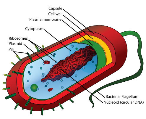
| Cell Structure | Description |
|---|---|
| Flagellum | Long projection(s) outside of the cell in some bacteria; aids in the motility |
| Pili | Small projections outside of the cell; aid in attachment |
| Capsule | A thick protective layer outside the cell wall of some bacteria |
| Cell wall | Outer layer of bacterial cells; more chemically complex than eukaryotic cell walls |
| Plasma Membrane | Phospholipid bilayer marking the outside of the cytoplasm |
| Cytoplasm | The fluid portion of the cell |
| Ribosome | Involved in protein synthesis |
| Nucleoid | Circular DNA found in the cytoplasm |
| Plasmid | Small loops of DNA found in some bacteria |
Eukaryotic Cells
Eukaryotic cells are cells that contain a nucleus. A typical eukaryotic cell is represented by the model below. Eukaryotic cells are usually larger than prokaryotic cells. They are found in some single-celled and all multicellular organisms. Organisms with eukaryotic cells are called eukaryotes, and they range from fungi to people.
Besides a nucleus, eukaryotic cells also contain other organelles. An organelle is a structure within the cytoplasm that performs a specific job in the cell. Organelles called mitochondria, for example, provide energy to the cell, and organelles called vacuoles store substances in the cell. Organelles allow eukaryotic cells to carry out more functions than prokaryotic cells can.
| Structure | Location | Description |
|---|---|---|
| Flagellum | Outside the cell | A projection used for locomotion in some eukaryotic cells |
| Plasma Membrane | Outer layer of cell | Phospholipid bilayer enclosing the cytoplasm |
| Cytoplasm | Bound by the plasma membrane | Entire region between the plasma membrane and the nuclear envelope, consisting of organelles suspended in the gel-like cytosol, the cytoskeleton, and various chemicals |
| Golgi Vesicles (Golgi Apparatus) |
Cytoplasm | A series of stacked membranes that sorts, tags, and packages lipids and proteins for distribution |
| Ribosomes | free-floating or on rough ER | Involved in protein synthesis |
| Rough Endoplasmic Reticulum | Cytoplasm | Interconnected membranous structures that are studded with ribosomes and engage in protein modification and phospholipid synthesis |
| Smooth Endoplasmic Reticulum | Cytoplasm | Interconnected membranous structures that have few or no ribosomes on its cytoplasmic surface and synthesize carbohydrates, lipids, and steroid hormones; detoxifies certain chemicals (like pesticides, preservatives, medications, and environmental pollutants), and stores calcium ions |
| Mitochondria | Cytoplasm | (singular = mitochondrion) cellular organelles responsible for carrying out cellular respiration, resulting in producing ATP, the cell’s main energy-carrying molecule |
| Peroxisome | Cytoplasm | The small, round organelle that contains hydrogen peroxide, and detoxifies many poisons |
| Lysosome | Cytoplasm | Organelle in an animal cell that functions as the cell’s digestive component; it breaks down proteins, polysaccharides, lipids, nucleic acids, and even worn-out organelles |
| Secretory Vesicle | Cytoplasm | Small, membrane-bound sac that functions in cellular storage and transport; its membrane is capable of fusing with the plasma membrane and the membranes of the endoplasmic reticulum and Golgi apparatus |
| Centrosome (with 2 centrioles) |
Cytoplasm | Region in animal cells made of two centrioles that serve as an organizing center for microtubules |
| Actin Filaments | Cytoskeleton | The cytoskeleton's narrowest element; it provides rigidity and shape to the cell and enables cellular movements |
| Intermediate Filaments | Cytoskeleton | Cytoskeletal component, comprised of several fibrous protein intertwined strands, that bears tension, supports cell-cell junctions, and anchors cells to extracellular structures |
| Microtubules | Cytoskeleton | The cytoskeleton’s widest element; provides a track along which vesicles move through the cell, pulls replicated chromosomes to opposite ends of a dividing cell, and is the structural element of centrioles |
| Cytoskeleton | Throughout cell | Protein fiber network that collectively maintains the cell’s shape, secures some organelles in specific positions and allows cytoplasm and vesicles to move within the cell |
| Nucleus | Cytoplasm | Cell organelle that houses the cell’s DNA and directs ribosome and protein synthesis |
| Nuclear Pore | Nucleus | Pores in the nuclear envelope allow substances to enter and exit the nucleus. |
| Nuclear Envelope | Nucleus | Double-membrane structure that constitutes the nucleus’ outermost portion |
| Chromatin | Nucleus | Protein-DNA complex that serves as the chromosomes’ building material |
| Nucleolus | Nucleus | Darkly staining body within the nucleus that is responsible for assembling ribosome subunits |

Review
- Explain why most cells are very small.
- Discuss variations in the form and function of cells.
- Compare and contrast prokaryotic and eukaryotic cells.
- True or False. Prokaryotic cells do not have mitochondria.
- True or False. Prokaryotic cells do not have DNA.
- True or False. All single-celled organisms are prokaryotes.
- Which was the first type of organism to evolve – eukaryotes or prokaryotes? Based on their structures, does this make sense to you? Explain your answer.
- Do human cells have organelles? Explain your answer.
- Which are usually larger – prokaryotic or eukaryotic cells? What do you think this means for their relative ability to take in needed substances and release wastes? Discuss your answer.
- DNA in eukaryotes is enclosed within the _______ ________.
- Name three different types of cells in humans.
- Which organelle provides energy in eukaryotic cells?
- What is the function of a vacuole in a cell?
Explore More
The video below explains why scientists believe endosymbiosis is the basis for complex cells.
Attributions
- Borrelia hermsii Bacteria by NIAI, public domain via Wikimedia Commons
- Cubes by Hana Zavadska; licensed CC BY-NC 3.0 via CK-12 Foundation
- Neuron by Wei-Chung Allen Le, et. al. from the PLOS article Dynamic Remodeling of Dendritic Arbors in GABAergic Interneurons of Adult Visual Cortex, CC BY 2.5 via Wikimedia Commons
- Spermatozoa public domain via Wikimedia Commons
- Neutrophil with anthrax by Volker Brinkmann, CC BY 2.5 via Wikimedia Commons
- Average prokaryote by LadyofHats, released into the public domain via Wikimedia Commons
- Animal cell by LadyofHats, released into the public domain via Wikimedia Commons
- Cell structures, endomembrane system, and cytoskeleton; for cell table adapted from Biology by BC Campus, CC BY 4.0
- Text adapted from Human Biology by CK-12 licensed CC BY-NC 3.0