Biology LibreTexts

23.2: Germinal Stage

  • Page ID
    22626
  • \( \newcommand{\vecs}[1]{\overset { \scriptstyle \rightharpoonup} {\mathbf{#1}} } \)

    \( \newcommand{\vecd}[1]{\overset{-\!-\!\rightharpoonup}{\vphantom{a}\smash {#1}}} \)

    \( \newcommand{\id}{\mathrm{id}}\) \( \newcommand{\Span}{\mathrm{span}}\)

    ( \newcommand{\kernel}{\mathrm{null}\,}\) \( \newcommand{\range}{\mathrm{range}\,}\)

    \( \newcommand{\RealPart}{\mathrm{Re}}\) \( \newcommand{\ImaginaryPart}{\mathrm{Im}}\)

    \( \newcommand{\Argument}{\mathrm{Arg}}\) \( \newcommand{\norm}[1]{\| #1 \|}\)

    \( \newcommand{\inner}[2]{\langle #1, #2 \rangle}\)

    \( \newcommand{\Span}{\mathrm{span}}\)

    \( \newcommand{\id}{\mathrm{id}}\)

    \( \newcommand{\Span}{\mathrm{span}}\)

    \( \newcommand{\kernel}{\mathrm{null}\,}\)

    \( \newcommand{\range}{\mathrm{range}\,}\)

    \( \newcommand{\RealPart}{\mathrm{Re}}\)

    \( \newcommand{\ImaginaryPart}{\mathrm{Im}}\)

    \( \newcommand{\Argument}{\mathrm{Arg}}\)

    \( \newcommand{\norm}[1]{\| #1 \|}\)

    \( \newcommand{\inner}[2]{\langle #1, #2 \rangle}\)

    \( \newcommand{\Span}{\mathrm{span}}\) \( \newcommand{\AA}{\unicode[.8,0]{x212B}}\)

    \( \newcommand{\vectorA}[1]{\vec{#1}}      % arrow\)

    \( \newcommand{\vectorAt}[1]{\vec{\text{#1}}}      % arrow\)

    \( \newcommand{\vectorB}[1]{\overset { \scriptstyle \rightharpoonup} {\mathbf{#1}} } \)

    \( \newcommand{\vectorC}[1]{\textbf{#1}} \)

    \( \newcommand{\vectorD}[1]{\overrightarrow{#1}} \)

    \( \newcommand{\vectorDt}[1]{\overrightarrow{\text{#1}}} \)

    \( \newcommand{\vectE}[1]{\overset{-\!-\!\rightharpoonup}{\vphantom{a}\smash{\mathbf {#1}}}} \)

    \( \newcommand{\vecs}[1]{\overset { \scriptstyle \rightharpoonup} {\mathbf{#1}} } \)

    \( \newcommand{\vecd}[1]{\overset{-\!-\!\rightharpoonup}{\vphantom{a}\smash {#1}}} \)

    \(\newcommand{\avec}{\mathbf a}\) \(\newcommand{\bvec}{\mathbf b}\) \(\newcommand{\cvec}{\mathbf c}\) \(\newcommand{\dvec}{\mathbf d}\) \(\newcommand{\dtil}{\widetilde{\mathbf d}}\) \(\newcommand{\evec}{\mathbf e}\) \(\newcommand{\fvec}{\mathbf f}\) \(\newcommand{\nvec}{\mathbf n}\) \(\newcommand{\pvec}{\mathbf p}\) \(\newcommand{\qvec}{\mathbf q}\) \(\newcommand{\svec}{\mathbf s}\) \(\newcommand{\tvec}{\mathbf t}\) \(\newcommand{\uvec}{\mathbf u}\) \(\newcommand{\vvec}{\mathbf v}\) \(\newcommand{\wvec}{\mathbf w}\) \(\newcommand{\xvec}{\mathbf x}\) \(\newcommand{\yvec}{\mathbf y}\) \(\newcommand{\zvec}{\mathbf z}\) \(\newcommand{\rvec}{\mathbf r}\) \(\newcommand{\mvec}{\mathbf m}\) \(\newcommand{\zerovec}{\mathbf 0}\) \(\newcommand{\onevec}{\mathbf 1}\) \(\newcommand{\real}{\mathbb R}\) \(\newcommand{\twovec}[2]{\left[\begin{array}{r}#1 \\ #2 \end{array}\right]}\) \(\newcommand{\ctwovec}[2]{\left[\begin{array}{c}#1 \\ #2 \end{array}\right]}\) \(\newcommand{\threevec}[3]{\left[\begin{array}{r}#1 \\ #2 \\ #3 \end{array}\right]}\) \(\newcommand{\cthreevec}[3]{\left[\begin{array}{c}#1 \\ #2 \\ #3 \end{array}\right]}\) \(\newcommand{\fourvec}[4]{\left[\begin{array}{r}#1 \\ #2 \\ #3 \\ #4 \end{array}\right]}\) \(\newcommand{\cfourvec}[4]{\left[\begin{array}{c}#1 \\ #2 \\ #3 \\ #4 \end{array}\right]}\) \(\newcommand{\fivevec}[5]{\left[\begin{array}{r}#1 \\ #2 \\ #3 \\ #4 \\ #5 \\ \end{array}\right]}\) \(\newcommand{\cfivevec}[5]{\left[\begin{array}{c}#1 \\ #2 \\ #3 \\ #4 \\ #5 \\ \end{array}\right]}\) \(\newcommand{\mattwo}[4]{\left[\begin{array}{rr}#1 \amp #2 \\ #3 \amp #4 \\ \end{array}\right]}\) \(\newcommand{\laspan}[1]{\text{Span}\{#1\}}\) \(\newcommand{\bcal}{\cal B}\) \(\newcommand{\ccal}{\cal C}\) \(\newcommand{\scal}{\cal S}\) \(\newcommand{\wcal}{\cal W}\) \(\newcommand{\ecal}{\cal E}\) \(\newcommand{\coords}[2]{\left\{#1\right\}_{#2}}\) \(\newcommand{\gray}[1]{\color{gray}{#1}}\) \(\newcommand{\lgray}[1]{\color{lightgray}{#1}}\) \(\newcommand{\rank}{\operatorname{rank}}\) \(\newcommand{\row}{\text{Row}}\) \(\newcommand{\col}{\text{Col}}\) \(\renewcommand{\row}{\text{Row}}\) \(\newcommand{\nul}{\text{Nul}}\) \(\newcommand{\var}{\text{Var}}\) \(\newcommand{\corr}{\text{corr}}\) \(\newcommand{\len}[1]{\left|#1\right|}\) \(\newcommand{\bbar}{\overline{\bvec}}\) \(\newcommand{\bhat}{\widehat{\bvec}}\) \(\newcommand{\bperp}{\bvec^\perp}\) \(\newcommand{\xhat}{\widehat{\xvec}}\) \(\newcommand{\vhat}{\widehat{\vvec}}\) \(\newcommand{\uhat}{\widehat{\uvec}}\) \(\newcommand{\what}{\widehat{\wvec}}\) \(\newcommand{\Sighat}{\widehat{\Sigma}}\) \(\newcommand{\lt}{<}\) \(\newcommand{\gt}{>}\) \(\newcommand{\amp}{&}\) \(\definecolor{fillinmathshade}{gray}{0.9}\)
    Experienced Newborn

    This newborn baby is just starting out in life. They have their whole life ahead of them!

    Actually, that’s not really true. While most of their life is still ahead of them — including life stages of infancy, childhood, adolescence, and adulthood — this newborn baby is not just starting out in life. They are already nine months old, and what happened to them during those nine months will help shape the rest of their life. Some of the shortest — but most important — life stages occur before birth. These stages include the germinal, embryonic, and fetal stages. This concept focuses on the earliest of all human life stages: the germinal stage.

    Newborn baby
    Figure \(\PageIndex{1}\): newborn

    What Is the Germinal Stage?

    human-fertilization.png
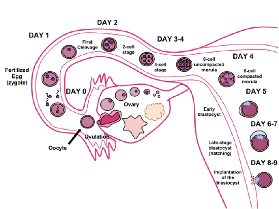
    Figure \(\PageIndex{2}\): The germinal stage of human development begins with fertilization in a Fallopian tube and ends with implantation in the uterus.

    The germinal stage of development is the first and shortest of the stages of the human lifespan. The main events in this stage of development are illustrated in Figure \(\PageIndex{2}\) and described in detail in the rest of this concept. The germinal stage lasts a total of eight to nine days. It begins in a Fallopian tube when an ovum is fertilized by a sperm to form a zygote (day 0). The germinal stage continues as the zygote undergoes several initial cell divisions to form a solid ball of cells called a morula (days 3-4). It then continues as the morula undergoes additional changes to become a hollow ball of cells called a blastocyst (days 5-7). The germinal stage ends when the blastocyst implants in the endometrium of the uterus (days 8-9). After implantation occurs, the blastocyst is called an embryo, and it will soon obtain nutrients from the mother’s blood via a temporary organ called the placenta. In the germinal stage, however, nutrients must be obtained from cell cytoplasm or secretions in the Fallopian tube or uterus.

    Processes in the Germinal Stage

    The germinal stage involves several different processes that change an egg and sperm first into a zygote, and then into an embryo. The processes include fertilization, cleavage, blastulation, and implantation.

    Fertilization

    egg fertilization

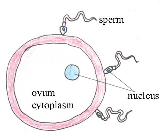
    Figure \(\PageIndex{3}\): This drawing represents sperm–egg interaction.

    Many sperm travel towards the egg due to chemical attraction. However, only one sperm will succeed in fertilizing the ovum (egg), by penetrating its cell membrane and depositing the genetic material into the egg, where the two nuclei fuse. The fertilized ovum (zygote) immediately becomes resistant to penetration by any other sperm arriving later. After fertilization occurs, the zygote remains in the fallopian tube for about 72 hours, and during this time it develops rapidly

    Cleavage

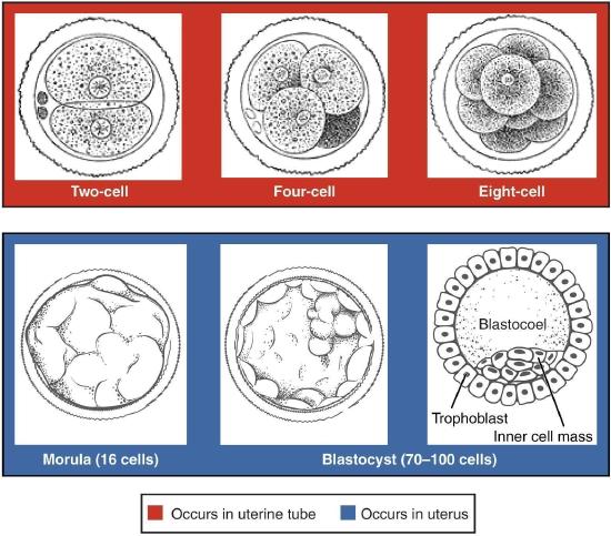
    By the second day after fertilization, the single-celled zygote undergoes mitosis to form two daughter cells. Mitosis continues to take place every 12 to 24 hours to produce the first four cells, then eight, and as many as sixteen cells by day 4. These early mitotic divisions are called cleavage. By day 4, the cells form a solid ball called a morula (see Figure \(\PageIndex{4}\)). Although cleavage results in more cells, the overall mass of cells making up the morula is still the same size as the initial zygote because the cells are confined within the zona pellucida. A large amount of cytoplasm in the original zygote becomes subdivided among the multiple cells of the morula.

    Blastulation

    Blastulation is the process of changing the morula into a blastocyst. It occurs from roughly day 5 to day 7 after fertilization. During blastulation, the morula changes from a solid ball of undifferentiated cells into a fluid-filled ball of differentiated cells, as shown in Figure \(\PageIndex{4}\). The major parts of the fully formed blastocyst are the embryoblast, trophoblast, and blastocoele.

    • The embryoblast (inner cell mass) consists of a mass of cells inside the blastocyst. These cells will eventually develop into the embryo.
    • The trophoblast is the outer cell layer of the blastocyst. Trophoblast cells will implant in the uterus and eventually develop into the placenta and other embryonic tissues.
    • The blastocoele is a cavity formed by the migration of embryoblast cells to one pole of the blastocyst. The blastocoele fills with fluid secreted by trophoblast cells.
    cleavage, morula, and blastula
    Figure \(\PageIndex{4}\): This image shows a morula consisting of eight cells on day 4 after fertilization. The image also shows a blastocyst consists of two types of differentiated cells (inner cell mass and trophoblast) and a fluid-filled cavity (blastocoele).

    Implantation

     Implantation in uterus
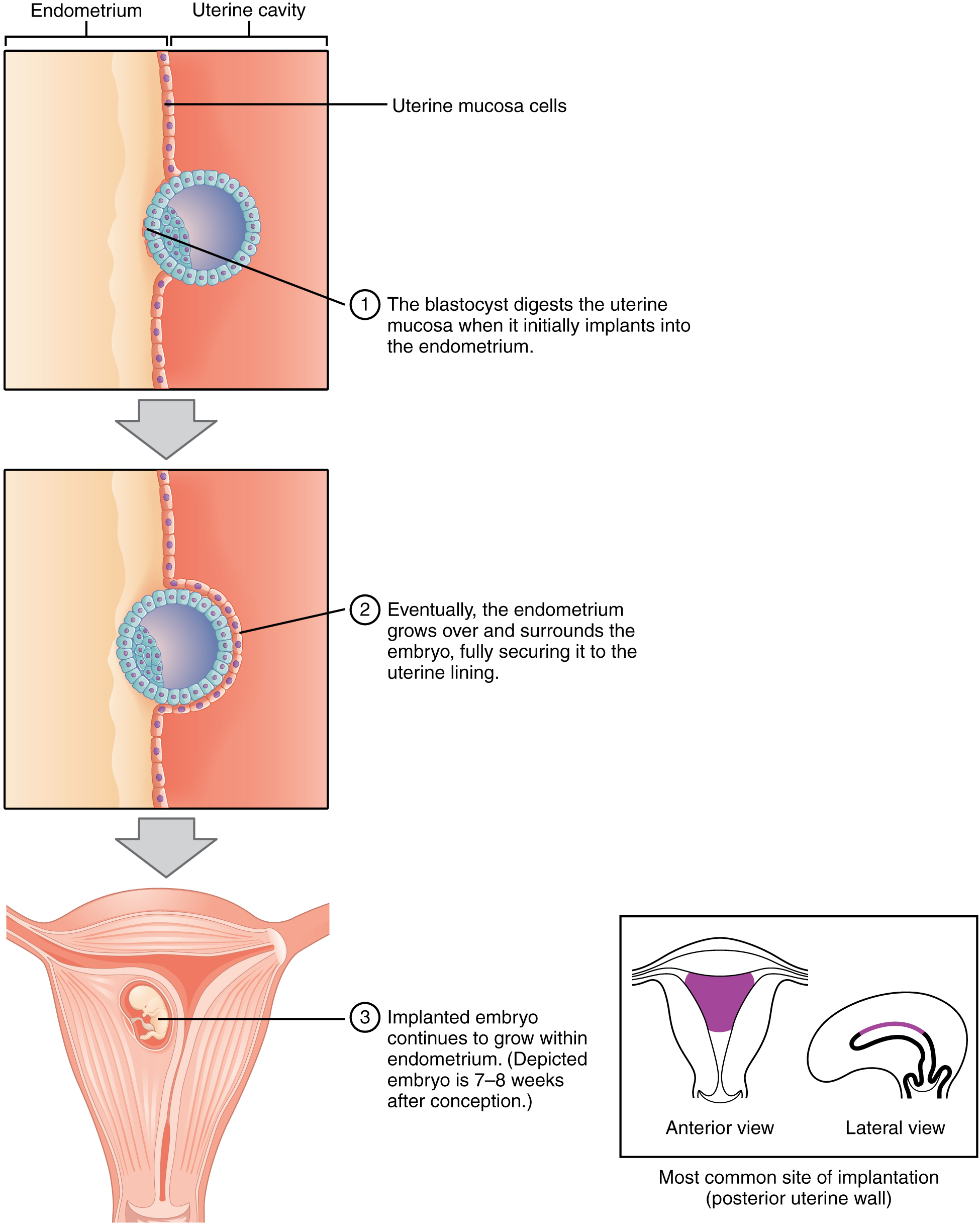
    Figure \(\PageIndex{5}\): The blastocyst digests the uterine mucosa. Eventually, the endometrium grows over the embryo. the embryo continues to grow until delivery. You can see a 7 to 8 weeks old embryo in the uterus.

    Around day 8 or 9 after fertilization, implantation begins. Implantation is the process in which a blastocyst becomes embedded in the endometrium of the uterus (see Figure \(\PageIndex{5}\)). Implantation is triggered by contact between the blastocyst and endometrium. In response to this contact, trophoblast cells start to proliferate. The trophoblast cells start secreting enzymes that digest the mucosa covering the endometrium. These changes allow finger-like projections (called villi) of the trophoblast to penetrate into the endometrium. The projections pull the blastocyst — now called an embryo — into the endometrium until it is fully covered by endometrial epithelium.

    Review

    1. Define the germinal stage of human development.
    2. Name four processes that occur during the germinal stage.
    3. Describe three processes that enable successful fertilization after ovulation occurs and sperm enter the Fallopian tube.
    4. What is cleavage? Where does it take place? What is its end result?
    5. What is blastulation? How does the morula change during this process?
    6. Identify the major parts of the blastocyst.
    7. Define implantation. When and how does implantation occur?
    8. List the stages of the developing human organism, in order, from fertilization to the end of implantation.
    9. Explain why cells in the zygote are smaller on day 3-4 after fertilization than they were on day 2.
    10. True or False: The zona pellucida disintegrates as the morula becomes a blastocyst.
    11. True or False: Some cells in the blastocyst do not become part of the embryo.
    12. Why do you think it is important that only one sperm fertilizes each egg? What mechanism helps ensure that this happens properly?
    13. Put the following events in order of when they occur during early human development, from earliest to latest:
      1. differentiation of cells
      2. cleavage
      3. implantation
      4. adhesion
    14. Which has the most cells?
      1. blastocoele
      2. embryoblast
      3. morula
      4. blastocyst

    Explore More

    bio.libretexts.org/link?17804#Explore_More

    Attributions

    1. Newborn by amsferguson, Pixabay license
    2. Human fertilization by Ttrue12, CC BY-SA 3.0 via Wikimedia Commons
    3. Sperm fertilization by CC BY-NC-SA 2.0 via Open Learn
    4. Embryonic development by OpenStax College, CC BY 3.0
    5. Implantation by OpenStax College, CC BY 3.0 via Wikimedia Commons
    6. Text adapted from Human Biology by CK-12 licensed CC BY-NC 3.0

    This page titled 23.2: Germinal Stage is shared under a CK-12 license and was authored, remixed, and/or curated by Suzanne Wakim & Mandeep Grewal via source content that was edited to the style and standards of the LibreTexts platform.

    CK-12 Foundation
    LICENSED UNDER
    CK-12 Foundation is licensed under CK-12 Curriculum Materials License