23.3: Embryonic Stage
- Last updated
- Save as PDF
- Page ID
- 22627
The Most Important Time in Your Life?
In many cultures, marriage — along with birth and death — is considered the most pivotal life event. For pioneering developmental biologist Lewis Wolpert, however, these life events are overrated. According to Wolpert, "It is not birth, marriage, or death, but gastrulation, which is truly the most important time in your life." Gastrulation is a major biological event that occurs early in the embryonic stage of human development.
Defining the Embryonic Stage
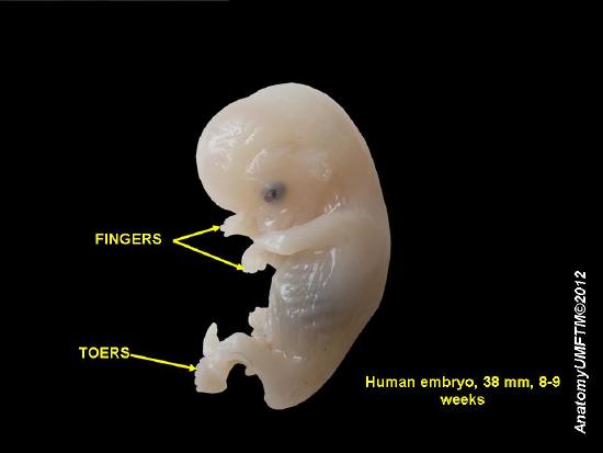
After a blastocyst implants in the uterus around the end of the first week after fertilization, its internal cell mass, which was called the embryoblast, is now known as the embryo. The embryonic stage lasts through the eighth week following fertilization, after which the embryo is called a fetus. The embryonic stage is short, lasting only about seven weeks in total, but developments that occur during this stage bring about enormous changes in the embryo. During the embryonic stage, the embryo becomes not only bigger but also much more complex. Figure \(\PageIndex{2}\) shows an eight to nine week old embryo. The embryo's finger, toes, head, eyes, and other structures are visible. It is no exaggeration to say that the embryonic stage lays the necessary groundwork for all of the remaining stages of life.
Embryonic Development
Starting in the second week after fertilization, the embryo starts to develop distinct cell layers, form the nervous system, make blood cells, and form many organs. By the end of the embryonic stage, most organs have started to form, although they will continue to develop and grow in the next stage (that of the fetus). As the embryo undergoes all of these changes, its cells continuously undergo mitosis, allowing the embryo to grow in size, as well as complexity.
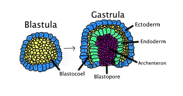
Figure \(\PageIndex{3}\): Blastula and Gastrula. The blastula is composed of one layer with a Blastocoel inside. Some cells of the outer layer fold into the Blastocoel to create a Blastopore. This invagination also gives rise to three germ layers. This diagram is color-coded. Ectoderm, blue. Endoderm, green. Blastocoel (the yolk sack), yellow. Archenteron (the gut), purple.
Gastrulation
Late in the second week after fertilization, gastrulation occurs when a blastula, made up of one layer, folds inward and enlarges to create a gastrula. A gastrula has 3 germ layers--the ectoderm, the mesoderm, and the endoderm. Some of the ectoderm cells from the blastula collapse inward and form the endoderm.
The final phase of gastrulation is the formation of the primitive gut that will eventually develop into the gastrointestinal tract. A tiny hole, called a blastopore, develops in one side of the embryo. The blastopore deepens and becomes the anus. The blastopore continues to tunnel through the embryo to the other side, where it forms an opening that will become the mouth. Whether this blastospore develops into a mouth or an anus determines whether the organism is a protostome or a deuterostome. With a functioning digestive tube, gastrulation is now complete.
Each of the three germ layers of the embryo will eventually give rise to different cells, tissues, and organs that make up the entire organism, which is illustrated in Figure \(\PageIndex{4}\). For example, the inner layer (the endoderm) will eventually form cells of many internal glands and organs, including the lungs, intestines, thyroid, pancreas, and bladder. The middle layer (the mesoderm) will form cells of the heart, blood, bones, muscles, and kidneys. The outer layer (the ectoderm) will form cells of the epidermis, nervous system, eyes, inner ears, and many connective tissues.
| Germ Layer | Gives rise to |
|---|---|
| Ectoderm | The epidermis, glands of the skin, some cranial bones, pituitary and adrenal medulla, the nervous system, the mouth between cheek and gums, the anus |
| Mesoderm | Connective tissues, bone, cartilage, blood endothelium of blood vessels, muscles, synovial membranes, serous membranes, kidneys, the lining of gonads |
| Endoderm | The lining of the airways and digestive system, except the moth and distal part of the digestive system. Digestive, endocrine, and adrenal cortex glands. |
Neurulation
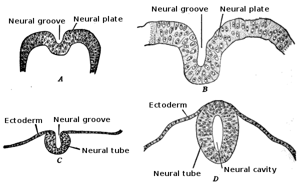
Following gastrulation, the next major development in the embryo is neurulation, which occurs during weeks three and four after fertilization. This is a process in which the embryo develops structures that will eventually become the nervous system. Neurulation is illustrated in Figure \(\PageIndex{4}\). It begins when a structure of differentiated cells called a neural plate forms from the ectoderm. The neural plate then starts to fold inward until its borders converge. The convergence of the neural plate borders also results in the formation of a neural tube. Most of the neural tube will eventually become the spinal cord. The neural tube also develops a bulge at one end, which will later become the brain.
Organogenesis
In addition to neurulation, gastrulation is followed by organogenesis, when organs develop within the newly formed germ layers. Most organs start to develop during the third to eighth weeks following fertilization. They will continue to develop and grow during the following fetal period.
The heart is the first functional organ to develop in the embryo. The primitive blood vessels start to develop in the mesoderm during the third week after fertilization. A couple of days later, the heart starts to form in the mesoderm when two endocardial tubes grow. The tubes migrate toward each other and fuse to form a single primitive heart tube. By about day 21 or 22, the tubular heart starts to beat and pump blood, even as it continues to develop. By day 23, the primitive heart has formed five distinct regions. These regions will develop into the chambers of the heart and the septa (walls) that separate them by the end of the eighth week after fertilization.
Other Developments in the Embryo
Several other major developments that occur during the embryonic stage are summarized chronologically below, starting with the fifth week after fertilization.
Week Five
By week five after fertilization, the embryo measures about 4 mm (0.16 in.) in length and has begun to curve into a C shape. During this week, the following developments take place:
- Grooves called pharyngeal arches form. These will develop into the face and neck.
- The inner ears begin to form.
- Arm buds are visible.
- The liver, pancreas, spleen, and gallbladder start to form.
Week Six
By week six after fertilization, the embryo measures about 8 mm (0.31 in.) in length. During the sixth week, some of the developments that occur include:
- The eyes and nose start to develop.
- Leg buds form and the hands form as flat paddles at the ends of the arms.
- The precursors of the kidneys begin to form.
- The stomach starts to develop.
Week Seven
By week seven, the embryo measures about 13 mm (0.51 in.) in length. During this week, some of the developments that take place include:
- The lungs begin to form.
- The arms and legs have lengthened, and the hands and feet have started to develop digits.
- The lymphatic system starts to develop.
- The primary prenatal development of the sex organs begins.
Week Eight
By week eight — which is the final week of the embryonic stage — the embryo measures about 20 mm (0.79 in.) in length. During this week, some of the developments that occur include:
- Nipples and hair follicles begin to develop.
- External ears start to form.
- The face takes on a human appearance.
- Fetal heartbeat and limb movements can be detected by ultrasound.
- All essential organs have at least started to form.
Genetic and Environmental Risks to Embryonic Development
The embryonic stage is a critical period of development. Events that occur in the embryo lay the foundation for virtually all of the body’s different cells, tissues, organs, and organ systems. Genetic defects or harmful environmental exposures during this stage are likely to have devastating effects on the developing organism. They may cause the embryo to die and be spontaneously aborted (also called a miscarriage). If the embryo survives and goes on to develop and grow as a fetus, it is likely to have birth defects.
Environmental exposures are known to have adverse effects on the embryo include:
- Alcohol consumption: Exposure of the embryo to alcohol from the mother’s blood can cause fetal alcohol spectrum disorder. Children born with this disorder may have cognitive deficits, developmental delays, behavioral issues, and distinctive facial features.
- Infection by rubella virus: In adults, rubella (German measles) is a relatively mild disease, but if the virus passes from an infected mother to her embryo, it may have severe consequences. The virus may cause fetal death, or result in a diversity of birth defects, such as heart defects, microcephaly (abnormally small head), vision and hearing problems, cognitive deficits, growth problems, and liver and spleen damage.
- Radiation from diagnostic X-rays or radiation therapy in the mother: Radiation may damage DNA and cause mutations in embryonic germ cells. When mutations occur at such an early stage of development, they are passed on to daughter cells in many tissues and organs, which is likely to have severe impacts on the offspring.
- Nutritional deficiencies: A maternal diet lacking certain nutrients may cause birth defects. The birth defect called spina bifida is caused by a lack of folate when the nervous system is first forming, which happens early in the embryonic stage. In this disorder, the neural tube does not close completely and may lead to paralysis below the affected region of the spinal cord.
Extraembryonic Structures
Several structures form simultaneously with the embryo. These structures help the embryo grow and develop. These extraembryonic structures include the placenta, chorion, yolk sac, and amnion.
Placenta
The placenta is a temporary organ that provides a connection between a developing embryo (and later the fetus) and the mother. It serves as a conduit from the maternal organism to the offspring for the transfer of nutrients, oxygen, antibodies, hormones, and other needed substances. It also passes waste products (such as urea and carbon dioxide) from the offspring to the mother’s blood for excretion from the body of the mother.
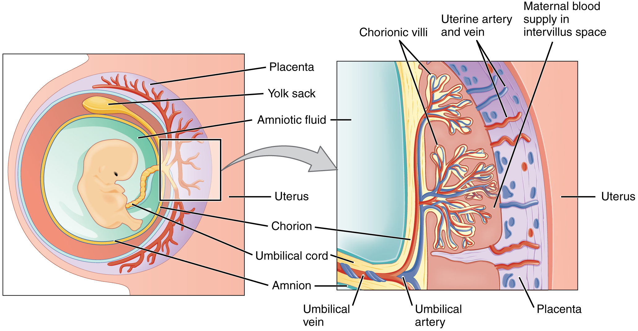
The placenta starts to develop after the blastocyst has implanted in the uterine lining. The placenta consists of both maternal and fetal tissues. The maternal portion of the placenta develops from the endometrial tissues lining the uterus. The fetal portion develops from the trophoblast, which forms a fetal membrane called the chorion (described below). Finger-like villi from the chorion penetrate the endometrium. The villi begin to branch and develop blood vessels from the embryo.
As shown in Figure \(\PageIndex{5}\), maternal blood flows into the spaces between the chorionic villi, allowing the exchange of substances between the fetal blood and the maternal blood without the two sources of blood actually intermixing. The embryo is joined to the fetal portion of the placenta by a narrow connecting stalk. This stalk develops into the umbilical cord, which contains two arteries and a vein. Blood from the fetus enters the placenta through the umbilical arteries, exchanges gases, and other substances with the mother’s blood, and travels back to the fetus through the umbilical vein.
Chorion, Yolk Sac, and Amnion
Besides the placenta, the chorion, yolk sac, and amnion also form around or near the developing embryo in the uterus. Their early development in the bilaminar embryonic disc is illustrated in Figure \(\PageIndex{5}\).
- Chorion: The chorion is a membrane formed by extraembryonic mesoderm and trophoblast. The chorion undergoes rapid proliferation and forms the chorionic villi. These villi invade the uterine lining and help form the fetal portion of the placenta.
- Yolk Sac: The yolk sac (or sack) is a membranous sac attached to the embryo and formed by cells of the hypoblast. The yolk sac provides nourishment to the early embryo. After the tubular heart forms and starts pumping blood during the third week after fertilization, the blood circulates through the yolk sac, where it absorbs nutrients before returning to the embryo. By the end of the embryonic stage, the yolk sac will have been incorporated into the primitive gut, and the embryo will obtain its nutrients from the mother’s blood via the placenta.
- Amnion: The amnion is a membrane that forms from extraembryonic mesoderm and ectoderm. It creates a sac, called the amniotic sac, around the embryo. By about the fourth or fifth week of embryonic development, amniotic fluid begins to accumulate within the amniotic sac. This fluid allows free movements of the fetus during the later stages of pregnancy and also helps cushion the fetus from potential injury.
Feature: My Human Body
Assume that you’ve been trying to conceive for many months and that you have just found out that you’re finally pregnant. You may be tempted to celebrate the good news with a champagne toast, but it’s not worth the risk. Alcohol can cross the placenta and enter the embryo’s (or fetus’s) blood. In essence, when a pregnant woman drinks alcohol, so does her unborn child. Alcohol in the embryo (or fetus) may cause many abnormalities in growth and development.
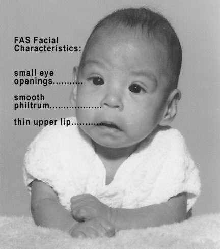
A child exposed to alcohol in utero may be born with a fetal alcohol spectrum disorder (FASD), the most severe of which is fetal alcohol syndrome (FAS). Signs and symptoms of FAS may include abnormal craniofacial appearance (Figure \(\PageIndex{6}\)), short height, low body weight, cognitive deficits, and behavioral problems, among others. The risk of FASDs and their severity if they occur depend on the amount and frequency of alcohol consumption, and also on the age of the embryo or fetus when the alcohol is consumed. Generally, greater consumption earlier in pregnancy is more detrimental. However, there is no known amount, frequency, or time at which drinking is known to be safe during pregnancy. The good news is that FASDs are completely preventable by abstaining from alcohol during pregnancy and while trying to conceive.
Review
- When does the embryonic stage occur?
- Name a few of the major developments that occur during the embryonic stage.
- What is the embryonic disc? When and how does it form?
- Define gastrulation. When does it occur?
- Identify the three embryonic germ layers. Give examples of specific cell types that originate in each germ layer.
- What happens during neurulation? When does it occur?
- Define organogenesis. When does organogenesis take place in the embryo?
- What is the first functional organ to develop in the embryo? When does this organ start to function?
- Identify some of the developments that take place during weeks five through eight of the embryonic stage.
- List three environmental exposures that may cause birth defects during the embryonic stage.
- Identify extraembryonic structures that form at the same time as the embryo and help the embryo grow and develop. Give a function of each structure.
- Put the following events in order of when they occur, from earliest to latest:
- formation of the neural tube
- formation of the three germ layers
- formation of the primitive streak
- incorporation of the yolk sac into the embryo
- True or False: The nervous system develops from the same germ layer as skin cells do.
- True or False: Leg buds are formed during gastrulation.
- What are two tissues produced by the hypoblast?
Explore More
Learn more about spina bifida here:
Attributions
- Wedding couple in Kandy Sri Lanka by Peter van der Sluijs, CC BY-SA 3.0 via Wikimedia Commons
- Human embryo by Anatomist 90, CC BY-SA 3.0 via Wikimedia Commons
- Blastula and Gastrula by Abigail Pyne, public domain via
- Neurulation by Stephen Walter Ranson, Public domain, via Wikimedia Commons
- Placenta development by OpenStax College, CC BY 3.0 via Wikimedia Commons
- FASD by Teresa Kellerman, CC BY-SA 3.0 via Wikimedia Commons
- Text adapted from Human Biology by CK-12 licensed CC BY-NC 3.0


