Biology LibreTexts

5.4: Plasma Membrane

  • Page ID
    22464
  • \( \newcommand{\vecs}[1]{\overset { \scriptstyle \rightharpoonup} {\mathbf{#1}} } \)

    \( \newcommand{\vecd}[1]{\overset{-\!-\!\rightharpoonup}{\vphantom{a}\smash {#1}}} \)

    \( \newcommand{\id}{\mathrm{id}}\) \( \newcommand{\Span}{\mathrm{span}}\)

    ( \newcommand{\kernel}{\mathrm{null}\,}\) \( \newcommand{\range}{\mathrm{range}\,}\)

    \( \newcommand{\RealPart}{\mathrm{Re}}\) \( \newcommand{\ImaginaryPart}{\mathrm{Im}}\)

    \( \newcommand{\Argument}{\mathrm{Arg}}\) \( \newcommand{\norm}[1]{\| #1 \|}\)

    \( \newcommand{\inner}[2]{\langle #1, #2 \rangle}\)

    \( \newcommand{\Span}{\mathrm{span}}\)

    \( \newcommand{\id}{\mathrm{id}}\)

    \( \newcommand{\Span}{\mathrm{span}}\)

    \( \newcommand{\kernel}{\mathrm{null}\,}\)

    \( \newcommand{\range}{\mathrm{range}\,}\)

    \( \newcommand{\RealPart}{\mathrm{Re}}\)

    \( \newcommand{\ImaginaryPart}{\mathrm{Im}}\)

    \( \newcommand{\Argument}{\mathrm{Arg}}\)

    \( \newcommand{\norm}[1]{\| #1 \|}\)

    \( \newcommand{\inner}[2]{\langle #1, #2 \rangle}\)

    \( \newcommand{\Span}{\mathrm{span}}\) \( \newcommand{\AA}{\unicode[.8,0]{x212B}}\)

    \( \newcommand{\vectorA}[1]{\vec{#1}}      % arrow\)

    \( \newcommand{\vectorAt}[1]{\vec{\text{#1}}}      % arrow\)

    \( \newcommand{\vectorB}[1]{\overset { \scriptstyle \rightharpoonup} {\mathbf{#1}} } \)

    \( \newcommand{\vectorC}[1]{\textbf{#1}} \)

    \( \newcommand{\vectorD}[1]{\overrightarrow{#1}} \)

    \( \newcommand{\vectorDt}[1]{\overrightarrow{\text{#1}}} \)

    \( \newcommand{\vectE}[1]{\overset{-\!-\!\rightharpoonup}{\vphantom{a}\smash{\mathbf {#1}}}} \)

    \( \newcommand{\vecs}[1]{\overset { \scriptstyle \rightharpoonup} {\mathbf{#1}} } \)

    \( \newcommand{\vecd}[1]{\overset{-\!-\!\rightharpoonup}{\vphantom{a}\smash {#1}}} \)

    \(\newcommand{\avec}{\mathbf a}\) \(\newcommand{\bvec}{\mathbf b}\) \(\newcommand{\cvec}{\mathbf c}\) \(\newcommand{\dvec}{\mathbf d}\) \(\newcommand{\dtil}{\widetilde{\mathbf d}}\) \(\newcommand{\evec}{\mathbf e}\) \(\newcommand{\fvec}{\mathbf f}\) \(\newcommand{\nvec}{\mathbf n}\) \(\newcommand{\pvec}{\mathbf p}\) \(\newcommand{\qvec}{\mathbf q}\) \(\newcommand{\svec}{\mathbf s}\) \(\newcommand{\tvec}{\mathbf t}\) \(\newcommand{\uvec}{\mathbf u}\) \(\newcommand{\vvec}{\mathbf v}\) \(\newcommand{\wvec}{\mathbf w}\) \(\newcommand{\xvec}{\mathbf x}\) \(\newcommand{\yvec}{\mathbf y}\) \(\newcommand{\zvec}{\mathbf z}\) \(\newcommand{\rvec}{\mathbf r}\) \(\newcommand{\mvec}{\mathbf m}\) \(\newcommand{\zerovec}{\mathbf 0}\) \(\newcommand{\onevec}{\mathbf 1}\) \(\newcommand{\real}{\mathbb R}\) \(\newcommand{\twovec}[2]{\left[\begin{array}{r}#1 \\ #2 \end{array}\right]}\) \(\newcommand{\ctwovec}[2]{\left[\begin{array}{c}#1 \\ #2 \end{array}\right]}\) \(\newcommand{\threevec}[3]{\left[\begin{array}{r}#1 \\ #2 \\ #3 \end{array}\right]}\) \(\newcommand{\cthreevec}[3]{\left[\begin{array}{c}#1 \\ #2 \\ #3 \end{array}\right]}\) \(\newcommand{\fourvec}[4]{\left[\begin{array}{r}#1 \\ #2 \\ #3 \\ #4 \end{array}\right]}\) \(\newcommand{\cfourvec}[4]{\left[\begin{array}{c}#1 \\ #2 \\ #3 \\ #4 \end{array}\right]}\) \(\newcommand{\fivevec}[5]{\left[\begin{array}{r}#1 \\ #2 \\ #3 \\ #4 \\ #5 \\ \end{array}\right]}\) \(\newcommand{\cfivevec}[5]{\left[\begin{array}{c}#1 \\ #2 \\ #3 \\ #4 \\ #5 \\ \end{array}\right]}\) \(\newcommand{\mattwo}[4]{\left[\begin{array}{rr}#1 \amp #2 \\ #3 \amp #4 \\ \end{array}\right]}\) \(\newcommand{\laspan}[1]{\text{Span}\{#1\}}\) \(\newcommand{\bcal}{\cal B}\) \(\newcommand{\ccal}{\cal C}\) \(\newcommand{\scal}{\cal S}\) \(\newcommand{\wcal}{\cal W}\) \(\newcommand{\ecal}{\cal E}\) \(\newcommand{\coords}[2]{\left\{#1\right\}_{#2}}\) \(\newcommand{\gray}[1]{\color{gray}{#1}}\) \(\newcommand{\lgray}[1]{\color{lightgray}{#1}}\) \(\newcommand{\rank}{\operatorname{rank}}\) \(\newcommand{\row}{\text{Row}}\) \(\newcommand{\col}{\text{Col}}\) \(\renewcommand{\row}{\text{Row}}\) \(\newcommand{\nul}{\text{Nul}}\) \(\newcommand{\var}{\text{Var}}\) \(\newcommand{\corr}{\text{corr}}\) \(\newcommand{\len}[1]{\left|#1\right|}\) \(\newcommand{\bbar}{\overline{\bvec}}\) \(\newcommand{\bhat}{\widehat{\bvec}}\) \(\newcommand{\bperp}{\bvec^\perp}\) \(\newcommand{\xhat}{\widehat{\xvec}}\) \(\newcommand{\vhat}{\widehat{\vvec}}\) \(\newcommand{\uhat}{\widehat{\uvec}}\) \(\newcommand{\what}{\widehat{\wvec}}\) \(\newcommand{\Sighat}{\widehat{\Sigma}}\) \(\newcommand{\lt}{<}\) \(\newcommand{\gt}{>}\) \(\newcommand{\amp}{&}\) \(\definecolor{fillinmathshade}{gray}{0.9}\)
    A Bag Full of Jell-O

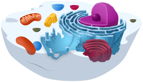
    This simple, cut-away model of an animal cell (Figure \(\PageIndex{1}\)) shows that a cell resembles a plastic bag full of Jell-O. Its basic structure is a plasma membrane filled with cytoplasm. Like Jell-O containing mixed fruit, the cytoplasm of the cell also contains various structures, such as a nucleus and other organelles. Your body is made up of trillions of cells, but all of them perform the same basic life functions. They all obtain and use energy, respond to the environment, and reproduce. How do your cells carry out these basic functions and keep themselves — and you — alive? To answer these questions, you need to know more about the structures that make up cells, starting with the plasma membrane.

    Animal Cell
    Figure \(\PageIndex{1}\): Animal cell model

    The plasma membrane is a structure that forms a barrier between the cytoplasm inside the cell and the environment outside the cell. Without the plasma membrane, there would be no cell. The membrane also protects and supports the cell and controls everything that enters and leaves it. It allows only certain substances to pass through while keeping others in or out. To understand how the plasma membrane controls what passes into or out of the cell, you need to know its basic structure.

    Phospholipid Bilayer

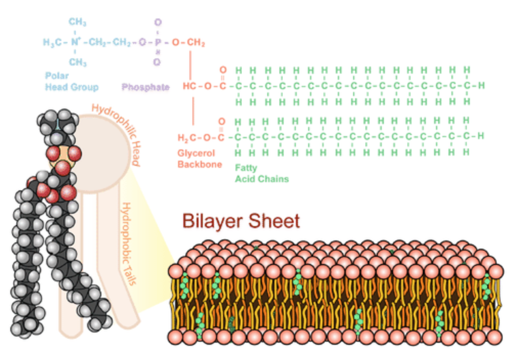
    The plasma membrane is composed mainly of phospholipids, which consist of fatty acids and alcohol. The phospholipids in the plasma membrane are arranged in two layers, called a phospholipid bilayer, with a hydrophobic, or water-hating, interior and a hydrophilic, or water-loving, exterior. Each phospholipid molecule has a head and two tails. The head “loves” water (hydrophilic) and the tails “fear” water (hydrophobic). The water-fearing tails are on the interior of the membrane, whereas the water-loving heads point outwards, toward either the cytoplasm or the fluid that surrounds the cell. The polar head group and fatty acid chains are attached by a 3-carbon glycerol unit. Figure \(\PageIndex{2}\) shows a single phospholipid next to a phospholipid bilayer.

    Molecules that are hydrophobic can easily pass through the plasma membrane if they are small enough because they are water-hating like the interior of the membrane. Molecules that are hydrophilic, on the other hand, cannot pass through the plasma membrane — at least not without help — because they are water-loving like the exterior of the membrane.

    phospholipids and  plasma membrane bilayer sheet; details in text above
    Figure \(\PageIndex{2}\): Phospholipid and a phospholipid Bilayer.

    Other Molecules in the Plasma Membrane

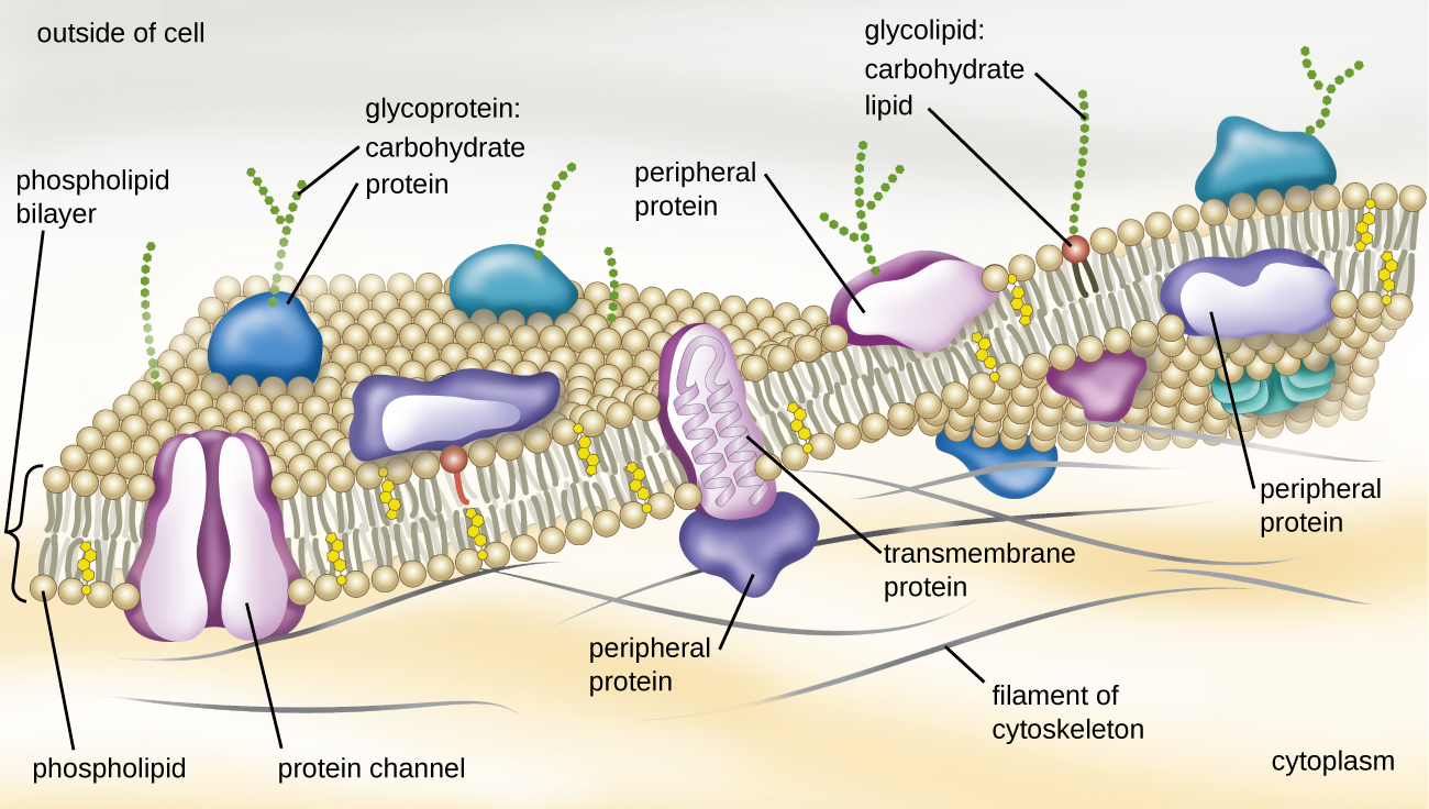
    The plasma membrane also contains other molecules, primarily other lipids and proteins. The green molecules in Figure \(\PageIndex{2}\), for example, are the lipid cholesterol. Molecules of the steroid lipid cholesterol help the plasma membrane keep its shape. (Figure \(\PageIndex{3}\)) shows the cholesterol molecules as yellow structures within the center of the phospholipid bilayer. Other structures shown in (Figure \(\PageIndex{3}\)):

    • Protein channels. These span the full membrane and have a space within them because they are used to transport materials into or out of the cell.
    • Transmembrane proteins. The root "trans" explains that these span (go "across") the membrane. Transmembrane proteins can have a variety of functions.
    • Peripheral proteins. These are found only on one side of the membrane. They can be found on either the cytoplasmic side or the outside of the membrane.
    • Glycoproteins. These consist of a protein in the plasma membrane with chains of carbohydrates projecting out of the cell.
    • Glycolipids. These are chains of carbohydrates attached directly to a lipid in the membrane. Both glycoproteins and glycolipids act as labels to identify the cell.
    • Filaments of cytoskeleton are found along the cytoplasmic side of the membrane and provide a scaffolding for the membrane.
    Plasma membrane and associated components; details in text above
    Figure \(\PageIndex{3}\): The figure illustrates the major components of the phospholipid bilayer.

    Additional Functions of the Plasma Membrane

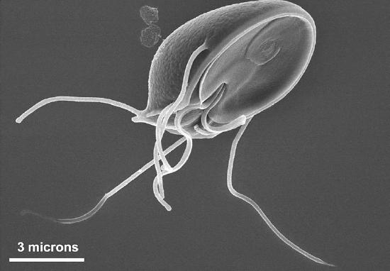
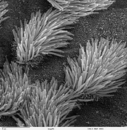
    The plasma membrane may have extensions, such as whip-like flagella or brush-like cilia, that give it other functions. In single-celled organisms, like those shown below, these membrane extensions may help the organisms move. In multicellular organisms, the extensions have different functions. For example, the cilia on human lung cells sweep foreign particles and mucus toward the mouth and nose.

    Giardia with long flagella
    bronchial cells showin cilia
    Figure \(\PageIndex{4}\): Flagella of Giardia (left) and cilia of human respiratory mucosa (right). Flagella and cilia are extensions of the plasma membrane of many cells.
    Feature: My Human Body

    If you smoke and need another reason to quit, here's a good one. We usually think of lung cancer as a major disease caused by smoking. But smoking can have devastating effects on the body's ability to protect itself from repeated, serious respiratory infections, such as bronchitis and pneumonia.

    Cilia are microscopic, hair-like projects on cells that line the respiratory, reproductive, and digestive systems. Cilia in the respiratory system line most of your airways where they have the job of trapping and removing dust, germs, and other foreign particles before they can make you sick. Cilia secrete mucus that traps particles, and they move in a continuous wave-like motion that sweeps the mucus and particles upward toward the throat, where they can be expelled from the body. When you are sick and cough up phlegm, that's what you are doing.

    Smoking prevents cilia from performing these important functions. Chemicals in tobacco smoke paralyze the cilia so they can't sweep mucus out of the airways and they also inhibit the cilia from producing mucus. Fortunately, these effects start to wear off soon after the last exposure to tobacco smoke. If you stop smoking, your cilia will return to normal. Even if prolonged smoking has destroyed cilia, they will regrow and resume functioning in a matter of months after you stop smoking.

    Review

    1. What are the general functions of the plasma membrane?
    2. Describe the phospholipid bilayer of the plasma membrane.
    3. Identify other molecules in the plasma membrane, and state their functions.
    4. Why do some cells have plasma membrane extensions such as flagella and cilia?
    5. Explain why hydrophilic molecules cannot easily pass through the cell membrane. What type of molecule in the cell membrane might help hydrophilic molecules pass through it?
    6. Which part of a phospholipid molecule in the plasma membrane is made of fatty acid chains? Is this part hydrophobic or hydrophilic?
    7. The two layers of phospholipids in the plasma membrane are called a phospholipid ____________.
    8. True or False. The flagella on your lung cells sweep foreign particles and mucus toward your mouth and nose.
    9. True or False. Small hydrophobic molecules can easily pass through the plasma membrane.
    10. True or False. The side of the cell membrane that faces the cytoplasm is hydrophilic.
    11. Steroid hormones can pass directly through cell membranes. Why do you think this is the case?
    12. Some antibiotics work by making holes in the plasma membrane of bacterial cells. How do you think this kills the cells?
    13. What is the name of the long, whip-like extensions of the plasma membrane that helps some single-celled organisms move?

    Explore More

    Watch the video below to learn the history of the discovery of cell membranes' structure.

    Attributions

    1. Animal cell model by Kevin Song, dedicated CC0 via Wikimedia Commons
    2. Phospholipid bilayer by LadyofHats, CC BY-NC 3.0 for CK-12 Foundation
    3. Plasma membrane by CNX OpenStax, licensed CC BY 4.0 via Wikimedia Commons
    4. Giardia by CDC/ Dr. Stan Erlandsen, public domain via Wikimedia Commons
      1. Bronchial cells by Charles Daghlian, released into the public domain via Wikimedia Commons
    5. Text adapted from Human Biology by CK-12 licensed CC BY-NC 3.0

    This page titled 5.4: Plasma Membrane is shared under a CK-12 license and was authored, remixed, and/or curated by Suzanne Wakim & Mandeep Grewal via source content that was edited to the style and standards of the LibreTexts platform.

    CK-12 Foundation
    LICENSED UNDER
    CK-12 Foundation is licensed under CK-12 Curriculum Materials License