
1.35: Helminth Parasites
- Page ID
- 79460

\( \newcommand{\vecs}[1]{\overset { \scriptstyle \rightharpoonup} {\mathbf{#1}} } \)
\( \newcommand{\vecd}[1]{\overset{-\!-\!\rightharpoonup}{\vphantom{a}\smash {#1}}} \)
\( \newcommand{\dsum}{\displaystyle\sum\limits} \)
\( \newcommand{\dint}{\displaystyle\int\limits} \)
\( \newcommand{\dlim}{\displaystyle\lim\limits} \)
\( \newcommand{\id}{\mathrm{id}}\) \( \newcommand{\Span}{\mathrm{span}}\)
( \newcommand{\kernel}{\mathrm{null}\,}\) \( \newcommand{\range}{\mathrm{range}\,}\)
\( \newcommand{\RealPart}{\mathrm{Re}}\) \( \newcommand{\ImaginaryPart}{\mathrm{Im}}\)
\( \newcommand{\Argument}{\mathrm{Arg}}\) \( \newcommand{\norm}[1]{\| #1 \|}\)
\( \newcommand{\inner}[2]{\langle #1, #2 \rangle}\)
\( \newcommand{\Span}{\mathrm{span}}\)
\( \newcommand{\id}{\mathrm{id}}\)
\( \newcommand{\Span}{\mathrm{span}}\)
\( \newcommand{\kernel}{\mathrm{null}\,}\)
\( \newcommand{\range}{\mathrm{range}\,}\)
\( \newcommand{\RealPart}{\mathrm{Re}}\)
\( \newcommand{\ImaginaryPart}{\mathrm{Im}}\)
\( \newcommand{\Argument}{\mathrm{Arg}}\)
\( \newcommand{\norm}[1]{\| #1 \|}\)
\( \newcommand{\inner}[2]{\langle #1, #2 \rangle}\)
\( \newcommand{\Span}{\mathrm{span}}\) \( \newcommand{\AA}{\unicode[.8,0]{x212B}}\)
\( \newcommand{\vectorA}[1]{\vec{#1}} % arrow\)
\( \newcommand{\vectorAt}[1]{\vec{\text{#1}}} % arrow\)
\( \newcommand{\vectorB}[1]{\overset { \scriptstyle \rightharpoonup} {\mathbf{#1}} } \)
\( \newcommand{\vectorC}[1]{\textbf{#1}} \)
\( \newcommand{\vectorD}[1]{\overrightarrow{#1}} \)
\( \newcommand{\vectorDt}[1]{\overrightarrow{\text{#1}}} \)
\( \newcommand{\vectE}[1]{\overset{-\!-\!\rightharpoonup}{\vphantom{a}\smash{\mathbf {#1}}}} \)
\( \newcommand{\vecs}[1]{\overset { \scriptstyle \rightharpoonup} {\mathbf{#1}} } \)
\(\newcommand{\longvect}{\overrightarrow}\)
\( \newcommand{\vecd}[1]{\overset{-\!-\!\rightharpoonup}{\vphantom{a}\smash {#1}}} \)
\(\newcommand{\avec}{\mathbf a}\) \(\newcommand{\bvec}{\mathbf b}\) \(\newcommand{\cvec}{\mathbf c}\) \(\newcommand{\dvec}{\mathbf d}\) \(\newcommand{\dtil}{\widetilde{\mathbf d}}\) \(\newcommand{\evec}{\mathbf e}\) \(\newcommand{\fvec}{\mathbf f}\) \(\newcommand{\nvec}{\mathbf n}\) \(\newcommand{\pvec}{\mathbf p}\) \(\newcommand{\qvec}{\mathbf q}\) \(\newcommand{\svec}{\mathbf s}\) \(\newcommand{\tvec}{\mathbf t}\) \(\newcommand{\uvec}{\mathbf u}\) \(\newcommand{\vvec}{\mathbf v}\) \(\newcommand{\wvec}{\mathbf w}\) \(\newcommand{\xvec}{\mathbf x}\) \(\newcommand{\yvec}{\mathbf y}\) \(\newcommand{\zvec}{\mathbf z}\) \(\newcommand{\rvec}{\mathbf r}\) \(\newcommand{\mvec}{\mathbf m}\) \(\newcommand{\zerovec}{\mathbf 0}\) \(\newcommand{\onevec}{\mathbf 1}\) \(\newcommand{\real}{\mathbb R}\) \(\newcommand{\twovec}[2]{\left[\begin{array}{r}#1 \\ #2 \end{array}\right]}\) \(\newcommand{\ctwovec}[2]{\left[\begin{array}{c}#1 \\ #2 \end{array}\right]}\) \(\newcommand{\threevec}[3]{\left[\begin{array}{r}#1 \\ #2 \\ #3 \end{array}\right]}\) \(\newcommand{\cthreevec}[3]{\left[\begin{array}{c}#1 \\ #2 \\ #3 \end{array}\right]}\) \(\newcommand{\fourvec}[4]{\left[\begin{array}{r}#1 \\ #2 \\ #3 \\ #4 \end{array}\right]}\) \(\newcommand{\cfourvec}[4]{\left[\begin{array}{c}#1 \\ #2 \\ #3 \\ #4 \end{array}\right]}\) \(\newcommand{\fivevec}[5]{\left[\begin{array}{r}#1 \\ #2 \\ #3 \\ #4 \\ #5 \\ \end{array}\right]}\) \(\newcommand{\cfivevec}[5]{\left[\begin{array}{c}#1 \\ #2 \\ #3 \\ #4 \\ #5 \\ \end{array}\right]}\) \(\newcommand{\mattwo}[4]{\left[\begin{array}{rr}#1 \amp #2 \\ #3 \amp #4 \\ \end{array}\right]}\) \(\newcommand{\laspan}[1]{\text{Span}\{#1\}}\) \(\newcommand{\bcal}{\cal B}\) \(\newcommand{\ccal}{\cal C}\) \(\newcommand{\scal}{\cal S}\) \(\newcommand{\wcal}{\cal W}\) \(\newcommand{\ecal}{\cal E}\) \(\newcommand{\coords}[2]{\left\{#1\right\}_{#2}}\) \(\newcommand{\gray}[1]{\color{gray}{#1}}\) \(\newcommand{\lgray}[1]{\color{lightgray}{#1}}\) \(\newcommand{\rank}{\operatorname{rank}}\) \(\newcommand{\row}{\text{Row}}\) \(\newcommand{\col}{\text{Col}}\) \(\renewcommand{\row}{\text{Row}}\) \(\newcommand{\nul}{\text{Nul}}\) \(\newcommand{\var}{\text{Var}}\) \(\newcommand{\corr}{\text{corr}}\) \(\newcommand{\len}[1]{\left|#1\right|}\) \(\newcommand{\bbar}{\overline{\bvec}}\) \(\newcommand{\bhat}{\widehat{\bvec}}\) \(\newcommand{\bperp}{\bvec^\perp}\) \(\newcommand{\xhat}{\widehat{\xvec}}\) \(\newcommand{\vhat}{\widehat{\vvec}}\) \(\newcommand{\uhat}{\widehat{\uvec}}\) \(\newcommand{\what}{\widehat{\wvec}}\) \(\newcommand{\Sighat}{\widehat{\Sigma}}\) \(\newcommand{\lt}{<}\) \(\newcommand{\gt}{>}\) \(\newcommand{\amp}{&}\) \(\definecolor{fillinmathshade}{gray}{0.9}\)- Identify and name the following helminth parasites in microscopic samples: Enterobius vermicularis, Dipylidium caninum, and Schistosoma sp.
- Name the diseases caused by the following helminth parasites: Enterobius vermicularis, Dipylidium caninum, and Schistosoma sp.
- Explain how the following helminth parasites are transmitted to humans (or dogs/cats for D. caninum): Enterobius vermicularis, Dipylidium caninum, and Schistosoma sp.
- Interpret the life cycles of the following helminth parasites: Enterobius vermicularis, Dipylidium caninum, and Schistosoma sp.
- Examine the following helminth parasites using a microscope and illustrate and label the specimens: Enterobius vermicularis, Dipylidium caninum, and Schistosoma sp.
Enterobius vermicularis (Causes Enterobiasis)
Introduction to Enterobiasis
The pinworm, Enterobius vermicularis (en’tur-o’bee-us/vur-mick-yoo-lair’is), is thought to be the most common helminth parasite in temperate regions with a high level of sanitation. While children are more commonly infected than adults, pinworm infestations are found in all ages and socioeconomic groups. Humans are the only host of Enterobius vermicularis.
The life cycle of Enterobius vermicularis is 4-6 weeks. Adults worms inhabit the cecum (part of the large intestine/colon) and adjacent portions of the large and small intestine of the host. The male pinworm is inconspicuous, about 2 to 5 mm in length, and no more than 0.2 mm in width. The female may reach a length of 13 mm (1/2 inch) and a maximum width of 0.5 mm The female is distinguished by a long, thin, sharply pointed tail, which gives rise to the common name “pinworm”. The gravid females, containing 11,000 to 15,000 eggs, migrate down the intestinal tract to the anus where they deposit their eggs. Enterobius ova, having a sticky albumin coating, adhere to the perianal region. The eggs mature quickly and are infective within a few hours after they are deposited.

Figure 1: (A) Image of adult female Enterobius vermicularis. (B) Image of E. vermicularis eggs. (C) Diagram of the anatomy of male and female E. vermicularis worms. (D) Side-by-side image of male and female E. vermicularis worms. (E) Image of male E. vermicularis worm.
Recovery of eggs from the perianal region provides the most efficient method of diagnosis of pinworm infection. Repeated examinations on consecutive days are necessary because of the irregular migrations of the gravid female worms. Since pinworm eggs are generally deposited at night, the examination should be made in the early morning hours before the patient has washed or defecated. Typically, Graham’s Scotch Tape Method is employed. The sticky side of cellophane tape is applied to the anal area of the patient. The tape is then pressed firmly on a microscope slide, sticky side down. The slide is examined under the low power of a microscope for the presence of pinworm eggs. The eggs are identified by their flattened oval (asymmetrical) shape and possibly the presence of a well-developed embryo.
Migration of the female worm may cause pruritus ani (anal itching). The local itching may cause insomnia and restlessness in children or adults who are infected, as the worms migrate from the anus during the resting hours. In about one-third of infected children, eggs may be obtained from beneath the fingernails. Reinfection of the patient by hand-to-mouth transmission (from scratching the perianal area or handling contaminated fomites) is common and makes control of the parasite difficult. Enterobius ova can survive for 2-3 weeks outside the body in high humidity and moderate temperatures. If towels or linens are shared, these eggs can be passed to others. It is recommended that all members of the infected household be treated simultaneously. Infection of others through contaminated bedding, clothing, bath tubs, toilet seats, dry dust, and air-borne eggs may serve to infect persons at some distance. The prescription drug, mebendazole, is used to treat pinworm. There are over-the-counter drugs available for treatment of pinworms also. Vermox, antiminth, and Povan (pyrvinium pamoate) are acceptable treatments. Patients using Povan should be forewarned that it colors the stool a bright red color!
E. vermicularis occurs worldwide, with infections occurring most frequently in school- or preschool-children and in crowded conditions.
Clinical Presentation of Enterobiasis
Enterobiasis is frequently asymptomatic. The most typical symptom is perianal pruritus, especially at night, which may lead to excoriations and bacterial superinfection. Occasionally, invasion of the female genital tract with vulvovaginitis and pelvic or peritoneal granulomas can occur. Other symptoms include, teeth grinding, enuresia, insomnia, anorexia, irritability, and abdominal pain, which can mimic appendicitis. E. vermicularis larvae are often found within the appendix on appendectomy, but the role of this nematode in appendicitis remains controversial. Very rare instances of eosinophilic colitis associated with E. vermicularis larvae have been reported.
Life Cycle of Enterobius vermicularis

Figure 2: Life cycle of Enterobius vermicularis. See the table below for explanation of this life cycle.
 |
Gravid adult female Enterobius vermicularis deposit eggs on perianal folds. |
 |
Infection occurs via self-inoculation (transferring eggs to the mouth with hands that have scratched the perianal area) or through exposure to eggs in the environment (e.g. contaminated surfaces, clothes, bed linens, etc.). |
 |
Following ingestion of infective eggs, the larvae hatch in the small intestine... |
 |
...and the adults establish themselves in the colon, usually in the cecum. |
| The time interval from ingestion of infective eggs to oviposition by the adult females is about one month. At full maturity adult females measure 8 to 13 mm, and adult males 2 to 5 mm; the adult life span is about two months. | |
 |
Gravid females migrate nocturnally outside the anus and oviposit while crawling on the skin of the perianal area. |
 |
The larvae contained inside the eggs develop (the eggs become infective) in 4 to 6 hours under optimal conditions. |
| Rarely, eggs may become airborne and be inhaled and swallowed. Retroinfection, or the migration of newly hatched larvae from the anal skin back into the rectum, may occur but the frequency with which this happens is unknown. |
Laboratory Instructions: Enterobius vermicularis
- Examine eggs of E. vermicularis (use 400x or 1000x magnification).
- Carefully illustrate the egg sample as you see it in the microscope.
- Examine an adult worm of E. vermicularis.
- Carefully illustrate the adult worm sample as you see it in the microscope. Label the following in your illustration:
- "male" or "female" (as appropriate)
- "mouth"
- "cephalic expansion"
- "pharynx"
- "end bulb"
- "intestine"
- if the worm is a male:
- "testis"
- "spicule"
- "cloaca"
- if the worm is a female:
- "vulva"
- "posterior uterus"
Results & Questions: Enterobius vermicularis
E. vermicularis eggs

E. vermicularis adult helminth
- Carefully illustrate the egg sample as you see it in the microscope.
- Carefully illustrate the adult worm sample as you see it in the microscope. Label the following in your illustration:
- "male" or "female" (as appropriate)
- "mouth"
- "cephalic expansion"
- "pharynx"
- "end bulb"
- "intestine"
- if the worm is a male:
- "testis"
- "spicule"
- "cloaca"
- if the worm is a female:
- "vulva"
- "posterior uterus"
- What disease is caused by E. vermicularis?
- Where does E. vermicularis live inside its human host?
- How can infection with E. vermicularis be diagnosed?
- Give at least three symptoms of enterobiasis.
- Can enterobiasis be asymptomatic?
- Where geographically in the world does E. vermicularis infection occur?
- What are three similarities of the male and female E. vermicularis worms?
- What are four differences between the male and female E. vermicularis worms?
Dipylidium caninum (Dog Tapeworm)
Introduction to Dipylidium caninum
Dipylidium caninum is a common tapeworm of dogs and cats, but is occasionally found in humans. It has many common names including the “flea tapeworm”, “cucumber tapeworm”, and “double-pored tapeworm”. Geographic distribution of D. caninum is worldwide. This tapeworm is ubiquitous and common among pet dogs and cats. Human infection is rare, but has been reported from every inhabited continent.
Tapeworms are flatworms in the phylum Platyhelminthes. These helminths have a scolex (its "head") that is equipped with suckers and hooks, but no mouth. Tapeworms absorb nutrients through their tegument (their "skin"). The scolex and its suckers and hooks are used for attachment to the host intestine. Behind the scolex, a tapeworm is composed of segments called proglottids. The proglottids closest to the scolex are the smallest in size and are considered immature proglottids, that is, they lack functional reproductive organs and they lack eggs. New proglottids are produced just behind the scolex and neck of the tapeworm. As new proglottids are formed, the older proglottids will be pushed further from the scolex. As proglottids age, they will ultimately become larger and produce mature proglottids that contain mature ovaries and testes. Yes, that is correct, a tapeworm's mature proglottids contain both male and female genitalia, and are therefore capable of conducting sexual reproduction alone (a second worm is not involved in sexual reproduction in this case). The largest proglottids, and those most distant from the scolex, are called gravid proglottids. These gravid proglottids are distinct because sexual reproduction has already occurred and these proglottids contain a multitude of eggs. Gravid proglottids on the end of the tapeworm can break off and be released from its host in feces to release the eggs and continue the life cycle.

Figure 3: (Left) Overall anatomy of a tapeworm. (Right) Anatomy of Dipyldidium caninum mature proglottid with its genital pores, ovaries, testes, vaginas, sperm ducts, and uterus labeled.
Diagnosis is made by demonstrating the typical proglottids or egg packets in the stool or the environment. Although concentration methods are usually not necessary due to the large size of the proglottids, flotation methods may miss infections as egg packets are heavy and may fail to float.
Clinical Presentation of the Dog Tapeworm
Most infections with Dipylidium caninum are asymptomatic. Pets may exhibit behavior to relieve anal pruritis (such as scraping anal region across grass or carpeting). Mild gastrointestinal disturbances may occur. The most striking feature in animals and children consists of the passage of proglottids. These can be found in the perianal region, in the feces, on diapers, and occasionally on floor covering and furniture. The proglottids are motile when freshly passed and may be mistaken for maggots or fly larvae.
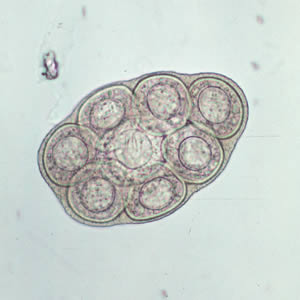
Figure 4: Dipylidium caninum egg packet containing 8 visible eggs.
Life Cycle of Dipylidium caninum

Figure 5: Dipylidium caninum life cycle. See the table below for details about this life cycle.
 |
Gravid proglottids are passed intact in the feces or emerge from the perianal region of the host. |
 |
In the environment, the proglottids disintegrate and release egg packets, which are also occasionally found free in the feces. |
 |
The intermediate host (most often larval stages of the dog or cat flea Ctenocephalides spp.) ingests egg packets, and the oncosphere within is released into the larval flea’s intestine. The oncosphere penetrates the intestinal wall, invades the insect’s hemocoel (body cavity), and develops into a cysticercoid. |
 |
The cysticercoid remains in the flea as it matures from a larva into an adult. |
 |
The vertebrate host becomes infected by ingesting the adult flea containing the cysticercoid. |
 |
In the small intestine of the vertebrate host, the cysticercoid develops into the adult tapeworm after about one month. The adult tapeworms (measuring up to 60 cm in length and 3 mm in width) reside in the small intestine of the host, where they each attach by their scolex. |
| Gravid, double-pored proglottids detach from the strobila (body) and are shed in the feces. | |
 |
Humans also acquire infection by ingesting the cysticercoid contaminated flea. Children are most frequently infected, possibly due to close contact with flea-infested pets. |
Laboratory Instructions: Dipylidium caninum
- Examine eggs of D. caninum (use 400x or 1000x magnification).
- Carefully illustrate the egg sample as you see it in the microscope.
- Examine an adult worm of D. caninum. The specimen will be separated into the following segments: scolex with neck and immature proglottids, mature proglottids, and gravid proglottids.
- Carefully illustrate the three segments of the adult worm sample as you see it in the microscope. Label the following in your illustration:
- the smallest sample (scolex, neck, and immature proglottids):
- "scolex"
- "suckers"
- "hooks"
- "neck"
- "immature proglottids"
- intermediate-sized sample (mature proglottids)
- "mature proglottid"
- "genital pore"
- "ovary"
- "testes"
- "uterus"
- largest sample (gravid proglottids)
- "gravid proglottid"
- "eggs"
- the smallest sample (scolex, neck, and immature proglottids):
Results & Questions: Dipylidium caninum
D. caninum eggs

D. caninum adult helminth: scolex & immature proglottids
D. caninum adult helminth: mature proglottids
D. caninum adult helminth: gravid proglottids
- Carefully illustrate the egg sample as you see it in the microscope.
- Carefully illustrate the three segments of the adult worm sample as you see it in the microscope. Label the following in your illustration:
- the smallest sample (scolex, neck, and immature proglottids):
- "scolex"
- "suckers"
- "hooks"
- "neck"
- "immature proglottids"
- intermediate-sized sample (mature proglottids)
- "mature proglottid"
- "genital pore"
- "ovary"
- "testes"
- "uterus"
- largest sample (gravid proglottids)
- "gravid proglottid"
- "eggs"
- the smallest sample (scolex, neck, and immature proglottids):
- Explain how immature proglottids become mature proglottids.
- Explain how mature proglottids become gravid proglottids.
- Explain how eggs are released from the host body to continue the life cycle.
- What is the function of the scolex?
- How does a tapeworm get its nutrients?
- How does a tapeworm conduct sexual reproduction?
- Give three signs/symptoms of D. caninum infection.
- What species is/are the definitive host(s) of D. caninum?
- What species is/are the intermediate host(s) of D. caninum?
Schistosoma sp. (Cause of Schistosomiasis)
Introduction to Schistosoma sp. (shis’to-so’muh)
There are several species of Schistosoma that cause disease in humans. Each species is unique to a particular area in the world. Since the United States does not have the specific host snails needed for its life cycle, infections are not contracted directly, yet many immigrants and travelers to endemic areas are infected. An estimated 236 million people require preventative drug treatment per year, out of which 105 million people were reported to have been treated. Preventative treatment, which should be repeated for a number of years, will reduce and prevent morbidity.
Infection occurs when your skin comes in contact with contaminated freshwater in which certain types of snails that carry schistosomes are living. Individuals infected with Schistosoma excrete the ova in the feces. Upon contact with water, the ova (which look like a cartoon talk bubble) develop into miracidia which enters the snail. Maturation in the snail results in the production of the free swimming cercaria. When the cercaria contacts the skin of a person who is in the water, it secretes enzymes that enable it to burrow through unbroken skin. The Schistosoma are then carried in the bloodstream to the liver or urinary bladder where they mature into an adult. Adults can live up to seven years. Schistosoma is dioecious – the male and female are separate animals. If both male and female are present, fertilization occurs, and ova are produced. The cycle then continues in a new host. Schistosoma infection may result in destruction of the liver, lungs, and/or urinary system. This is second most devastating parasitic disease. (Malaria is the most common). Praziquantel is an effective treatment.
Birds in the United States suffer from a Schistosoma infection. The Schistosoma parasite that infects birds has an intermediate snail host that is predominantly found in water in the Great Lakes and on the east coast. Sometimes when the free swimming cercaria of this bird parasite encounters a human swimming in the water, it will attempt to burrow through the skin of the human host instead of the normal bird host. Since humans are not the correct host for this parasite, the cercaria are unable to penetrate the human skin and instead, become embedded in the skin. This causes a painful, itchy inflammatory dermatitis called “swimmer’s itch”.
Geographic regions where schistosomiasis occurs include:
- Africa: contact with any freshwater in southern and sub-Saharan Africa–including the great lakes and rivers as well as smaller bodies of water– should be considered a risk for schistosomiasis transmission. Transmission also occurs in the Mahgreb region of North Africa and the Nile River valley in Egypt and Sudan .
- South America: Brazil, Suriname, Venezuela
- Caribbean: Dominican Republic, Guadeloupe, Martinique, Saint Lucia (risk in Caribbean is very low)
- The Middle East: Iran, Iraq, Saudi Arabia, Yemen
- Southern China
- Parts of Southeast Asia and the Philippines, Laos
- A recent focus of ongoing transmission has been identified in Corsica.
Clinical Presentation of the Schistosomiasis
Within days after becoming infected, you may develop a rash or itchy skin. Fever, chills, cough, and muscle aches can begin within 1-2 months of infection. Most people have no symptoms at this early phase of infection.
When adult worms are present, the eggs that are produced usually travel to the intestine, liver or bladder, causing inflammation or scarring. Children who are repeatedly infected can develop anemia, malnutrition, and learning difficulties. After years of infection, the parasite can also damage the liver, intestine, lungs, and bladder. Rarely, eggs are found in the brain or spinal cord and can cause seizures, paralysis, or spinal cord inflammation.
Symptoms of schistosomiasis are caused by the body’s reaction to the eggs produced by worms, not by the worms themselves.
Life Cycle of Schistosoma sp.

Figure 6: Life cycle of Schistosoma sp. See the table below for details about this life cycle.
|
Schistosoma eggs are eliminated with feces or urine, depending on species. |
|
Under appropriate conditions the eggs hatch and release miracidia... |
|
...which swim and penetrate specific snail intermediate hosts. |
|
The stages in the snail include two generations of sporocysts... |
|
...and the production of cercariae. |
|
Upon release from the snail, the infective cercariae swim, penetrate the skin of the human host... |
|
...and shed their forked tails, becoming schistosomulae. |
  |
The schistosomulae migrate via venous circulation to lungs, then to the heart, and then develop in the liver, exiting the liver via the portal vein system when mature, |
 |
Male and female adult worms copulate and reside in the mesenteric venules, the location of which varies by species (with some exceptions). |
 |
For instance, S. japonicum is more frequently found in the superior mesenteric veins draining the small intestine... |
 |
...and S. mansoni occurs more often in the inferior mesenteric veins draining the large intestine. |
 |
However, both species can occupy either location and are capable of moving between sites. S. intercalatum and S. guineensis also inhabit the inferior mesenteric plexus but lower in the bowel than S. mansoni. S. haematobium most often inhabits in the vesicular and pelvic venous plexus of the bladder... |
|
...but it can also be found in the rectal venules. The females (size ranges from 7–28 mm, depending on species) deposit eggs in the small venules of the portal and perivesical systems. The eggs are moved progressively toward the lumen of the intestine (S. mansoni,S. japonicum, S. mekongi, S. intercalatum/guineensis) and of the bladder and ureters (S. haematobium), and are eliminated with feces or urine, respectively |
Laboratory Instructions: Schistosoma sp.
- Examine eggs of Schistosoma sp. (use 400x or 1000x magnification).
- Carefully illustrate the egg sample as you see it in the microscope.
- Examine an adult worm of Schistosoma sp.
- Carefully illustrate the adult worm sample as you see it in the microscope.
Results & Questions: Schistosoma sp.
Schistosoma sp. eggs
Schistosoma sp. adult helminth

- Carefully illustrate the egg sample as you see it in the microscope.
- Carefully illustrate the adult worm sample as you see it in the microscope.
- What disease does Schistosoma sp. cause?
- How do people become infected with Schistosoma sp.?
- What is the definitive host species of Schistosoma sp.?
- What is the intermediate host species of Schistosoma sp.?
- List four species of Schistosoma that causes schistosomiasis in humans.
- Give at least five symptoms of schistosomiasis.
- Where in the human body to adult worms live?
- How does Schistosoma sp. leave a human to continue its life cycle?
- What areas of the world can humans contract schistosomiasis?
Attributions
- Chapter Image: Schistosoma mansoni2.jpg by Waisberg at English Wikipedia is in the public domain
- Centers for Disease Control. “Dipylidium caninum.” In the public domain. Use of CDC material, including any links to the materials on the CDC, ATSDR or HHS websites, does not imply endorsement by CDC, ATSDR, HHS or the United States Government of this page, this textbook, the author, or the institution. The material is otherwise available on the agency website for no charge.
- Centers for Disease Control. “Enterobiasis.” In the public domain. Use of CDC material, including any links to the materials on the CDC, ATSDR or HHS websites, does not imply endorsement by CDC, ATSDR, HHS or the United States Government of this page, this textbook, the author, or the institution. The material is otherwise available on the agency website for no charge.
- Centers for Disease Control. “Schistosomiasis.” In the public domain. Use of CDC material, including any links to the materials on the CDC, ATSDR or HHS websites, does not imply endorsement by CDC, ATSDR, HHS or the United States Government of this page, this textbook, the author, or the institution. The material is otherwise available on the agency website for no charge.
- Enterobius vermicularis 1.jpg by Danvasilis is licensed under CC BY-SA 3.0
- Red Mountain Microbiology by Jill Raymond Ph.D.; Graham Boorse, Ph.D.; and Anne Mason M.S. is licensed under CC BY-NC 4.0
- TaeniaPisiformisLabeledProglottid.jpg by Dr. Michael Hildreth is licensed under CC BY-SA 3.0
- Tapeworm (PSF).png by Pearson Scott Foresman is in the public domain