
1.16: Eukaryotic Cells
- Page ID
- 79446

\( \newcommand{\vecs}[1]{\overset { \scriptstyle \rightharpoonup} {\mathbf{#1}} } \)
\( \newcommand{\vecd}[1]{\overset{-\!-\!\rightharpoonup}{\vphantom{a}\smash {#1}}} \)
\( \newcommand{\dsum}{\displaystyle\sum\limits} \)
\( \newcommand{\dint}{\displaystyle\int\limits} \)
\( \newcommand{\dlim}{\displaystyle\lim\limits} \)
\( \newcommand{\id}{\mathrm{id}}\) \( \newcommand{\Span}{\mathrm{span}}\)
( \newcommand{\kernel}{\mathrm{null}\,}\) \( \newcommand{\range}{\mathrm{range}\,}\)
\( \newcommand{\RealPart}{\mathrm{Re}}\) \( \newcommand{\ImaginaryPart}{\mathrm{Im}}\)
\( \newcommand{\Argument}{\mathrm{Arg}}\) \( \newcommand{\norm}[1]{\| #1 \|}\)
\( \newcommand{\inner}[2]{\langle #1, #2 \rangle}\)
\( \newcommand{\Span}{\mathrm{span}}\)
\( \newcommand{\id}{\mathrm{id}}\)
\( \newcommand{\Span}{\mathrm{span}}\)
\( \newcommand{\kernel}{\mathrm{null}\,}\)
\( \newcommand{\range}{\mathrm{range}\,}\)
\( \newcommand{\RealPart}{\mathrm{Re}}\)
\( \newcommand{\ImaginaryPart}{\mathrm{Im}}\)
\( \newcommand{\Argument}{\mathrm{Arg}}\)
\( \newcommand{\norm}[1]{\| #1 \|}\)
\( \newcommand{\inner}[2]{\langle #1, #2 \rangle}\)
\( \newcommand{\Span}{\mathrm{span}}\) \( \newcommand{\AA}{\unicode[.8,0]{x212B}}\)
\( \newcommand{\vectorA}[1]{\vec{#1}} % arrow\)
\( \newcommand{\vectorAt}[1]{\vec{\text{#1}}} % arrow\)
\( \newcommand{\vectorB}[1]{\overset { \scriptstyle \rightharpoonup} {\mathbf{#1}} } \)
\( \newcommand{\vectorC}[1]{\textbf{#1}} \)
\( \newcommand{\vectorD}[1]{\overrightarrow{#1}} \)
\( \newcommand{\vectorDt}[1]{\overrightarrow{\text{#1}}} \)
\( \newcommand{\vectE}[1]{\overset{-\!-\!\rightharpoonup}{\vphantom{a}\smash{\mathbf {#1}}}} \)
\( \newcommand{\vecs}[1]{\overset { \scriptstyle \rightharpoonup} {\mathbf{#1}} } \)
\(\newcommand{\longvect}{\overrightarrow}\)
\( \newcommand{\vecd}[1]{\overset{-\!-\!\rightharpoonup}{\vphantom{a}\smash {#1}}} \)
\(\newcommand{\avec}{\mathbf a}\) \(\newcommand{\bvec}{\mathbf b}\) \(\newcommand{\cvec}{\mathbf c}\) \(\newcommand{\dvec}{\mathbf d}\) \(\newcommand{\dtil}{\widetilde{\mathbf d}}\) \(\newcommand{\evec}{\mathbf e}\) \(\newcommand{\fvec}{\mathbf f}\) \(\newcommand{\nvec}{\mathbf n}\) \(\newcommand{\pvec}{\mathbf p}\) \(\newcommand{\qvec}{\mathbf q}\) \(\newcommand{\svec}{\mathbf s}\) \(\newcommand{\tvec}{\mathbf t}\) \(\newcommand{\uvec}{\mathbf u}\) \(\newcommand{\vvec}{\mathbf v}\) \(\newcommand{\wvec}{\mathbf w}\) \(\newcommand{\xvec}{\mathbf x}\) \(\newcommand{\yvec}{\mathbf y}\) \(\newcommand{\zvec}{\mathbf z}\) \(\newcommand{\rvec}{\mathbf r}\) \(\newcommand{\mvec}{\mathbf m}\) \(\newcommand{\zerovec}{\mathbf 0}\) \(\newcommand{\onevec}{\mathbf 1}\) \(\newcommand{\real}{\mathbb R}\) \(\newcommand{\twovec}[2]{\left[\begin{array}{r}#1 \\ #2 \end{array}\right]}\) \(\newcommand{\ctwovec}[2]{\left[\begin{array}{c}#1 \\ #2 \end{array}\right]}\) \(\newcommand{\threevec}[3]{\left[\begin{array}{r}#1 \\ #2 \\ #3 \end{array}\right]}\) \(\newcommand{\cthreevec}[3]{\left[\begin{array}{c}#1 \\ #2 \\ #3 \end{array}\right]}\) \(\newcommand{\fourvec}[4]{\left[\begin{array}{r}#1 \\ #2 \\ #3 \\ #4 \end{array}\right]}\) \(\newcommand{\cfourvec}[4]{\left[\begin{array}{c}#1 \\ #2 \\ #3 \\ #4 \end{array}\right]}\) \(\newcommand{\fivevec}[5]{\left[\begin{array}{r}#1 \\ #2 \\ #3 \\ #4 \\ #5 \\ \end{array}\right]}\) \(\newcommand{\cfivevec}[5]{\left[\begin{array}{c}#1 \\ #2 \\ #3 \\ #4 \\ #5 \\ \end{array}\right]}\) \(\newcommand{\mattwo}[4]{\left[\begin{array}{rr}#1 \amp #2 \\ #3 \amp #4 \\ \end{array}\right]}\) \(\newcommand{\laspan}[1]{\text{Span}\{#1\}}\) \(\newcommand{\bcal}{\cal B}\) \(\newcommand{\ccal}{\cal C}\) \(\newcommand{\scal}{\cal S}\) \(\newcommand{\wcal}{\cal W}\) \(\newcommand{\ecal}{\cal E}\) \(\newcommand{\coords}[2]{\left\{#1\right\}_{#2}}\) \(\newcommand{\gray}[1]{\color{gray}{#1}}\) \(\newcommand{\lgray}[1]{\color{lightgray}{#1}}\) \(\newcommand{\rank}{\operatorname{rank}}\) \(\newcommand{\row}{\text{Row}}\) \(\newcommand{\col}{\text{Col}}\) \(\renewcommand{\row}{\text{Row}}\) \(\newcommand{\nul}{\text{Nul}}\) \(\newcommand{\var}{\text{Var}}\) \(\newcommand{\corr}{\text{corr}}\) \(\newcommand{\len}[1]{\left|#1\right|}\) \(\newcommand{\bbar}{\overline{\bvec}}\) \(\newcommand{\bhat}{\widehat{\bvec}}\) \(\newcommand{\bperp}{\bvec^\perp}\) \(\newcommand{\xhat}{\widehat{\xvec}}\) \(\newcommand{\vhat}{\widehat{\vvec}}\) \(\newcommand{\uhat}{\widehat{\uvec}}\) \(\newcommand{\what}{\widehat{\wvec}}\) \(\newcommand{\Sighat}{\widehat{\Sigma}}\) \(\newcommand{\lt}{<}\) \(\newcommand{\gt}{>}\) \(\newcommand{\amp}{&}\) \(\definecolor{fillinmathshade}{gray}{0.9}\)- Differentiate between eukaryotic cell and prokaryotic cell structures.
- Identify the structures and functions of components of eukaryotic cells.
- Name the categories of microorganisms that are eukaryotic.
- Provide a description that differentiates each type of eukaryotic microorganism from the other types of eukaryotic microorganisms.
- Examine specimens of different types of eukaryotic microorganisms and identify structures of these microorganisms, especially the nucleus.
Eukaryotic Cells
The cells of eukaryotic organisms have several distinguishing characteristics. Above all, eukaryotic cells are defined by the presence of a nucleus surrounded by a complex nuclear membrane. Also, eukaryotic cells are characterized by the presence of membrane-bound organelles in the cytoplasm. Organelles such as mitochondria, the endoplasmic reticulum (ER), Golgi apparatus, lysosomes, and peroxisomes are held in place by the cytoskeleton, an internal network that supports transport of intracellular components and helps maintain cell shape. The genome of eukaryotic cells is packaged in multiple, rod-shaped chromosomes as opposed to the single, circular-shaped chromosome that characterizes most prokaryotic cells.

Figure 1: An illustration of a generalized, single-celled eukaryotic organism. Note that cells of eukaryotic organisms vary greatly in terms of structure and function, and a particular cell may not have all of the structures shown here.
Table 1: A comparison of prokaryotic cells and eukaryotic cells.
| Summary of Cell Structures | |||
|---|---|---|---|
| Prokaryotes | Eukaryotes | ||
| Bacteria | Archaea | ||
| size | ~0.5–1 μm | ~0.5–1 μm | ~5–20 μm |
| surface area-to-volume ratio | High | High | Low |
| nucleus | No | No | Yes |
| genome characteristics |
|
|
|
| cell division | Binary fission | Binary fission | Mitosis, meiosis |
| membrane lipid composition |
|
|
|
| cell wall composition |
|
|
|
| motility structures | Rigid spiral flagella composed of flagellin | Rigid spiral flagella composed of archaeal flagellins | Flexible flagella and cilia composed of microtubules |
| membrane-bound organelles | No | No | Yes |
| endomembrane system | No | No | Yes (ER, Golgi, lysosomes) |
| ribosomes | 70S | 70S |
|
Table 2: Functions of Eukaryotic Cell Structures.
| Cell Structure | Function |
|---|---|
| cell wall | structure outside of the plasma membrane in some types of species; maintains cell shape |
| centrioles / centrosome | organizes microtubules (particularly important during cell division to move the chromosomes and separate them correctly into the two daughter cells) |
| chloroplast | conducts photosynthesis |
| chromatin | DNA with its associated proteins |
| cilia | external-facing protein fibers that wave to move a cell |
| cytoplasm | semi-fluid surrounding cellular structures with dissolved molecules; location of many cellular metabolic reactions |
| cytoskeleton | microtubules, intermediate filaments, and microfilaments (provides a cell shape, structure, can be used for cell movement or moving materials around the cell) |
| flagellum / flagella | a whip-like tail that moves a cell |
| Golgi complex / apparatus / body | modifies proteins and packages them into vesicles for transport to their destinations |
| lysosome | digests food and waste materials |
| mitochondria / mitochondrion | makes ATP (an energy-rich molecule) using nutrients |
| nuclear membrane / nuclear envelope | membrane enclosing the nucleus; protein-lined pores allow materials to move in and out of the nucleus |
| nuclear pore | protein-lined pores allow materials to move in and out of the nucleus |
| nucleolus | a condensed region within the cell nucleus where ribosomes are formed |
| nucleus | contains chromatin, a nuclear envelope, and a nucleolus |
| peroxisomes | metabolizes oxygen-containing waste |
| ribosomes | makes proteins |
| rough endoplasmic reticulum | membranes associated with ribosomes where the ribosomes make membrane proteins and proteins for secretion out of the cell |
| smooth endoplasmic reticulum | membranes without ribosomes; make lipids; detoxification |
Microbes with Eukaryotic Cells

Figure 2: Eukaryotic cells come in a variety of cell shapes. (a) Spheroid Chromulina (a species of algae). (b) Fusiform shaped Trypanosoma (a parasitic protozoan shown with red blood cells of its host). (c) Bell-shaped Vorticella (a free-living protozoan). (d) Ovoid Paramecium (a free-living protozoan). (e) Ring-shaped Plasmodium ovale (a parasitic protozoan living inside a red blood cell of its host). (credit a: modification of work by NOAA; credit b, e: modification of work by Centers for Disease Control and Prevention)
Eukaryotic organisms include protozoans, algae, fungi, plants, and animals. Some eukaryotic cells are independent, single-celled microorganisms, whereas others are part of multicellular organisms.
Eukaryotic microbes are an extraordinarily diverse group, including species with a wide range of life cycles, morphological specializations, and nutritional needs. Although more diseases are caused by viruses and bacteria than by microscopic eukaryotes, these eukaryotes are responsible for some diseases of great public health importance. For example, the protozoal disease malaria was responsible for 584,000 deaths worldwide (primarily children in Africa) in 2013, according to the World Health Organization (WHO). The protozoan parasite Giardia causes a diarrheal illness (giardiasis) that is easily transmitted through contaminated water supplies. In the United States, Giardia is the most common human intestinal parasite. Although it may seem surprising, parasitic worms are included within the study of microbiology because identification depends on observation of microscopic adult worms or eggs. Even in developed countries, these worms are important parasites of humans and of domestic animals. There are fewer fungal pathogens, but these are important causes of illness, as well. On the other hand, fungi have been important in producing antimicrobial substances such as penicillin.
Protozoa
Protozoa are nonphotosynthetic, motile eukaryotic organisms that are always unicellular. Historically, these microorganisms have been classified as protozoa because they were "animal like" unicellular eukaryotic organisms that did not fit with other eukaryotic taxonomic groupings (plants, fungi, or animals).
Protozoans inhabit a wide variety of habitats, both aquatic and terrestrial. Many are free-living, while others are parasitic, carrying out a life cycle within a host or hosts and potentially causing illness. There are also beneficial symbionts that provide metabolic services to their hosts. During the feeding and growth part of their life cycle, they are called trophozoites; these feed on small particulate food sources such as bacteria. While some types of protozoa exist exclusively in the trophozoite form, others can develop from trophozoite to an encapsulated cyst stage when environmental conditions are too harsh for the trophozoite. A cyst is a cell with a protective wall, and the process by which a trophozoite becomes a cyst is called encystment. When conditions become more favorable, these cysts are triggered by environmental cues to become active again through excystment.

Figure 3: (a) A scanning electron micrograph shows many Giardia parasites in the trophozoite, or feeding stage, in a gerbil intestine. (b) An individual trophozoite of G. lamblia, visualized here in a scanning electron micrograph. This waterborne protozoan causes severe diarrhea when ingested. (credit a, b: modification of work by Centers for Disease Control and Prevention)
Protozoans have a variety of reproductive mechanisms. Some protozoans reproduce asexually and others reproduce sexually; still others are capable of both sexual and asexual reproduction. In protozoans, asexual reproduction occurs by binary fission, budding, or schizogony. In schizogony, the nucleus of a cell divides multiple times before the cell divides into many smaller cells. The products of schizogony are called merozoites and they are stored in structures known as schizonts. Protozoans may also reproduce sexually, which increases genetic diversity and can lead to complex life cycles. Protozoans can produce haploid gametes that fuse through syngamy. However, they can also exchange genetic material by joining to exchange DNA in a process called conjugation. This is a different process than the conjugation that occurs in bacteria. The term protist conjugation refers to a true form of eukaryotic sexual reproduction between two cells of different mating types. It is found in ciliates, a group of protozoans, and is described later in this subsection.
All protozoans have a plasma membrane, or plasmalemma, and some have bands of protein just inside the membrane that add rigidity, forming a structure called the pellicle. Some protists, including protozoans, have distinct layers of cytoplasm under the membrane. In these protists, the outer gel layer (with microfilaments of actin) is called the ectoplasm. Inside this layer is a sol (fluid) region of cytoplasm called the endoplasm. These structures contribute to complex cell shapes in some protozoans, whereas others (such as amoebas) have more flexible shapes.
Figure 4: Amoeba motion involves formation of pseudopodia ("false feet"). These extensions enable the cell to creep along in a liquid environmental and also means that the shape of the cell is constantly changing.
Different groups of protozoans have specialized feeding structures. They may have a specialized structure for taking in food through phagocytosis, called a cytostome, and a specialized structure for the exocytosis of wastes called a cytoproct. Oral grooves leading to cytostomes are lined with hair-like cilia to sweep in food particles. Protozoans are heterotrophic. Protozoans that are holozoic ingest whole food particles through phagocytosis. Forms that are saprozoic ingest small, soluble food molecules.
Many protists have whip-like flagella or hair-like cilia made of microtubules that can be used for locomotion. Other protists use cytoplasmic extensions known as pseudopodia (“false feet”) to attach the cell to a surface; they then allow cytoplasm to flow into the extension, thus moving themselves forward.
Protozoans have a variety of unique organelles and sometimes lack organelles found in other cells. Some have contractile vacuoles, organelles that can be used to move water out of the cell for osmotic regulation (salt and water balance). Mitochondria may be absent in parasites or altered to kinetoplastids (modified mitochondria) or hydrogenosomes.
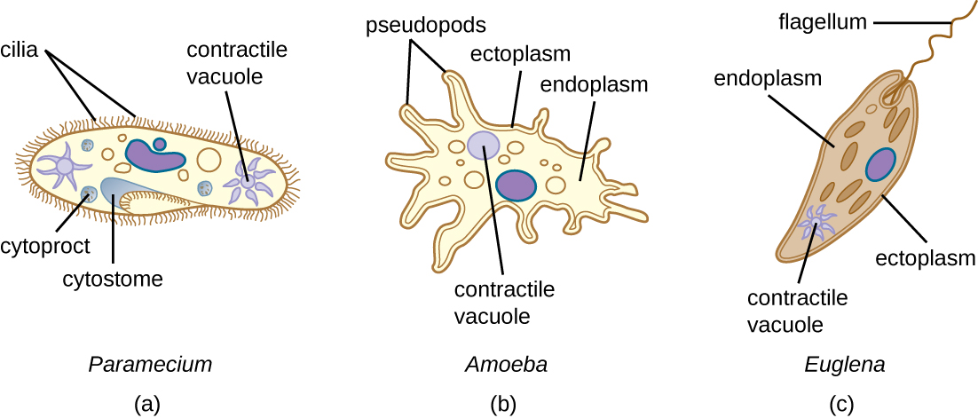
Figure 5: (a) Paramecium spp. have hair-like appendages called cilia for locomotion. (b) Amoeba spp. use lobe-like pseudopodia to anchor the cell to a solid surface and pull forward. (c) Euglena spp. use a whip-like structure called a flagellum to propel the cell.
Figure 6: A free-living ciliated cell with contractile vacuole pulsating to regulate water and salt balance in the cell.
Algae
The algae are autotrophic eukaryotes that can be unicellular or multicellular and are distinct from other eukaryotic groupings (plants, fungi, and animals). These organisms are found in the supergroups Chromalveolata (dinoflagellates, diatoms, golden algae, and brown algae) and Archaeplastida (red algae and green algae). They are important ecologically and environmentally because they are responsible for the production of approximately 70% of the oxygen and organic matter in aquatic environments. Some types of algae, even those that are microscopic, are regularly eaten by humans and other animals. Additionally, algae are the source for agar, agarose, and carrageenan, solidifying agents used in laboratories and in food production. Although algae are typically not pathogenic, some produce toxins. Harmful algal blooms, which occur when algae grow quickly and produce dense populations, can produce high concentrations of toxins that impair liver and nervous-system function in aquatic animals and humans.

Figure 7: Some of the diversity of algae. (a) These large multicellular kelps are members of the brown algae. Note the “leaves” and “stems” that make them appear similar to green plants. (b) This is a species of red algae that is also multicellular. (c) The green alga Halimeda incrassata, shown here growing on the sea floor in shallow water, appears to have plant-like structures, but is not a true plant. (d) Bioluminesence, visible in the cresting wave in this picture, is a phenomenon of certain dinoflagellates. (e) Diatoms (pictured in this micrograph) produce silicaceous tests (skeletons) that form diatomaceous earths. (f) Colonial green algae, like volvox in these three micrographs, exhibit simple cooperative associations of cells. (credit a, e: modification of work by NOAA; credit b: modification of work by Ed Bierman; credit c: modification of work by James St. John; credit d: modification of work by “catalano82”/Flickr; credit f: modification of work by Dr. Ralf Wagner)
Like protozoans, algae often have complex cell structures. For instance, algal cells can have one or more chloroplasts that contain structures called pyrenoids to synthesize and store starch. The chloroplasts themselves differ in their number of membranes, indicative of secondary or rare tertiary endosymbiotic events. Primary chloroplasts have two membranes—one from the original cyanobacteria that the ancestral eukaryotic cell engulfed, and one from the plasma membrane of the engulfing cell. Chloroplasts in some lineages appear to have resulted from secondary endosymbiosis, in which another cell engulfed a green or red algal cell that already had a primary chloroplast within it. The engulfing cell destroyed everything except the chloroplast and possibly the cell membrane of its original cell, leaving three or four membranes around the chloroplast. Different algal groups have different pigments, which are reflected in common names such as red algae, brown algae, and green algae.
Some algae, the seaweeds, are macroscopic and may be confused with plants. Seaweeds can be red, brown, or green, depending on their photosynthetic pigments. Green algae, in particular, share some important similarities with land plants; however, there are also important distinctions. For example, seaweeds do not have true tissues or organs like plants do. Additionally, seaweeds do not have a waxy cuticle to prevent desiccation. Algae can also be confused with cyanobacteria, photosynthetic bacteria that bear a resemblance to algae; however, cyanobacteria are prokaryotes.
Algae have a variety of life cycles. Reproduction may be asexual by mitosis or sexual using gametes.
Fungi
The fungi comprise a diverse group of organisms that are heterotrophic and typically saprozoic (consume dead organic matter). In addition to the well-known macroscopic fungi (such as mushrooms and molds), many unicellular yeasts and spores of macroscopic fungi are microscopic. For this reason, fungi are included within the field of microbiology.
Fungi are important to humans in a variety of ways. Both microscopic and macroscopic fungi have medical relevance, with some pathogenic species that can cause mycoses (illnesses caused by fungi). Some pathogenic fungi are opportunistic, meaning that they mainly cause infections when the host’s immune defenses are compromised and do not normally cause illness in healthy individuals. Fungi are important in other ways. They act as decomposers in the environment, and they are critical for the production of certain foods such as cheeses. Fungi are also major sources of antibiotics, such as penicillin from the fungus Penicillium.
Fungi have well-defined characteristics that set them apart from other organisms. Most multicellular fungal bodies, commonly called molds, are made up of filaments called hyphae. Hyphae can form a tangled network called a mycelium and form the thallus (body) of fleshy fungi. Hyphae that have walls between the cells are called septate hyphae; hyphae that lack walls and cell membranes between the cells are called nonseptate or coenocytic hyphae).

Figure 8: Multicellular fungi (molds) form hyphae, which may be septate or nonseptate. Unicellular fungi (yeasts) cells form pseudohyphae from individual yeast cells.
In contrast to molds, yeasts are unicellular fungi. The budding yeasts reproduce asexually by budding off a smaller daughter cell; the resulting cells may sometimes stick together as a short chain or pseudohypha.
Some fungi are dimorphic, having more than one appearance during their life cycle. These dimorphic fungi may be able to appear as yeasts or molds, which can be important for infectivity. They are capable of changing their appearance in response to environmental changes such as nutrient availability or fluctuations in temperature, growing as a mold, for example, at 25 °C (77 °F), and as yeast cells at 37 °C (98.6 °F). This ability helps dimorphic fungi to survive in diverse environments. Two examples of dimorphic yeasts are the human pathogens Histoplasma capsulatum and Candida albicans. H. capsulatum causes the lung disease histoplasmosis, and C. albicans is associated with vaginal yeast infections, oral thrush, and candidiasis of the skin.

Figure 9: Histoplasma capsulatum is a dimorphic fungus that grows in soil exposed to bird feces or bat feces (guano) (top left). It can change forms to survive at different temperatures. In the outdoors, it typically grows as a mycelium (as shown in the micrograph, bottom left), but when the spores are inhaled (right), it responds to the high internal temperature of the body (37 °C [98.6 °F]) by turning into a yeast that can multiply in the lungs, causing the chronic lung disease histoplasmosis. (credit: modification of work by Centers for Disease Control and Prevention)
There are notable unique features in fungal cell walls and membranes. Fungal cell walls contain chitin, as opposed to the cellulose found in the cell walls of plants and many protists. Additionally, whereas animals have cholesterol in their cell membranes, fungal cell membranes have different sterols called ergosterols. Ergosterols are often exploited as targets for antifungal drugs.
Fungal life cycles are unique and complex. Fungi reproduce sexually either through cross- or self-fertilization. Haploid fungi form hyphae that have gametes at the tips. Two different mating types (represented as “+ type” and “– type”) are involved. The cytoplasms of the + and – type gametes fuse (in an event called plasmogamy), producing a cell with two distinct nuclei (a dikaryotic cell). Later, the nuclei fuse (in an event called karyogamy) to create a diploid zygote. The zygote undergoes meiosis to form spores that germinate to start the haploid stage, which eventually creates more haploid mycelia. Depending on the taxonomic group, these sexually produced spores are known as zygospores (in Zygomycota), ascospores (in Ascomycota), or basidiospores (in Basidiomycota).
Fungi may also exhibit asexual reproduction by mitosis, mitosis with budding, fragmentation of hyphae, and formation of asexual spores by mitosis. These spores are specialized cells that, depending on the organism, may have unique characteristics for survival, reproduction, and dispersal. Fungi exhibit several types of asexual spores and these can be important in classification.
Helminths
Parasitic helminths are animals that are often included within the study of microbiology because many species of these worms are identified by their microscopic eggs and larvae. There are two major groups of parasitic helminths: the roundworms (Nematoda) and flatworms (Platyhelminthes). Of the many species that exist in these groups, about half are parasitic and some are important human pathogens. As animals, they are multicellular and have organ systems. However, the parasitic species often have limited digestive tracts, nervous systems, and locomotor abilities. Parasitic forms may have complex reproductive cycles with several different life stages and more than one type of host. Some are monoecious, having both male and female reproductive organs in a single individual, while others are dioecious, each having either male or female reproductive organs.
Nematoda (Roundworms)
Phylum Nematoda (the roundworms) is a diverse group containing more than 15,000 species, of which several are important human parasites. These unsegmented worms have a full digestive system even when parasitic. Some are common intestinal parasites, and their eggs can sometimes be identified in feces or around the anus of infected individuals. Ascaris lumbricoides is the largest nematode intestinal parasite found in humans; females may reach lengths greater than 1 meter. A. lumbricoides is also very widespread, even in developed nations, although it is now a relatively uncommon problem in the United States. It may cause symptoms ranging from relatively mild (such as a cough and mild abdominal pain) to severe (such as intestinal blockage and impaired growth).
Figure 10: Enterobius vermicularis is a type of parasitic roundworm.
Of all nematode infections in the United States, pinworm (caused by Enterobius vermicularis) is the most common. Pinworm causes sleeplessness and itching around the anus, where the female worms lay their eggs during the night. Toxocara canis and T. cati are nematodes found in dogs and cats, respectively, that can be transmitted to humans, causing toxocariasis. Antibodies to these parasites have been found in approximately 13.9% of the U.S. population, suggesting that exposure is common.7 Infection can cause larval migrans, which can result in vision loss and eye inflammation, or fever, fatigue, coughing, and abdominal pain, depending on whether the organism infects the eye or the viscera. Another common nematode infection is hookworm, which is caused by Necator americanus (the New World or North American hookworm) and Ancylostoma duodenale (the Old World hookworm). Symptoms of hookworm infection can include abdominal pain, diarrhea, loss of appetite, weight loss, fatigue, and anemia.
Trichinellosis, also called trichinosis, caused by Trichinella spiralis, is contracted by consuming undercooked meat, which releases the larvae and allows them to encyst in muscles. Infection can cause fever, muscle pains, and digestive system problems; severe infections can lead to lack of coordination, breathing and heart problems, and even death. Finally, heartworm in dogs and other animals is caused by the nematode Dirofilaria immitis, which is transmitted by mosquitoes. Symptoms include fatigue and cough; when left untreated, death may result.
Platyhelminths (Flatworms)
Phylum Platyhelminthes (the platyhelminths) are flatworms. This group includes the flukes, tapeworms, and the turbellarians, which include planarians. The flukes and tapeworms are medically important parasites.
The flukes (trematodes) are nonsegmented flatworms that have an oral sucker (and sometimes a second ventral sucker) and attach to the inner walls of intestines, lungs, large blood vessels, or the liver. Trematodes have complex life cycles, often with multiple hosts. Several important examples are the liver flukes (Clonorchis and Opisthorchis), the intestinal fluke (Fasciolopsis buski), and the oriental lung fluke (Paragonimus westermani). Schistosomiasis is a serious parasitic disease, considered second in the scale of its impact on human populations only to malaria. The parasites Schistosoma mansoni, S. haematobium, and S. japonicum, which are found in freshwater snails, are responsible for schistosomiasis. Immature forms burrow through the skin into the blood. They migrate to the lungs, then to the liver and, later, other organs. Symptoms include anemia, malnutrition, fever, abdominal pain, fluid buildup, and sometimes death.

Figure 11: Phylum Platyhelminthes is divided into four classes. (a) Class Turbellaria includes the Bedford’s flatworm (Pseudobiceros bedfordi), which is about 8–10 cm long. (b) The parasitic class Monogenea includes Dactylogyrus spp. Worms in this genus are commonly called gill flukes. The specimen pictured here is about 0.2 mm long and has two anchors, indicated by arrows, that it uses to latch onto the gills of host fish. (c) The Trematoda class includes the common liver fluke Fasciola hepatica and the giant liver fluke Fascioloides magna (right). The F. magna specimen shown here is about 7 cm long. (d) Class Cestoda includes tapeworms such as Taenia saginata, which infects both cattle and humans and can reach lengths of 4–10 meters; the specimen shown here is about 4 meters long. (credit c: modification of work by “Flukeman”/Wikimedia Commons)
The other medically important group of platyhelminths are commonly known as tapeworms (cestodes) and are segmented flatworms that may have suckers or hooks at the scolex (head region). Tapeworms use these suckers or hooks to attach to the wall of the small intestine. The body of the worm is made up of segments called proglottids that contain reproductive structures; these detach when the gametes are fertilized, releasing gravid proglottids with eggs. Tapeworms often have an intermediate host that consumes the eggs, which then hatch into a larval form called an oncosphere. The oncosphere migrates to a particular tissue or organ in the intermediate host, where it forms cysticerci. After being eaten by the definitive host, the cysticerci develop into adult tapeworms in the host's digestive system. Taenia saginata (the beef tapeworm) and T. solium (the pork tapeworm) enter humans through ingestion of undercooked, contaminated meat. The adult worms develop and reside in the intestine, but the larval stage may migrate and be found in other body locations such as skeletal and smooth muscle. The beef tapeworm is relatively benign, although it can cause digestive problems and, occasionally, allergic reactions. The pork tapeworm can cause more serious problems when the larvae leave the intestine and colonize other tissues, including those of the central nervous system. Diphylobothrium latum is the largest human tapeworm and can be ingested in undercooked fish. It can grow to a length of 15 meters. Echinococcus granulosus, the dog tapeworm, can parasitize humans and uses dogs as an important host.
Laboratory Activities
Eukaryotic Cell Structures

|
A: ______________________________ B: ______________________________ C: ______________________________ D: ______________________________ E: ______________________________ F: ______________________________ G: ______________________________ H: ______________________________ |
I: ______________________________ J: ______________________________ K: ______________________________ L: ______________________________ M: ______________________________ N: ______________________________ O: ______________________________ |
- Examine the eukaryotic cell model and name the cell structures labeled with letters A-O.
- Match the descriptions with the corresponding eukaryotic cell structures.
|
A. nucleolus B. cytoplasm C. ribosomes D. nuclear membrane E. rough endoplasmic reticulum F. plasma membrane G. cytoskeleton H. lysosome I. peroxisome J. nucleus K. nuclear pore L. Golgi complex M. chloroplast N. mitochondria O. cilia P. flagella Q. cell wall R. smooth endoplasmic reticulum S. chromatin |
Examine Microbes that have Eukaryotic Cells
Protozoa
Your instructor will provide you with a slide of a species of protozoan. Examine the protozoan with the microscope, make an illustration in the location below, label the nucleus of the protozoan cell, and label any additional cell structures you can identify.

Algae
Your instructor will provide you with a slide of a species of algae. Examine the algae with the microscope, make an illustration in the location below, label the nucleus of the algal cell, and label any additional cell structures you can identify.
Fungi
Your instructor will provide you with a slide of a species of fungus. Examine the fungus with the microscope, make an illustration in the location below, label the nucleus of the fungal cell, and label any additional cell structures you can identify.
Helminth
Your instructor will provide you with a slide of a species of helminth. Examine the helminth with the microscope, make an illustration in the location below and label any structures you can identify.
Attributions
- Biology 2e by OpenStax is licensed under CC BY 4.0
- Chapter Image: Microscopic image of yeasts.tif by Molnarova.Lucia is licensed under CC BY-SA 4.0
- Microbiology by OpenStax is licensed under CC BY 4.0