7.26D: Phase Display
- Page ID
- 9513
\( \newcommand{\vecs}[1]{\overset { \scriptstyle \rightharpoonup} {\mathbf{#1}} } \)
\( \newcommand{\vecd}[1]{\overset{-\!-\!\rightharpoonup}{\vphantom{a}\smash {#1}}} \)
\( \newcommand{\dsum}{\displaystyle\sum\limits} \)
\( \newcommand{\dint}{\displaystyle\int\limits} \)
\( \newcommand{\dlim}{\displaystyle\lim\limits} \)
\( \newcommand{\id}{\mathrm{id}}\) \( \newcommand{\Span}{\mathrm{span}}\)
( \newcommand{\kernel}{\mathrm{null}\,}\) \( \newcommand{\range}{\mathrm{range}\,}\)
\( \newcommand{\RealPart}{\mathrm{Re}}\) \( \newcommand{\ImaginaryPart}{\mathrm{Im}}\)
\( \newcommand{\Argument}{\mathrm{Arg}}\) \( \newcommand{\norm}[1]{\| #1 \|}\)
\( \newcommand{\inner}[2]{\langle #1, #2 \rangle}\)
\( \newcommand{\Span}{\mathrm{span}}\)
\( \newcommand{\id}{\mathrm{id}}\)
\( \newcommand{\Span}{\mathrm{span}}\)
\( \newcommand{\kernel}{\mathrm{null}\,}\)
\( \newcommand{\range}{\mathrm{range}\,}\)
\( \newcommand{\RealPart}{\mathrm{Re}}\)
\( \newcommand{\ImaginaryPart}{\mathrm{Im}}\)
\( \newcommand{\Argument}{\mathrm{Arg}}\)
\( \newcommand{\norm}[1]{\| #1 \|}\)
\( \newcommand{\inner}[2]{\langle #1, #2 \rangle}\)
\( \newcommand{\Span}{\mathrm{span}}\) \( \newcommand{\AA}{\unicode[.8,0]{x212B}}\)
\( \newcommand{\vectorA}[1]{\vec{#1}} % arrow\)
\( \newcommand{\vectorAt}[1]{\vec{\text{#1}}} % arrow\)
\( \newcommand{\vectorB}[1]{\overset { \scriptstyle \rightharpoonup} {\mathbf{#1}} } \)
\( \newcommand{\vectorC}[1]{\textbf{#1}} \)
\( \newcommand{\vectorD}[1]{\overrightarrow{#1}} \)
\( \newcommand{\vectorDt}[1]{\overrightarrow{\text{#1}}} \)
\( \newcommand{\vectE}[1]{\overset{-\!-\!\rightharpoonup}{\vphantom{a}\smash{\mathbf {#1}}}} \)
\( \newcommand{\vecs}[1]{\overset { \scriptstyle \rightharpoonup} {\mathbf{#1}} } \)
\(\newcommand{\longvect}{\overrightarrow}\)
\( \newcommand{\vecd}[1]{\overset{-\!-\!\rightharpoonup}{\vphantom{a}\smash {#1}}} \)
\(\newcommand{\avec}{\mathbf a}\) \(\newcommand{\bvec}{\mathbf b}\) \(\newcommand{\cvec}{\mathbf c}\) \(\newcommand{\dvec}{\mathbf d}\) \(\newcommand{\dtil}{\widetilde{\mathbf d}}\) \(\newcommand{\evec}{\mathbf e}\) \(\newcommand{\fvec}{\mathbf f}\) \(\newcommand{\nvec}{\mathbf n}\) \(\newcommand{\pvec}{\mathbf p}\) \(\newcommand{\qvec}{\mathbf q}\) \(\newcommand{\svec}{\mathbf s}\) \(\newcommand{\tvec}{\mathbf t}\) \(\newcommand{\uvec}{\mathbf u}\) \(\newcommand{\vvec}{\mathbf v}\) \(\newcommand{\wvec}{\mathbf w}\) \(\newcommand{\xvec}{\mathbf x}\) \(\newcommand{\yvec}{\mathbf y}\) \(\newcommand{\zvec}{\mathbf z}\) \(\newcommand{\rvec}{\mathbf r}\) \(\newcommand{\mvec}{\mathbf m}\) \(\newcommand{\zerovec}{\mathbf 0}\) \(\newcommand{\onevec}{\mathbf 1}\) \(\newcommand{\real}{\mathbb R}\) \(\newcommand{\twovec}[2]{\left[\begin{array}{r}#1 \\ #2 \end{array}\right]}\) \(\newcommand{\ctwovec}[2]{\left[\begin{array}{c}#1 \\ #2 \end{array}\right]}\) \(\newcommand{\threevec}[3]{\left[\begin{array}{r}#1 \\ #2 \\ #3 \end{array}\right]}\) \(\newcommand{\cthreevec}[3]{\left[\begin{array}{c}#1 \\ #2 \\ #3 \end{array}\right]}\) \(\newcommand{\fourvec}[4]{\left[\begin{array}{r}#1 \\ #2 \\ #3 \\ #4 \end{array}\right]}\) \(\newcommand{\cfourvec}[4]{\left[\begin{array}{c}#1 \\ #2 \\ #3 \\ #4 \end{array}\right]}\) \(\newcommand{\fivevec}[5]{\left[\begin{array}{r}#1 \\ #2 \\ #3 \\ #4 \\ #5 \\ \end{array}\right]}\) \(\newcommand{\cfivevec}[5]{\left[\begin{array}{c}#1 \\ #2 \\ #3 \\ #4 \\ #5 \\ \end{array}\right]}\) \(\newcommand{\mattwo}[4]{\left[\begin{array}{rr}#1 \amp #2 \\ #3 \amp #4 \\ \end{array}\right]}\) \(\newcommand{\laspan}[1]{\text{Span}\{#1\}}\) \(\newcommand{\bcal}{\cal B}\) \(\newcommand{\ccal}{\cal C}\) \(\newcommand{\scal}{\cal S}\) \(\newcommand{\wcal}{\cal W}\) \(\newcommand{\ecal}{\cal E}\) \(\newcommand{\coords}[2]{\left\{#1\right\}_{#2}}\) \(\newcommand{\gray}[1]{\color{gray}{#1}}\) \(\newcommand{\lgray}[1]{\color{lightgray}{#1}}\) \(\newcommand{\rank}{\operatorname{rank}}\) \(\newcommand{\row}{\text{Row}}\) \(\newcommand{\col}{\text{Col}}\) \(\renewcommand{\row}{\text{Row}}\) \(\newcommand{\nul}{\text{Nul}}\) \(\newcommand{\var}{\text{Var}}\) \(\newcommand{\corr}{\text{corr}}\) \(\newcommand{\len}[1]{\left|#1\right|}\) \(\newcommand{\bbar}{\overline{\bvec}}\) \(\newcommand{\bhat}{\widehat{\bvec}}\) \(\newcommand{\bperp}{\bvec^\perp}\) \(\newcommand{\xhat}{\widehat{\xvec}}\) \(\newcommand{\vhat}{\widehat{\vvec}}\) \(\newcommand{\uhat}{\widehat{\uvec}}\) \(\newcommand{\what}{\widehat{\wvec}}\) \(\newcommand{\Sighat}{\widehat{\Sigma}}\) \(\newcommand{\lt}{<}\) \(\newcommand{\gt}{>}\) \(\newcommand{\amp}{&}\) \(\definecolor{fillinmathshade}{gray}{0.9}\)- Assess the uses of phage display technology
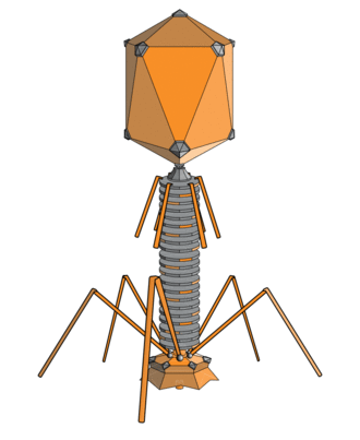
A phage or bacteriophage is a virus capable of infecting a bacterial cell, and may cause lysis to its host cell. Bacteriophages have a specific affinity for bacteria. They are made of an outer protein coat or capsid that encloses the genetic material (which can be RNA or DNA, about 5,000 to 500,000 nucleotides in length). They inject their genetic material into the bacterium following infection. When the strain is virulent, all the synthesis of the host’s DNA, RNA and proteins ceases. The phage genome is then used to direct the synthesis of phage nucleic acids and proteins using the host’s transcriptional and translational apparatus. When the sub-components of the phage are produced, they self-assemble to form new phage particles. The new phages produce lysozyme that ruptures the cell wall of the host, leading to the release of the new phages, each ready to invade other bacterial cells. This inherent property of phages is the basis for the phage display technology.

Phage display technology is the process of inserting new genetic material into a phage gene. The bacteria process the new gene so that a new protein or peptide is made. This protein or peptide is exposed on the phage surface. Phage display begins by inserting a diverse set of genes into the phage genome with each phage receiving a different gene. The modified gene contains an added segment (an antibody, small protein, or peptide), which is to be expressed on the surface of the phage. Each phage receives only one gene, so each expresses a single protein or peptide. A collection of phage displaying a population of related but diverse proteins or peptides is called a library. The related proteins keep most of the physical and chemical properties of their parent protein. The library is then exposed to an immobilized target. It is anticipated that some members of the library will bind to the target through an interaction between the displayed molecule and the target itself. After the phage is given the chance to bind to a target, the immobilized target is washed to remove phage that did not bind. Replicating the bound phage in bacteria increases the amount of phage several million-fold overnight, providing enough material for sequencing. Sequencing of the phage DNA tells the identity of the peptide that binds the target. Phage libraries are screened for binding to synthetic or native targets.
Phage display technology is advantageous in many applications including selection of inhibitors for the active and allosteric sites of enzymes, receptor agonists and antagonists, and G-protein binding modulatory peptides. Phage display is also used in epitope mapping and analysis of protein-protein interactions. The specific molecules isolated from phage libraries can be used in therapeutic target validation, drug design and vaccine development.
LICENSES AND ATTRIBUTIONS
CC LICENSED CONTENT, SPECIFIC ATTRIBUTION
- Two-hybrid screening. Provided by: Wikipedia. Located at: en.Wikipedia.org/wiki/Two-hybrid_screening. License: CC BY-SA: Attribution-ShareAlike
- Proteinu2013protein interaction. Provided by: Wikipedia. Located at: en.Wikipedia.org/wiki/Protein%E2%80%93protein_interaction. License: CC BY-SA: Attribution-ShareAlike
- Mass spectrometry. Provided by: Wikipedia. Located at: en.Wikipedia.org/wiki/Mass_spectrometry. License: CC BY-SA: Attribution-ShareAlike
- Affinity chromatography. Provided by: Wikipedia. Located at: en.Wikipedia.org/wiki/Affinit...chromatography. License: CC BY-SA: Attribution-ShareAlike
- mass spectrometry. Provided by: Wiktionary. Located at: en.wiktionary.org/wiki/mass_spectrometry. License: CC BY-SA: Attribution-ShareAlike
- File:Two hybrid assay.svg - Wikipedia, the free encyclopedia. Provided by: Wikipedia. Located at: en.Wikipedia.org/w/index.php?...say.svg&page=1. License: CC BY-SA: Attribution-ShareAlike
- Green fluorescent protein. Provided by: Wikipedia. Located at: en.Wikipedia.org/wiki/Green_fluorescent_protein. License: CC BY-SA: Attribution-ShareAlike
- Reporter gene. Provided by: Wikipedia. Located at: en.Wikipedia.org/wiki/Reporter_gene. License: CC BY-SA: Attribution-ShareAlike
- Fluorescent microscopy. Provided by: Wikipedia. Located at: en.Wikipedia.org/wiki/Fluorescent_microscopy. License: CC BY-SA: Attribution-ShareAlike
- Fluorescence microscopy. Provided by: Wikipedia. Located at: en.Wikipedia.org/wiki/Fluores...e%20microscopy. License: CC BY-SA: Attribution-ShareAlike
- Spectroscopy. Provided by: Wikipedia. Located at: en.Wikipedia.org/wiki/Spectroscopy. License: CC BY-SA: Attribution-ShareAlike
- File:Two hybrid assay.svg - Wikipedia, the free encyclopedia. Provided by: Wikipedia. Located at: en.Wikipedia.org/w/index.php?...say.svg&page=1. License: CC BY-SA: Attribution-ShareAlike
- Reporter gene. Provided by: Wikipedia. Located at: en.Wikipedia.org/wiki/File:Reporter_gene.png. License: CC BY-SA: Attribution-ShareAlike
- GFPneuron. Provided by: Wikimedia. Located at: commons.wikimedia.org/wiki/File:GFPneuron.png. License: CC BY: Attribution
- Multiplex polymerase chain reaction. Provided by: Wikipedia. Located at: en.Wikipedia.org/wiki/Multipl...chain_reaction. License: CC BY-SA: Attribution-ShareAlike
- Real-time polymerase chain reaction. Provided by: Wikipedia. Located at: en.Wikipedia.org/wiki/Real-ti...chain_reaction. License: CC BY-SA: Attribution-ShareAlike
- oligonucleotide. Provided by: Wikipedia. Located at: en.Wikipedia.org/wiki/oligonucleotide. License: CC BY-SA: Attribution-ShareAlike
- agarose gel electrophoresis. Provided by: Wikipedia. Located at: en.Wikipedia.org/wiki/agarose...lectrophoresis. License: CC BY-SA: Attribution-ShareAlike
- File:Two hybrid assay.svg - Wikipedia, the free encyclopedia. Provided by: Wikipedia. Located at: en.Wikipedia.org/w/index.php?...say.svg&page=1. License: CC BY-SA: Attribution-ShareAlike
- Reporter gene. Provided by: Wikipedia. Located at: en.Wikipedia.org/wiki/File:Reporter_gene.png. License: CC BY-SA: Attribution-ShareAlike
- GFPneuron. Provided by: Wikimedia. Located at: commons.wikimedia.org/wiki/File:GFPneuron.png. License: CC BY: Attribution
- G-Storm thermal cycler. Provided by: Wikipedia. Located at: en.Wikipedia.org/wiki/File:G-...mal_cycler.jpg. License: CC BY-SA: Attribution-ShareAlike
- Molecular Beacons. Provided by: Wikipedia. Located at: en.Wikipedia.org/wiki/File:Mo...ar_Beacons.jpg. License: CC BY-SA: Attribution-ShareAlike
- Phage display. Provided by: Wikipedia. Located at: en.Wikipedia.org/wiki/Phage_display. License: CC BY-SA: Attribution-ShareAlike
- lysozyme. Provided by: Wiktionary. Located at: en.wiktionary.org/wiki/lysozyme. License: CC BY-SA: Attribution-ShareAlike
- sequencing. Provided by: Wiktionary. Located at: en.wiktionary.org/wiki/sequencing. License: CC BY-SA: Attribution-ShareAlike
- File:Two hybrid assay.svg - Wikipedia, the free encyclopedia. Provided by: Wikipedia. Located at: en.Wikipedia.org/w/index.php?title=File:Two_hybrid_assay.svg&page=1. License: CC BY-SA: Attribution-ShareAlike
- Reporter gene. Provided by: Wikipedia. Located at: en.Wikipedia.org/wiki/File:Reporter_gene.png. License: CC BY-SA: Attribution-ShareAlike
- GFPneuron. Provided by: Wikimedia. Located at: commons.wikimedia.org/wiki/File:GFPneuron.png. License: CC BY: Attribution
- G-Storm thermal cycler. Provided by: Wikipedia. Located at: en.Wikipedia.org/wiki/File:G-...mal_cycler.jpg. License: CC BY-SA: Attribution-ShareAlike
- Molecular Beacons. Provided by: Wikipedia. Located at: en.Wikipedia.org/wiki/File:Mo...ar_Beacons.jpg. License: CC BY-SA: Attribution-ShareAlike
- File:PhageExterior.svg - Wikipedia, the free encyclopedia. Provided by: Wikipedia. Located at: en.Wikipedia.org/w/index.php?...ior.svg&page=1. License: CC BY-SA: Attribution-ShareAlike
Key Points
- A phage, short for bacteriophage, is a virus that reproduces itself in bacteria.
- Phage display technology introduces genes into the phage’s genome which encoded proteins would be presented on the surface of the phage.
- Displayed proteins are tested for binding affinity against target molecules immobilized on a platform.
Key Terms
- lysozyme: enzyme that damages bacterial cell wall.
- sequencing: the process of reading the nucleotide bases in a DNA molecule.


