
5.11: Vesicles and Vacuoles, Lysosomes, and Peroxisomes
- Page ID
- 68569
Vesicles and Vacuoles
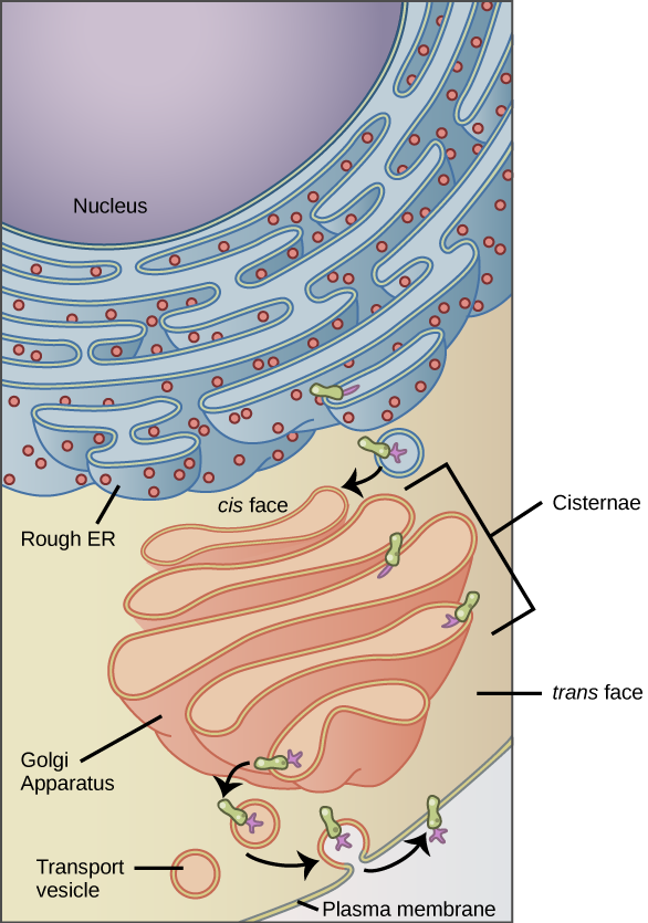
Vesicles and vacuoles are membrane-bound sacs that function in storage and transport. Vacuoles are somewhat larger than vesicles, and the membrane of a vacuole does not fuse with the membranes of other cellular components. Vesicles can fuse with other membranes within the cell system (Figure \(\PageIndex{1}\)). Additionally, enzymes within plant vacuoles can break down macromolecules.
The Central Vacuole (plants)
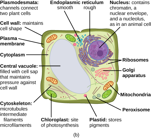
Previously, we mentioned vacuoles as essential components of plant cells. If you look at Figure \(\PageIndex{2}\), you will see that plant cells each have a large, central vacuole that occupies most of the cell.
The central vacuole plays a key role in regulating the cell’s concentration of water in changing environmental conditions. In plant cells, the liquid inside the central vacuole provides turgor pressure, which is the outward pressure caused by the fluid inside the cell. Have you ever noticed that if you forget to water a plant for a few days, it wilts? That is because as the water concentration in the soil becomes lower than the water concentration in the plant, water moves out of the central vacuoles and cytoplasm and into the soil. As the central vacuole shrinks, it leaves the cell wall unsupported. This loss of support to the cell walls of a plant results in the wilted appearance. Additionally, this fluid has a very bitter taste, which discourages consumption by insects and animals. The central vacuole also functions to store proteins in developing seed cells.
Lysosome
In animal cells, the lysosomes are the cell’s “garbage disposal.” Digestive enzymes within the lysosomes aid the breakdown of proteins, polysaccharides, lipids, nucleic acids, and even worn-out organelles. In single-celled eukaryotes, lysosomes are important for digestion of the food they ingest and the recycling of organelles. These enzymes are active at a much lower pH (more acidic) than those located in the cytoplasm. Many reactions that take place in the cytoplasm could not occur at a low pH, thus the advantage of compartmentalizing the eukaryotic cell into organelles is apparent.
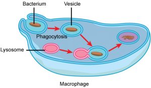
Lysosomes also use their hydrolytic enzymes to destroy disease-causing organisms that might enter the cell. A good example of this occurs in a group of white blood cells called macrophages, which are part of your body’s immune system. In a process known as phagocytosis, a section of the plasma membrane of the macrophage invaginates (folds in) and engulfs a pathogen. The invaginated section, with the pathogen inside, then pinches itself off from the plasma membrane and becomes a vesicle. The vesicle fuses with a lysosome. The lysosome’s hydrolytic enzymes then destroy the pathogen (Figure \(\PageIndex{3}\)).
Lysosomes are basically small bags of membrane containing enzymes, so they look structurally similar to a small vacuole.
Peroxisomes
Peroxisomes are small, round organelles enclosed by single membranes (so again, they look similar to small vacuoles). They carry out oxidation reactions that break down fatty acids and amino acids. They also detoxify many poisons that may enter the body. Alcohol is detoxified by peroxisomes in liver cells. A byproduct of these oxidation reactions is hydrogen peroxide, H2O2, which is contained within the peroxisomes to prevent the chemical from causing damage to cellular components outside of the organelle. Hydrogen peroxide is safely broken down by peroxisomal enzymes into water and oxygen.
References
Unless otherwise noted, images on this page are licensed under CC-BY 4.0 by OpenStax.
Text adapted from: OpenStax, Concepts of Biology. OpenStax CNX. May 18, 2016 http://cnx.org/contents/b3c1e1d2-839...9a8aafbdd@9.10