7.7: Mitosis vs. Meiosis and Disorders
- Page ID
- 17046
\( \newcommand{\vecs}[1]{\overset { \scriptstyle \rightharpoonup} {\mathbf{#1}} } \)
\( \newcommand{\vecd}[1]{\overset{-\!-\!\rightharpoonup}{\vphantom{a}\smash {#1}}} \)
\( \newcommand{\id}{\mathrm{id}}\) \( \newcommand{\Span}{\mathrm{span}}\)
( \newcommand{\kernel}{\mathrm{null}\,}\) \( \newcommand{\range}{\mathrm{range}\,}\)
\( \newcommand{\RealPart}{\mathrm{Re}}\) \( \newcommand{\ImaginaryPart}{\mathrm{Im}}\)
\( \newcommand{\Argument}{\mathrm{Arg}}\) \( \newcommand{\norm}[1]{\| #1 \|}\)
\( \newcommand{\inner}[2]{\langle #1, #2 \rangle}\)
\( \newcommand{\Span}{\mathrm{span}}\)
\( \newcommand{\id}{\mathrm{id}}\)
\( \newcommand{\Span}{\mathrm{span}}\)
\( \newcommand{\kernel}{\mathrm{null}\,}\)
\( \newcommand{\range}{\mathrm{range}\,}\)
\( \newcommand{\RealPart}{\mathrm{Re}}\)
\( \newcommand{\ImaginaryPart}{\mathrm{Im}}\)
\( \newcommand{\Argument}{\mathrm{Arg}}\)
\( \newcommand{\norm}[1]{\| #1 \|}\)
\( \newcommand{\inner}[2]{\langle #1, #2 \rangle}\)
\( \newcommand{\Span}{\mathrm{span}}\) \( \newcommand{\AA}{\unicode[.8,0]{x212B}}\)
\( \newcommand{\vectorA}[1]{\vec{#1}} % arrow\)
\( \newcommand{\vectorAt}[1]{\vec{\text{#1}}} % arrow\)
\( \newcommand{\vectorB}[1]{\overset { \scriptstyle \rightharpoonup} {\mathbf{#1}} } \)
\( \newcommand{\vectorC}[1]{\textbf{#1}} \)
\( \newcommand{\vectorD}[1]{\overrightarrow{#1}} \)
\( \newcommand{\vectorDt}[1]{\overrightarrow{\text{#1}}} \)
\( \newcommand{\vectE}[1]{\overset{-\!-\!\rightharpoonup}{\vphantom{a}\smash{\mathbf {#1}}}} \)
\( \newcommand{\vecs}[1]{\overset { \scriptstyle \rightharpoonup} {\mathbf{#1}} } \)
\( \newcommand{\vecd}[1]{\overset{-\!-\!\rightharpoonup}{\vphantom{a}\smash {#1}}} \)
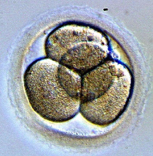
\(\newcommand{\avec}{\mathbf a}\) \(\newcommand{\bvec}{\mathbf b}\) \(\newcommand{\cvec}{\mathbf c}\) \(\newcommand{\dvec}{\mathbf d}\) \(\newcommand{\dtil}{\widetilde{\mathbf d}}\) \(\newcommand{\evec}{\mathbf e}\) \(\newcommand{\fvec}{\mathbf f}\) \(\newcommand{\nvec}{\mathbf n}\) \(\newcommand{\pvec}{\mathbf p}\) \(\newcommand{\qvec}{\mathbf q}\) \(\newcommand{\svec}{\mathbf s}\) \(\newcommand{\tvec}{\mathbf t}\) \(\newcommand{\uvec}{\mathbf u}\) \(\newcommand{\vvec}{\mathbf v}\) \(\newcommand{\wvec}{\mathbf w}\) \(\newcommand{\xvec}{\mathbf x}\) \(\newcommand{\yvec}{\mathbf y}\) \(\newcommand{\zvec}{\mathbf z}\) \(\newcommand{\rvec}{\mathbf r}\) \(\newcommand{\mvec}{\mathbf m}\) \(\newcommand{\zerovec}{\mathbf 0}\) \(\newcommand{\onevec}{\mathbf 1}\) \(\newcommand{\real}{\mathbb R}\) \(\newcommand{\twovec}[2]{\left[\begin{array}{r}#1 \\ #2 \end{array}\right]}\) \(\newcommand{\ctwovec}[2]{\left[\begin{array}{c}#1 \\ #2 \end{array}\right]}\) \(\newcommand{\threevec}[3]{\left[\begin{array}{r}#1 \\ #2 \\ #3 \end{array}\right]}\) \(\newcommand{\cthreevec}[3]{\left[\begin{array}{c}#1 \\ #2 \\ #3 \end{array}\right]}\) \(\newcommand{\fourvec}[4]{\left[\begin{array}{r}#1 \\ #2 \\ #3 \\ #4 \end{array}\right]}\) \(\newcommand{\cfourvec}[4]{\left[\begin{array}{c}#1 \\ #2 \\ #3 \\ #4 \end{array}\right]}\) \(\newcommand{\fivevec}[5]{\left[\begin{array}{r}#1 \\ #2 \\ #3 \\ #4 \\ #5 \\ \end{array}\right]}\) \(\newcommand{\cfivevec}[5]{\left[\begin{array}{c}#1 \\ #2 \\ #3 \\ #4 \\ #5 \\ \end{array}\right]}\) \(\newcommand{\mattwo}[4]{\left[\begin{array}{rr}#1 \amp #2 \\ #3 \amp #4 \\ \end{array}\right]}\) \(\newcommand{\laspan}[1]{\text{Span}\{#1\}}\) \(\newcommand{\bcal}{\cal B}\) \(\newcommand{\ccal}{\cal C}\) \(\newcommand{\scal}{\cal S}\) \(\newcommand{\wcal}{\cal W}\) \(\newcommand{\ecal}{\cal E}\) \(\newcommand{\coords}[2]{\left\{#1\right\}_{#2}}\) \(\newcommand{\gray}[1]{\color{gray}{#1}}\) \(\newcommand{\lgray}[1]{\color{lightgray}{#1}}\) \(\newcommand{\rank}{\operatorname{rank}}\) \(\newcommand{\row}{\text{Row}}\) \(\newcommand{\col}{\text{Col}}\) \(\renewcommand{\row}{\text{Row}}\) \(\newcommand{\nul}{\text{Nul}}\) \(\newcommand{\var}{\text{Var}}\) \(\newcommand{\corr}{\text{corr}}\) \(\newcommand{\len}[1]{\left|#1\right|}\) \(\newcommand{\bbar}{\overline{\bvec}}\) \(\newcommand{\bhat}{\widehat{\bvec}}\) \(\newcommand{\bperp}{\bvec^\perp}\) \(\newcommand{\xhat}{\widehat{\xvec}}\) \(\newcommand{\vhat}{\widehat{\vvec}}\) \(\newcommand{\uhat}{\widehat{\uvec}}\) \(\newcommand{\what}{\widehat{\wvec}}\) \(\newcommand{\Sighat}{\widehat{\Sigma}}\) \(\newcommand{\lt}{<}\) \(\newcommand{\gt}{>}\) \(\newcommand{\amp}{&}\) \(\definecolor{fillinmathshade}{gray}{0.9}\)Figure \(\PageIndex{1}\) shows a tiny embryo just beginning to form. Once an egg is fertilized, the resulting single cell must divide many times to develop a fetus. Both mitosis and meiosis involve cell division; is this type of cell division an example of mitosis or meiosis? The answer is mitosis. With each division, you are making a genetically exact copy of the parent cell, which only happens through mitosis.

Mitosis vs. Meiosis
Both mitosis and meiosis result in eukaryotic cell division. The primary difference between these divisions is the differing goals of each process. The goal of mitosis is to produce two daughter cells that are genetically identical to the parent cell. Mitosis happens when you grow. You want all your new cells to have the same DNA as the previous cells. The goal of meiosis is to produce sperm or eggs, also known as gametes. The resulting gametes are not genetically identical to the parent cell. Gametes are haploid cells, with only half the DNA present in the diploid parent cell. This is necessary so that when a sperm and an egg combine at fertilization, the resulting zygote has the correct amount of DNA—not twice as much as the parents. The zygote then begins to divide through mitosis.
| Mitosis | Meiosis | |
|---|---|---|
| Purpose | To produce new cells | To produce gametes |
| Number of Cells Produced | 2 | 4 |
| Rounds of Cell Division | 1 | 2 |
| Haploid or Diploid | Diploid | Haploid |
| Are daughter cells identical to parent cells? | Yes | No |
| Are daughter cells identical to each other? | Yes | No |
Figure \(\PageIndex{2}\) shows a comparison of mitosis, meiosis, and binary fission.
- Binary fission occurs in bacterial. Note that bacterial cells have a single loop of DNA. The DNA of the cell is replicated. Each loop of DNA moves to the opposite side of the cell and the cell splits in half.
- Mitosis and Meiosis both occur in eukaryotic cells. In the example below the cell has 4 total chromosomes. These are replicated during the S phase.
- In mitosis, the chromosomes line up in the center of the cell. Then, sister chromatids separate and move to the opposite poles of the cell. The cell divides, producing two cells with 4 total chromosomes
- In meiosis, the homologous chromosomes line up in the center of the cell. Then each chromosome moves to opposite poles and the cell divides.
- Next, the chromosomes line up in the center of the cell. Then, sister chromatids separate and move to the opposite poles of the cell. This produces four cells with 2 chromosomes each. These are the gametes.
- Two gametes combine to form a zygote with 4 total chromosomes.
Chromosome Disorders
Changes in Chromosome Number
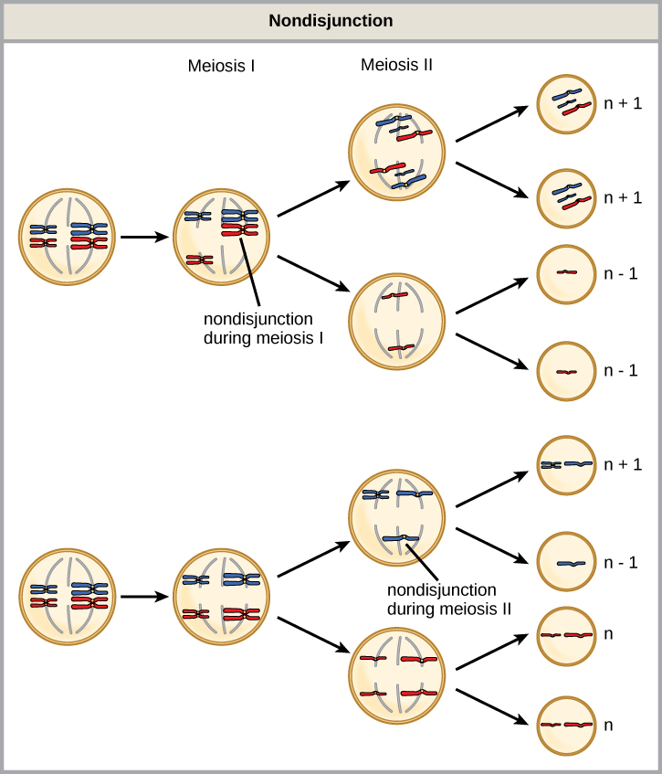
What would happen if an entire chromosome were missing or duplicated? What if a human had only 45 chromosomes? Or 47? This real possibility is usually due to mistakes during meiosis; the chromosomes do not fully separate from each other during sperm or egg formation. Specifically, nondisjunction occurs when homologous chromosomes or sister chromatids fail to separate during meiosis, resulting in an abnormal chromosome number. Nondisjunction may occur during meiosis I or meiosis II Most human atypical chromosome numbers result in the death of the developing embryo, often before a woman even realizes she is pregnant. Occasionally, a zygote with an extra chromosome can become a viable embryo and develop.
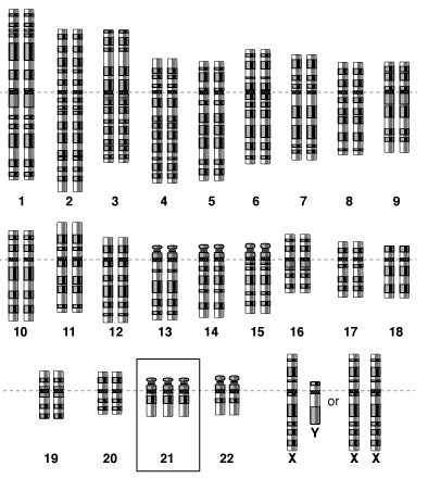
Trisomy is a state where humans have an extra autosome. That is, they have three of a particular chromosome instead of two. For example, trisomy 18 results from an extra chromosome 18, resulting in 47 total chromosomes. To identify the chromosome number (including an abnormal number), a sample of cells is removed from an individual or developing fetus. Metaphase chromosomes are photographed and a karyotype is produced. A karyotype will display any abnormalities in chromosome number or large chromosomal rearrangements. Trisomy 8, 9, 12, 13, 16, 18, and 21 have been identified in humans. Trisomy 16 is the most common trisomy in humans, occurring in more than 1% of pregnancies. This condition, however, usually results in spontaneous miscarriage in the first trimester. The most common trisomy in viable births is Trisomy 21.


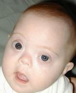
One of the most common chromosome abnormalities is Down syndrome, due to nondisjunction of chromosome 21 resulting in an extra complete chromosome 21, or part of chromosome 21 (Figure \(\PageIndex{5}\)). Down syndrome is the only autosomal trisomy where an affected individual may survive to adulthood. Individuals with Down syndrome often have some degree of mental and physical impairments and a specific facial appearance. With proper assistance, individuals with Down syndrome can become successful, contributing members of society. The risk of having a child with Down syndrome is significantly higher among women age 35 and older.

Review
- Define genetic disorder.
- What is nondisjunction? Why may it cause genetic disorders?
- Explain why genetic disorders caused by abnormal numbers of chromosomes most often involve the X chromosome.
- How is Down syndrome detected in utero?
- Compare and contrast genetic disorders and congenital disorders.
- Explain why parents that do not have Down syndrome can have a child with Down syndrome.
- What is the goal of mitosis? Or meiosis?
- How many cells are created from cytokinesis following mitosis? Following meiosis?
- Which process, mitosis or meiosis, creates genetically identical cells?
- "Gametes are haploid cells." What does this sentence mean?
Explore More
https://bio.libretexts.org/link?17046#Explore_MoreExplore More I
- Mitosis and Meiosis Simulation in the following video:
- What are homologous chromosomes?
- How does the location of specific genes compare between homologous chromosomes?
- What is the outcome of mitosis?
- What is a tetrad? Why are they an important feature of meiosis?
- How does meiosis differ between females and males?
Explore More II
Explore How Cells Divide and answer the following questions:
- How many daughter cells arise from mitosis? How many daughter cells are produced in meiosis?
- How does the attachment of spindle fibers differ between mitosis and meiosis I?
- Is anaphase I or anaphase II in meiosis more analogous to anaphase in mitosis? Explain your reasoning.
- How many steps are there in mitosis? How many steps are there in meiosis?
- How does interphase I of meiosis differ from interphase II of meiosis?
Attributions
- 4 cell embryo by Nina Sesina, CC BY-SA 4.0 via Wikimedia Commons
- Three growth types by domdomegg, CC BY-SA 4.0 via Wikimedia Commons
- Nondisjunction by OpenStax, CC BY 4.0
- Trisomy 21 by National Human Genome Research Institute, public domain via Wikimedia Commons
- Brushfield eyes by Erin Ryan, released into the public domain via Wikimedia Commons
- Text adapted from Human Biology by CK-12 licensed CC BY-NC 3.0


