
8.2: The Muscles of the Trunk
- Page ID
- 59402
Information
Figures \(\PageIndex{1}\) and \(\PageIndex{2}\) shows many of the muscles of the body’s trunk that you need to know, as well as some of the muscles of the arms and legs you will learn about in the next lab.
The deltoid muscles are the triangular muscles over each shoulder.
Some of the trunk muscles have been given nicknames by gym rats. For instance, the pecs are the pectoralis major muscles at each breast.
Lats are the latissimus dorsi muscle that covers most of the lower back with its lateral fibers.
The upper back is covered by the large trapezius muscle that is almost diamond-shaped as it extends from the neck, out to the shoulders, then tapers in midway down the back.
Obliques are the external oblique muscle whose fibers angle down as it covers both sides of the abdominal region. The external oblique muscle has two sets of fibers, which cover the left and right abdomen, that are connected by a wide aponeurosis sheet in the center of the abdomen. In most muscle models that aponeurosis sheet is cut away to reveal the rectus abdominis muscles below it.
What gym rats call the core muscles are three layers of muscle that sit over the abdomen. These layers are shown in Figure \(\PageIndex{3}\). The outer layer is the external oblique muscle, with its aponeurosis covering the medial abdomen. Under the external oblique are the internal obliques on the sides of the abdomen and the rectus abdominis muscle in-between the internal obliques. The fibers of the internal obliques run up at an angle, opposite in direction to the fiber angle of the external obliques. The rectus abdominis muscle is also known as the abs. The deepest layer has the transverse abdominis muscle, whose fibers run laterally. Its fibers are concentrated at the sides of the abdomen and, like the external oblique, has an aponeurosis covering the medial abdomen under the rectus abdominis.
Extending from the back and wrapping around the sides of the rib cage is the serratus anterior muscle. This muscle’s anterior edges are serrated like the teeth of a saw because this muscle’s origins are on ribs 1 through 8 and each serration is the attachment point to another rib. This muscle is shown in Figure \(\PageIndex{4}\).
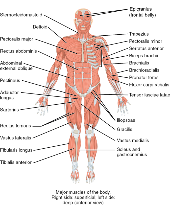
Figure \(\PageIndex{1}\): The major muscles of the body, anterior view. Anatomical right shows superficial muscles. Anatomical left shows deep muscles. (CC-BY-4.0, OpenStax, Human Anatomy)
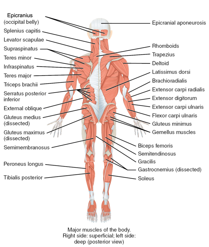
Figure \(\PageIndex{2}\): The major muscles of the body, posterior view. Anatomical right shows superficial muscles. Anatomical left shows deep muscles. (CC-BY-4.0, OpenStax, Human Anatomy)
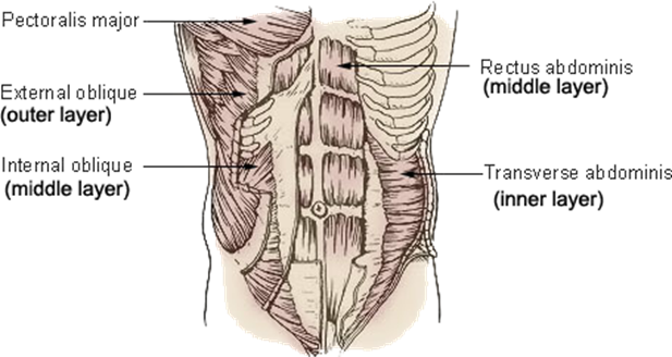
Figure \(\PageIndex{3}\): The three layers of muscles in the abdomen. (CC-BY-SA, Wikimedia)
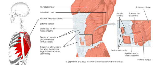
Figure \(\PageIndex{4}\): The external muscles of the body, lateral view. (CC-BY-4.0, OpenStax, Human Anatomy)
LAB 8 EXERCISE \(\PageIndex{1}\)
1. The following are muscles that move the pectoral girdle. For each, give its location and describe its action when it contracts.
|
Muscle |
Location |
Action when contracted |
|
Trapezius |
|
|
|
Pectoralis minor |
|
|
|
Serratus anterior |
|
|
2. The following are muscles that move the arm. For each, give its location and describe its action when it contracts.
|
Muscle |
Location |
Action when contracted |
|
Pectoralis major |
|
|
|
Latissimus dorsi |
|
|
|
Deltoid |
|
|
3. The following are muscles of the abdominal wall. For each, give its location and describe its action when it contracts.
|
Muscle |
Location |
Action when contracted |
|
Rectus abdominis |
|
|
|
External oblique |
|
|
|
Internal oblique |
|
|
|
Transversus abdominis |
|
|
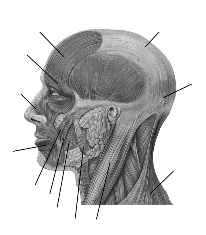
4. Label the indicated facial muscles in Figure \(\PageIndex{5}\). (CC-BY-SA, Wikimedia)
 |
|
LICENSES AND ATTRIBUTIONS
CC LICENSED CONTENT, ORIGINAL
A&P Labs. Authored by: Ross Whitwam. Provided by: Mississippi University for Women. Located at: http://www.muw.edu. License: CC BY-SA: Attribution-ShareAlike
CC LICENSED CONTENT, SHARED PREVIOUSLY
Figure \(\PageIndex{4}\). The external muscles of the body, lateral view.. Authored by: This image was made out of, or made from, content published in a BodyParts3D/Anatomography web site. . Provided by: BodyParts3D, u00a9 The Database Center for Life Science licensed under CC Attribution-Share Alike 2.1 Japan.. Located at: https://commons.wikimedia.org/wiki/F...es_lateral.png. License: CC BY-SA: Attribution- ShareAlike
CC LICENSED CONTENT, SPECIFIC ATTRIBUTION
Figure \(\PageIndex{1}\) The major muscles of the body, anterior view. Anatomical right shows superficial muscles. Anatomical left shows deep muscles.. Authored by: OpenStax College. Located at: https://cnx.org/resources/8a3b1231f3...s_of_Muscles.j pg. License: CC BY-SA: Attribution-ShareAlike
Figure \(\PageIndex{2}\). The major muscles of the body, posterior view. Anatomical right shows superficial muscles. Anatomical left shows deep muscles.. Authored by: OpenStax College. Located at: https://cnx.org/resources/8a3b1231f3...s_of_Muscles.j pg. License: CC BY-SA: Attribution-ShareAlike
Figure \(\PageIndex{4}\). The external muscles of the body, lateral view.. Authored by: OpenStax College. Located at: https://cnx.org/resources/33fa36d780...he_Abdomen.jpg. License: CC BY-SA: Attribution-ShareAlike
Figure \(\PageIndex{5}\). Facial muscles.. Authored by: Patrick J. Lynch. Located at: https://commons.wikimedia.org/wiki/F...ad_anatomy.jpg. License: CC BY-SA: Attribution-ShareAlike
PUBLIC DOMAIN CONTENT
Figure \(\PageIndex{3}\) The three layers of muscles in the abdomen.. Authored by: Arcadian. Located at: https://commons.wikimedia.org/wiki/F...nk_muscles.jpg. License: Public Domain: No Known Copyright