
8.1: The Muscles of the Head and Neck
- Page ID
- 59401
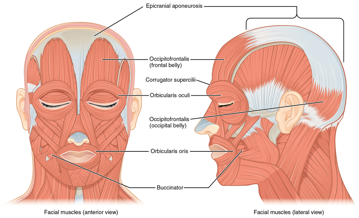
Figure \(\PageIndex{1}\) lists the muscles of the head and neck that you will need to know. A single platysma muscle is only shown in the lateral view of the head muscles in Figure 8.1. There are two platysma muscles, one on each side of the neck. Each is a broad sheet of a muscle that covers most of the anterior neck on that side of the body. The other anterior neck muscles are below them, and most models have the platysma muscles cut away to show the deeper muscles. The platysma muscles help pull down the lower jaw (mandible.)
Under the platysma are two sternocleidomastoid muscles. One on each side of the neck. These muscles have two origins, one on the sternum and the other on the clavicle. They insert on the mastoid process of the temporal bone. They can flex or extend the head, or can rotate the towards the shoulders.
The epicranius muscle is also very broad and covers most of the top of the head. The epicranius muscle includes a middle section which is all aponeurosis. The actual muscle tissue is only found over the forehead (the portion of the muscle called the epicranius frontalis; sometimes called the frontal belly of the epicranius) and the back of the head (the portion of the muscle called the epicranius occipitalis; sometimes called the occipital belly of the epicranius).
The buccinator muscles, one on each side of the face, compress the cheeks when contracted. The name is from the Latin for trumpet, which requires blowing air out of the cheeks to play, and also reflects the anatomical adjective for the cheek, buccal.
The two masseter muscles are also on each side of the face. They close the jaw when contracted. Its name is derived from the same Greek root as mastication, which means to chew.
The zygomaticus major muscles and the zygomaticus minor muscles are found on each side of the face both have their origins on the zygomatic bone. They both can change the shape of the mouth by elevating it.
LAB 8 EXERCISE 8-1
1. The following are muscles of facial expression. For each, give its location and describe its action when it contracts. Complete figure \(\PageIndex{1}\): above by adding in any muscles found in the table below.
|
Muscle |
Location |
Action when contracted |
|
Epicranius frontalis |
|
|
|
Epicranius occipitalis |
|
|
|
Orbicularis oculi |
|
|
|
Zygomaticus major |
|
|
|
Zygomaticus minor |
|
|
|
Buccinator |
|
|
|
Orbicularis oris |
|
|
|
Platysma |
|
|
|
Levator labii sup. |
|
|
|
Depressor labii inf. |
|
|
|
Levator anguli oris |
|
|
|
Depressor anguli oris |
|
|
2. The following are muscles of mastication. For each, give its location and describe its action when it contracts.
|
Muscle |
Location |
Action when contracted |
|
Masseter |
|
|
|
Temporalis |
|
|
|
Medial pterygoid |
|
|
|
Lateral pterygoid |
|
|
LICENSES AND ATTRIBUTIONS
CC LICENSED CONTENT, ORIGINAL
A&P Labs. Authored by: Ross Whitwam. Provided by: Mississippi University for Women. Located at: http://www.muw.edu. License: CC BY-SA: Attribution-ShareAlike
CC LICENSED CONTENT, SPECIFIC ATTRIBUTION
Figure \(\PageIndex{1}\). The muscles of the head.. Authored by: OpenStax College. Located at: https://cnx.org/resources/9b369a7466..._the_Muscles_o f_Facial_Expressions.jpg. License: CC BY-SA: Attribution-ShareAlike