
7.1: Meristem Morphology
- Page ID
- 93171

\( \newcommand{\vecs}[1]{\overset { \scriptstyle \rightharpoonup} {\mathbf{#1}} } \)
\( \newcommand{\vecd}[1]{\overset{-\!-\!\rightharpoonup}{\vphantom{a}\smash {#1}}} \)
\( \newcommand{\dsum}{\displaystyle\sum\limits} \)
\( \newcommand{\dint}{\displaystyle\int\limits} \)
\( \newcommand{\dlim}{\displaystyle\lim\limits} \)
\( \newcommand{\id}{\mathrm{id}}\) \( \newcommand{\Span}{\mathrm{span}}\)
( \newcommand{\kernel}{\mathrm{null}\,}\) \( \newcommand{\range}{\mathrm{range}\,}\)
\( \newcommand{\RealPart}{\mathrm{Re}}\) \( \newcommand{\ImaginaryPart}{\mathrm{Im}}\)
\( \newcommand{\Argument}{\mathrm{Arg}}\) \( \newcommand{\norm}[1]{\| #1 \|}\)
\( \newcommand{\inner}[2]{\langle #1, #2 \rangle}\)
\( \newcommand{\Span}{\mathrm{span}}\)
\( \newcommand{\id}{\mathrm{id}}\)
\( \newcommand{\Span}{\mathrm{span}}\)
\( \newcommand{\kernel}{\mathrm{null}\,}\)
\( \newcommand{\range}{\mathrm{range}\,}\)
\( \newcommand{\RealPart}{\mathrm{Re}}\)
\( \newcommand{\ImaginaryPart}{\mathrm{Im}}\)
\( \newcommand{\Argument}{\mathrm{Arg}}\)
\( \newcommand{\norm}[1]{\| #1 \|}\)
\( \newcommand{\inner}[2]{\langle #1, #2 \rangle}\)
\( \newcommand{\Span}{\mathrm{span}}\) \( \newcommand{\AA}{\unicode[.8,0]{x212B}}\)
\( \newcommand{\vectorA}[1]{\vec{#1}} % arrow\)
\( \newcommand{\vectorAt}[1]{\vec{\text{#1}}} % arrow\)
\( \newcommand{\vectorB}[1]{\overset { \scriptstyle \rightharpoonup} {\mathbf{#1}} } \)
\( \newcommand{\vectorC}[1]{\textbf{#1}} \)
\( \newcommand{\vectorD}[1]{\overrightarrow{#1}} \)
\( \newcommand{\vectorDt}[1]{\overrightarrow{\text{#1}}} \)
\( \newcommand{\vectE}[1]{\overset{-\!-\!\rightharpoonup}{\vphantom{a}\smash{\mathbf {#1}}}} \)
\( \newcommand{\vecs}[1]{\overset { \scriptstyle \rightharpoonup} {\mathbf{#1}} } \)
\(\newcommand{\longvect}{\overrightarrow}\)
\( \newcommand{\vecd}[1]{\overset{-\!-\!\rightharpoonup}{\vphantom{a}\smash {#1}}} \)
\(\newcommand{\avec}{\mathbf a}\) \(\newcommand{\bvec}{\mathbf b}\) \(\newcommand{\cvec}{\mathbf c}\) \(\newcommand{\dvec}{\mathbf d}\) \(\newcommand{\dtil}{\widetilde{\mathbf d}}\) \(\newcommand{\evec}{\mathbf e}\) \(\newcommand{\fvec}{\mathbf f}\) \(\newcommand{\nvec}{\mathbf n}\) \(\newcommand{\pvec}{\mathbf p}\) \(\newcommand{\qvec}{\mathbf q}\) \(\newcommand{\svec}{\mathbf s}\) \(\newcommand{\tvec}{\mathbf t}\) \(\newcommand{\uvec}{\mathbf u}\) \(\newcommand{\vvec}{\mathbf v}\) \(\newcommand{\wvec}{\mathbf w}\) \(\newcommand{\xvec}{\mathbf x}\) \(\newcommand{\yvec}{\mathbf y}\) \(\newcommand{\zvec}{\mathbf z}\) \(\newcommand{\rvec}{\mathbf r}\) \(\newcommand{\mvec}{\mathbf m}\) \(\newcommand{\zerovec}{\mathbf 0}\) \(\newcommand{\onevec}{\mathbf 1}\) \(\newcommand{\real}{\mathbb R}\) \(\newcommand{\twovec}[2]{\left[\begin{array}{r}#1 \\ #2 \end{array}\right]}\) \(\newcommand{\ctwovec}[2]{\left[\begin{array}{c}#1 \\ #2 \end{array}\right]}\) \(\newcommand{\threevec}[3]{\left[\begin{array}{r}#1 \\ #2 \\ #3 \end{array}\right]}\) \(\newcommand{\cthreevec}[3]{\left[\begin{array}{c}#1 \\ #2 \\ #3 \end{array}\right]}\) \(\newcommand{\fourvec}[4]{\left[\begin{array}{r}#1 \\ #2 \\ #3 \\ #4 \end{array}\right]}\) \(\newcommand{\cfourvec}[4]{\left[\begin{array}{c}#1 \\ #2 \\ #3 \\ #4 \end{array}\right]}\) \(\newcommand{\fivevec}[5]{\left[\begin{array}{r}#1 \\ #2 \\ #3 \\ #4 \\ #5 \\ \end{array}\right]}\) \(\newcommand{\cfivevec}[5]{\left[\begin{array}{c}#1 \\ #2 \\ #3 \\ #4 \\ #5 \\ \end{array}\right]}\) \(\newcommand{\mattwo}[4]{\left[\begin{array}{rr}#1 \amp #2 \\ #3 \amp #4 \\ \end{array}\right]}\) \(\newcommand{\laspan}[1]{\text{Span}\{#1\}}\) \(\newcommand{\bcal}{\cal B}\) \(\newcommand{\ccal}{\cal C}\) \(\newcommand{\scal}{\cal S}\) \(\newcommand{\wcal}{\cal W}\) \(\newcommand{\ecal}{\cal E}\) \(\newcommand{\coords}[2]{\left\{#1\right\}_{#2}}\) \(\newcommand{\gray}[1]{\color{gray}{#1}}\) \(\newcommand{\lgray}[1]{\color{lightgray}{#1}}\) \(\newcommand{\rank}{\operatorname{rank}}\) \(\newcommand{\row}{\text{Row}}\) \(\newcommand{\col}{\text{Col}}\) \(\renewcommand{\row}{\text{Row}}\) \(\newcommand{\nul}{\text{Nul}}\) \(\newcommand{\var}{\text{Var}}\) \(\newcommand{\corr}{\text{corr}}\) \(\newcommand{\len}[1]{\left|#1\right|}\) \(\newcommand{\bbar}{\overline{\bvec}}\) \(\newcommand{\bhat}{\widehat{\bvec}}\) \(\newcommand{\bperp}{\bvec^\perp}\) \(\newcommand{\xhat}{\widehat{\xvec}}\) \(\newcommand{\vhat}{\widehat{\vvec}}\) \(\newcommand{\uhat}{\widehat{\uvec}}\) \(\newcommand{\what}{\widehat{\wvec}}\) \(\newcommand{\Sighat}{\widehat{\Sigma}}\) \(\newcommand{\lt}{<}\) \(\newcommand{\gt}{>}\) \(\newcommand{\amp}{&}\) \(\definecolor{fillinmathshade}{gray}{0.9}\)By the end of this lesson you will be able to:
- Differentiate between primary growth from apical meristems and secondary growth from lateral meristems.
- Describe two types of lateral meristems and the types of tissues that are derived from these meristems.
Primary growth from meristems
You’ll recall that the apical meristem is the site of cell division and new cell production at the tips of the plant stems and roots. The cells that make up the meristem are undergoing mitotic cell division to produce more cells. These new cells result in growth and development of plant tissues. (If you haven’t previously studied mitosis, you’ll have the opportunity to do so during this class.) For now it is sufficient to know that mitosis is the process of cell division where one plant cell divides into two identical cells.
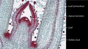
Above is a micrograph of a coleus shoot tip. You can see the dome of the apical meristem at the very tip of the shoot surrounded by leaf primordia (rudimentary leaves). On the far left and far right are the cells of two growing leaves. You can see a trace of vascular tissue on the left leaf near the “L” of the leaf label. There is another red stained area called the axillary bud, which we’ve studied previously. The axillary bud is another very small shoot tip with a meristematic area. Axillary buds are found at a node and typically occur where a leaf petiole attaches to a stem. The axillary buds in this stage of growth are inactive, but in time may begin active cell division and develop into new branches off of the main stem.
The coleus micrograph is clearly stem tissue because you can see leaves and leaf primordia, so where are the nodes and internodes? The region where the leaves are attached, and where you find the axillary buds, is a node. Above this is the internode, and at the top where you find the leaf primordia is another node.
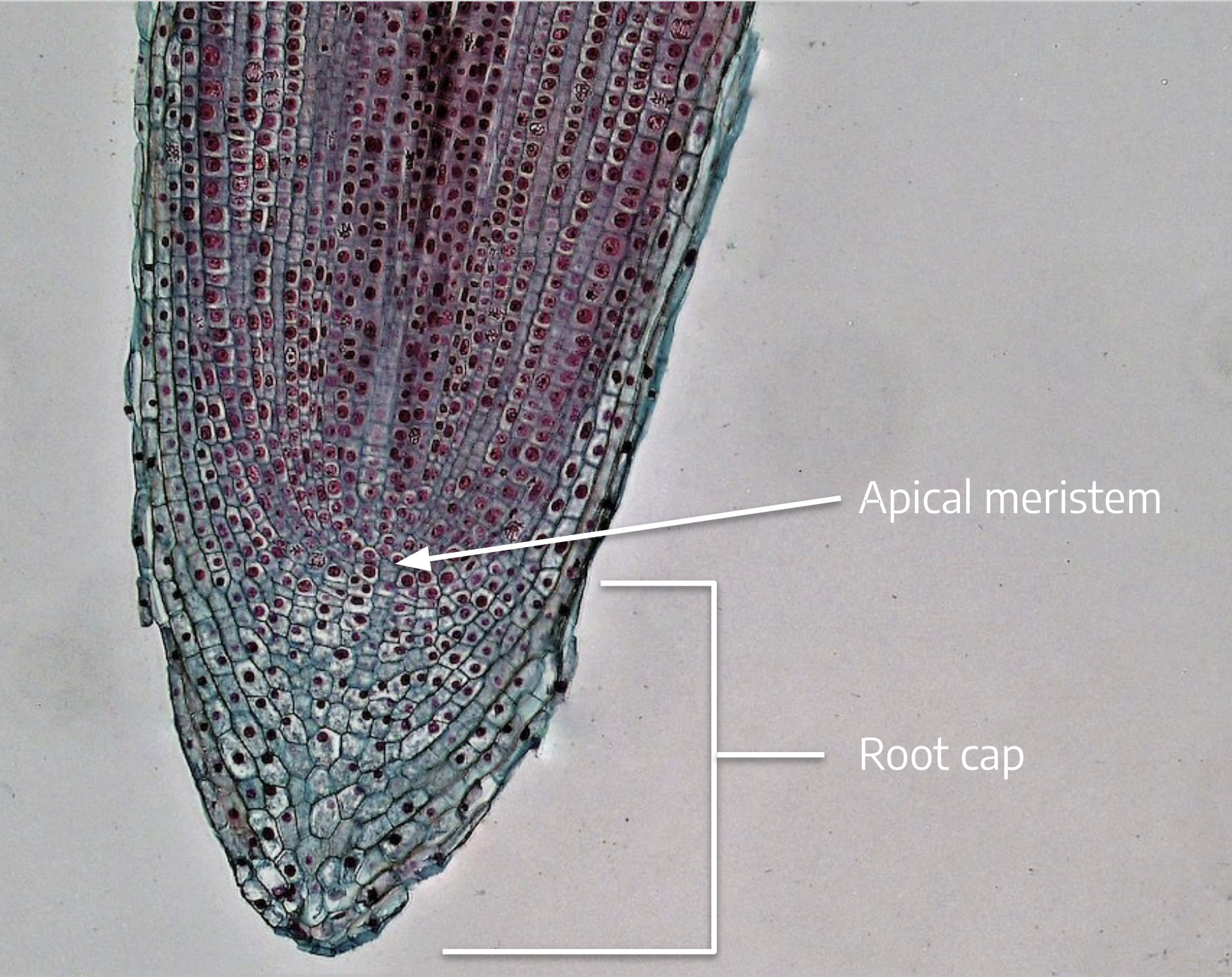
The root meristem looks very different from the shoot apical meristem. Recall that, unlike branches that develop at nodes, lateral roots are formed adventitiously, as the result of meristematic activity in the pericycle cells of the root’s vascular system in the zone of maturation. We don’t see a node-internode structure like we saw with the coleus shoot tip.
When meristem cells divide, whether in the shoot or the root, one of the two resulting sister cells typically continues to be a meristem cell. The other sister cell divides a few more times and then differentiates into dermal, cortex, or vascular tissue in the stem or root. Meristem cells that remain meristematic are called initials because they continue to divide, producing new cells. The other sister cells that divide once or twice more and then differentiate are called derivative cells. The xylem and phloem tissues that result from differentiation of derivative cells are called primary xylem and primary phloem, where the word “primary” signals that the cells originated from cell divisions of the apical meristem.
To reiterate, young stems and roots have primary xylem and primary phloem that formed as a result of differentiation of derivative cells. Primary xylem and primary phloem cells trace back to an apical meristem.
Earlier you learned the arrangement of the vascular tissues in monocot and dicot stems and roots. Remember that mitotic cell divisions in the apical meristem result in lengthening of the root or shoot through production of new cells plus the elongation of those cells. With a few exceptions, this is the only type of growth — growth that is initiated by cell division in the apical meristem — you’ll find in monocots. Dicots, however, have another type of growth — from a different type of meristem — that results in thickening of the stem.
- If shown a micrograph of an apical meristem, how would you determine whether it is from a root or a shoot?
- What happens to the initial cell mentioned in the question above? Does it continue to divide?
Secondary growth (thickening): Introducing lateral meristems
Watch this video for a closer look at apical and lateral meristems (2:26)
Unlike annual herbaceous plants that only survive for one growing season, and whose stem and root cells trace back to cell divisions of the apical meristems, woody plants and shrubs are perennial dicots that have the capacity for secondary growth and can survive from year to year.
Some annual herbaceous dicot plants, like tomatoes, can have secondary growth, but for now let’s consider those the exceptions and focus on perennial dicot woody plants. Secondary growth is the result of activity by a special type of meristem called a lateral meristem. As with apical meristems, lateral meristems are made up of cells that undergo mitotic cell division. Mitosis in lateral meristems results in lateral growth (thickening of the stem or root) and adds to the girth of a plant rather than its length. Remember that length is the outcome of cell division in the apical meristem plus elongation of those cells. Girth or thickening is the result of lateral meristems. We’ll learn about two types of lateral meristems: vascular cambium, and cork cambium.
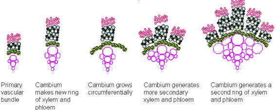
Vascular cambium

Let’s start with the vascular cambium.
The three drawings on the right show a cross section of a stem for an imaginary woody dicot. The top drawing illustrates the stem early in the first year of growth, and shows the vascular cylinders arranged in a ring around the stem. The phloem is oriented to the outside, the xylem to the inside. A thin layer of parenchyma cells between the xylem and phloem has differentiated into the fascicular cambium (fascicular refers to bundles, in this case, cambium in the vascular bundles). The fascicular cambium is meristematic and can divide to produce new phloem toward the outside and new xylem to the inside. The new xylem and phloem produced by the cambium are called 2o (secondary) xylem and 2o phloem. Recall that the original xylem and phloem that differentiated from the apical meristem’s derivative cells are called the 1o (primary) xylem and 1o phloem.
The middle drawing is of the same stem later in the year. The cortex (cortical) parenchyma cells that lay between the vascular cylinders directly in line with the fascicular cambium begin to differentiate into a type of cambium called interfascicular cambium (cambium between the bundles). This is symbolized by the line connecting the vascular cylinders. This cambium is also meristematic, and produces 2o xylem and 2o phloem.
The cross section on the bottom illustrates the stem in its second or third year of growth, when there is a noticeable buildup of 2o xylem and 2o phloem with remnants of 1o xylem and 1o phloem.
In summary, the vascular cambium is a lateral meristem formed by differentiation of parenchyma cells located between the primary xylem and phloem into fascicular cambium, followed by differentiation of cortical parenchyma between the vascular cylinders into interfascicular cambium. After a few years of secondary growth, fascicular and interfascicular cambium can no longer be distinguished, and it is all simply known as vascular cambium. This layer of cambium runs vertically (assuming that the stem is oriented vertically) and parallel to the surface of the woody stem.
The illustration below shows how the cambium divides to produce 2o xylem and 2o phloem, with the outside of the stem toward the top of the page. Frame #1 shows a single cambium cell (C). This cell divides mitotically (M) to form two cambium daughter cells (Frame #2). Frame #3 shows that the cambium cell on the top differentiates (D) into a phloem cell (P-toward the outside) and the other cambium cell divides mitotically (M).
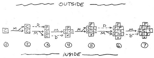
This type of cell division, in which new cells are formed either to the outside or inside, and the cell wall that separates the two new cells is parallel to the outside of the stem, is called periclinal division. Periclinal division by the cambium makes new cells that add girth to the plant. The cells that are added subsequently differentiate into xylem and phloem depending on their location to the outside or inside of the cambium. The meristem needs to divide periclinally to add girth to the plant stem.
In Frame #4, pay particular attention to a different type of cell division, where the cambium cell has divided so that the wall between the two cells is perpendicular to the outside of the stem. This is called anticlinal division. The meristem occasionally needs to divide anticlinally because as the stem is growing in girth, the diameter of the ring of vascular cambium must expand to keep up, or it will split into pieces and no longer form a continuous ring around the stem. Frame #4 also shows that the cambium cell to the inside has differentiated into xylem (X). In Frame #5, the two cambial cells that formed from anticlinal division now each divide periclinally.
Watch this video for a demonstration of periclinal and anticlinal division (3:50)
- How might you recognize a plant that has secondary growth?
- In a perennial woody dicot, how do the discrete vascular bundles found in the new seedling stem become continuous rings of xylem and phloem in the three-year-old woody stem?
- Explain what is happening in Frames #6 and #7 in the drawing above. Note that there are two important changes: differentiation and anticlinal division.
Cork cambium
Let’s look at the second type of lateral meristem, cork cambium. Cork (called “phellem” in this image) provides a protective covering around the expanding trunk of the woody plant.
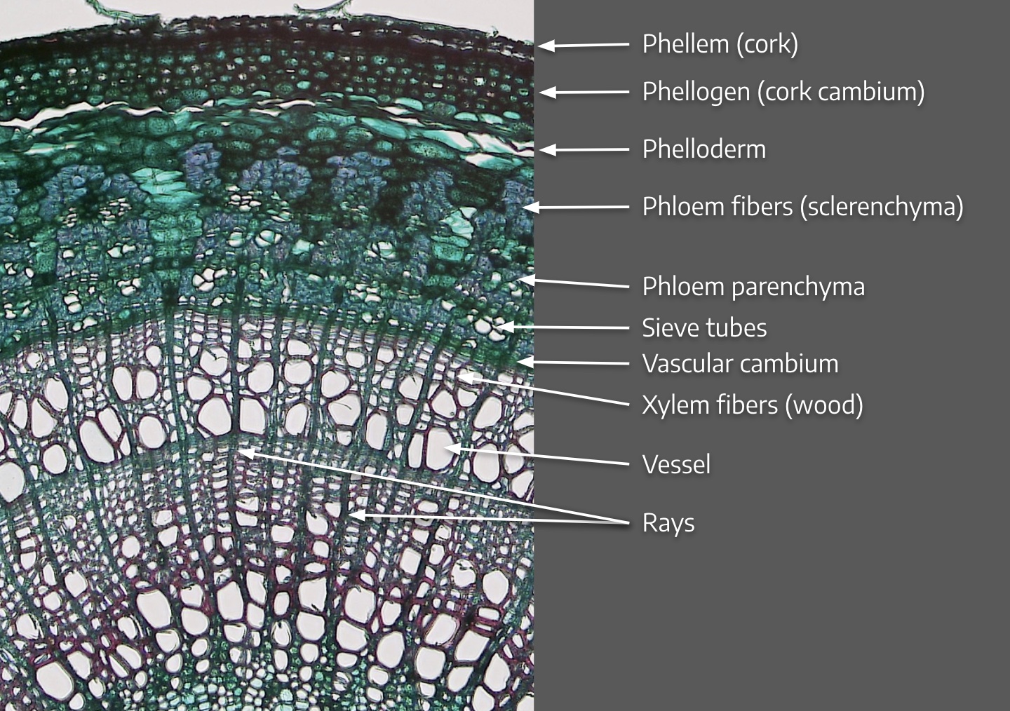
Cork develops in plants with secondary growth after the initiation of secondary xylem and phloem and the expansion of the stem and root’s girth. Cortex parenchyma cells next to the epidermis of the young stem differentiate into the cork cambium (also called phellogen), which is meristematic. The cork cambium lays down some new cells toward the inside called phelloderm, but lays down most of its new cells to the outside, and these derivatives of the cork cambium differentiate into the cork cells. The cork cells are lined with a waxy substance called suberin (we first saw this substance in conjunction with the Casparian strip around endodermis cells) that make the cells impermeable to water and gases. Breaks in the cork cells, called lenticels, allow gas and water exchange. You can see these lenticels in corks from wine bottles (wine corks are made from the thick cork of the Cork Oak, Quercus suber). Cork cells die when they mature. These cells replace the protective function provided by epidermis in young roots and stems. Cork cambium and its derivatives (phelloderm and cork) are called the periderm.
Botanically speaking, the word pb_glossary id=”1215″]”bark”[/pb_glossary] refers to all of the tissues exterior of the vascular cambium. So bark includes:
- Primary and secondary phloem
- Phelloderm if present
- Cork cambium
- Cork
Look again at the list above and note that the phloem is part of the bark. This is often overlooked!
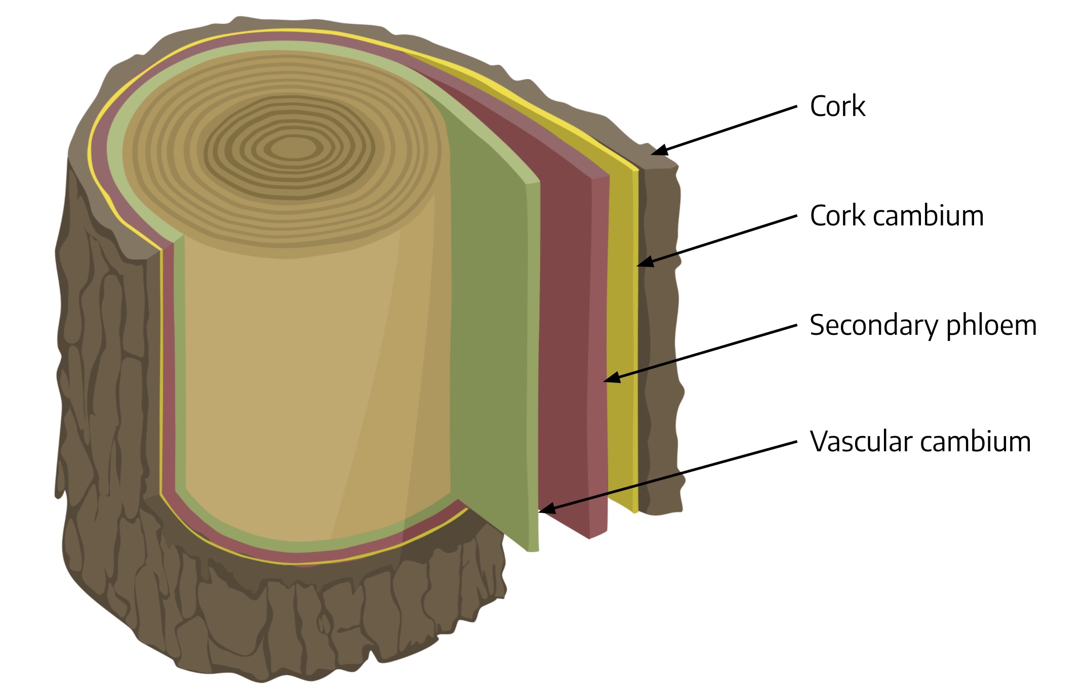
This illustration summarizes the layers introduced above, but omits the phelloderm. If you were to add a label for the phelloderm, where would it be? In mature trunks the phelloderm is quite a thin layer in the bark though, particularly relative to the cork layer, so that is probably why it is absent in this illustration.
If you peel bark from a living tree, you are exposing the surface of the vascular cambium of the tree, and on the inside of the bark will be the phloem. Peeling off bark will kill the plant because you are removing phloem, which disrupts the ability of the plant to move sugars through the trunk of the tree, exposes the tree to moisture loss and predator invasion, and kills the vascular cambium through desiccation. Killing the vascular cambium halts production of new xylem which will subsequently interfere with water transpiration up the stem. This is what happens when rodents girdle the trunks of young fruit trees (left) just below the snow line as they feed on the tender tissues over winter.

A tree’s annual rings are found in the secondary xylem. In the spring, the newly produced xylem cells have thin walls and are large in diameter so they can accommodate the abundance of soil moisture that is typical of April and May.

As the growing season progresses and soil moisture levels are depleted, the cell walls of newly produced xylem are thicker and the diameter of the xylem cells diminishes. In the next spring, the newly produced xylem cells are once again thin-walled and large. The distinct contrast between the small-celled late summer wood from the previous year and the large-celled spring wood from the following year is noticeable, and this line of demarcation between late summer and spring xylem is called the annual ring. In the image to the right you can see the darker xylem, called heartwood, and the lighter xylem, called sapwood. Heartwood is older xylem that is clogged with resins that darken the cells and limit their ability to transport water. Sapwood is younger, resin-free, and still functioning to conduct water up the trunk.
There isn’t nearly as much phloem as xylem in a woody plant. Phloem isn’t produced as rapidly as xylem, and is crushed between the vascular and cork cambium layers, so we don’t see annual rings in phloem.
Watch this video for a look at what makes up bark (2:38)
- Describe the origin of annual rings. If a woody dicot is growing in a tropical climate where the weather is the same day in and day out, will you find annual rings in the wood?
- What is the most exterior cell layer in an herbaceous stem called? What is the most exterior layer of cells in a five-year-old woody perennial plant stem called.
- If some kids in your neighborhood get hold of a little hatchet and chop off thin slices of bark all the way around the base of one of your trees, what will happen to the tree? Why?
What about roots?
Roots of woody perennial dicots also have vascular and cork cambium. The vascular cambium arises from parenchyma cells lying between the xylem and phloem in roots, just as in stems. The illustration below shows the initiation of the vascular cambium in a young root. It looks like a trace of white between the primary xylem in the middle and the primary phloem in each of the four lobes of the vascular cylinder. With age, the cambium encircles the root and produces (secondary) xylem to the inside, and (secondary) phloem to outside, again just as in stems. Older roots with secondary growth also have a periderm similar to that found in the stem that replaces the function of the epidermis. If you’ve ever tried to dig in the soil surrounding a mature tree, you know that roots are as tough and woody as the branches above ground.
Here is a diagram to help you visualize lateral meristems including cell division.

The next diagram shows a vascular bundle as it grows by dividing both periclinally and anticlinally.
Ross Koning at Eastern Connecticut State University has a helpful site regarding secondary plant growth (optional reading) with descriptions and images that explain secondary growth in the stem vascular column, in the stem dermal tissues, and in the roots. L. Rost from U.C.-Davis has this nice explanation of secondary growth in roots (more optional reading).