
3.3.2: Internal Anatomy of the Primary Stem
- Page ID
- 37048

\( \newcommand{\vecs}[1]{\overset { \scriptstyle \rightharpoonup} {\mathbf{#1}} } \)
\( \newcommand{\vecd}[1]{\overset{-\!-\!\rightharpoonup}{\vphantom{a}\smash {#1}}} \)
\( \newcommand{\dsum}{\displaystyle\sum\limits} \)
\( \newcommand{\dint}{\displaystyle\int\limits} \)
\( \newcommand{\dlim}{\displaystyle\lim\limits} \)
\( \newcommand{\id}{\mathrm{id}}\) \( \newcommand{\Span}{\mathrm{span}}\)
( \newcommand{\kernel}{\mathrm{null}\,}\) \( \newcommand{\range}{\mathrm{range}\,}\)
\( \newcommand{\RealPart}{\mathrm{Re}}\) \( \newcommand{\ImaginaryPart}{\mathrm{Im}}\)
\( \newcommand{\Argument}{\mathrm{Arg}}\) \( \newcommand{\norm}[1]{\| #1 \|}\)
\( \newcommand{\inner}[2]{\langle #1, #2 \rangle}\)
\( \newcommand{\Span}{\mathrm{span}}\)
\( \newcommand{\id}{\mathrm{id}}\)
\( \newcommand{\Span}{\mathrm{span}}\)
\( \newcommand{\kernel}{\mathrm{null}\,}\)
\( \newcommand{\range}{\mathrm{range}\,}\)
\( \newcommand{\RealPart}{\mathrm{Re}}\)
\( \newcommand{\ImaginaryPart}{\mathrm{Im}}\)
\( \newcommand{\Argument}{\mathrm{Arg}}\)
\( \newcommand{\norm}[1]{\| #1 \|}\)
\( \newcommand{\inner}[2]{\langle #1, #2 \rangle}\)
\( \newcommand{\Span}{\mathrm{span}}\) \( \newcommand{\AA}{\unicode[.8,0]{x212B}}\)
\( \newcommand{\vectorA}[1]{\vec{#1}} % arrow\)
\( \newcommand{\vectorAt}[1]{\vec{\text{#1}}} % arrow\)
\( \newcommand{\vectorB}[1]{\overset { \scriptstyle \rightharpoonup} {\mathbf{#1}} } \)
\( \newcommand{\vectorC}[1]{\textbf{#1}} \)
\( \newcommand{\vectorD}[1]{\overrightarrow{#1}} \)
\( \newcommand{\vectorDt}[1]{\overrightarrow{\text{#1}}} \)
\( \newcommand{\vectE}[1]{\overset{-\!-\!\rightharpoonup}{\vphantom{a}\smash{\mathbf {#1}}}} \)
\( \newcommand{\vecs}[1]{\overset { \scriptstyle \rightharpoonup} {\mathbf{#1}} } \)
\(\newcommand{\longvect}{\overrightarrow}\)
\( \newcommand{\vecd}[1]{\overset{-\!-\!\rightharpoonup}{\vphantom{a}\smash {#1}}} \)
\(\newcommand{\avec}{\mathbf a}\) \(\newcommand{\bvec}{\mathbf b}\) \(\newcommand{\cvec}{\mathbf c}\) \(\newcommand{\dvec}{\mathbf d}\) \(\newcommand{\dtil}{\widetilde{\mathbf d}}\) \(\newcommand{\evec}{\mathbf e}\) \(\newcommand{\fvec}{\mathbf f}\) \(\newcommand{\nvec}{\mathbf n}\) \(\newcommand{\pvec}{\mathbf p}\) \(\newcommand{\qvec}{\mathbf q}\) \(\newcommand{\svec}{\mathbf s}\) \(\newcommand{\tvec}{\mathbf t}\) \(\newcommand{\uvec}{\mathbf u}\) \(\newcommand{\vvec}{\mathbf v}\) \(\newcommand{\wvec}{\mathbf w}\) \(\newcommand{\xvec}{\mathbf x}\) \(\newcommand{\yvec}{\mathbf y}\) \(\newcommand{\zvec}{\mathbf z}\) \(\newcommand{\rvec}{\mathbf r}\) \(\newcommand{\mvec}{\mathbf m}\) \(\newcommand{\zerovec}{\mathbf 0}\) \(\newcommand{\onevec}{\mathbf 1}\) \(\newcommand{\real}{\mathbb R}\) \(\newcommand{\twovec}[2]{\left[\begin{array}{r}#1 \\ #2 \end{array}\right]}\) \(\newcommand{\ctwovec}[2]{\left[\begin{array}{c}#1 \\ #2 \end{array}\right]}\) \(\newcommand{\threevec}[3]{\left[\begin{array}{r}#1 \\ #2 \\ #3 \end{array}\right]}\) \(\newcommand{\cthreevec}[3]{\left[\begin{array}{c}#1 \\ #2 \\ #3 \end{array}\right]}\) \(\newcommand{\fourvec}[4]{\left[\begin{array}{r}#1 \\ #2 \\ #3 \\ #4 \end{array}\right]}\) \(\newcommand{\cfourvec}[4]{\left[\begin{array}{c}#1 \\ #2 \\ #3 \\ #4 \end{array}\right]}\) \(\newcommand{\fivevec}[5]{\left[\begin{array}{r}#1 \\ #2 \\ #3 \\ #4 \\ #5 \\ \end{array}\right]}\) \(\newcommand{\cfivevec}[5]{\left[\begin{array}{c}#1 \\ #2 \\ #3 \\ #4 \\ #5 \\ \end{array}\right]}\) \(\newcommand{\mattwo}[4]{\left[\begin{array}{rr}#1 \amp #2 \\ #3 \amp #4 \\ \end{array}\right]}\) \(\newcommand{\laspan}[1]{\text{Span}\{#1\}}\) \(\newcommand{\bcal}{\cal B}\) \(\newcommand{\ccal}{\cal C}\) \(\newcommand{\scal}{\cal S}\) \(\newcommand{\wcal}{\cal W}\) \(\newcommand{\ecal}{\cal E}\) \(\newcommand{\coords}[2]{\left\{#1\right\}_{#2}}\) \(\newcommand{\gray}[1]{\color{gray}{#1}}\) \(\newcommand{\lgray}[1]{\color{lightgray}{#1}}\) \(\newcommand{\rank}{\operatorname{rank}}\) \(\newcommand{\row}{\text{Row}}\) \(\newcommand{\col}{\text{Col}}\) \(\renewcommand{\row}{\text{Row}}\) \(\newcommand{\nul}{\text{Nul}}\) \(\newcommand{\var}{\text{Var}}\) \(\newcommand{\corr}{\text{corr}}\) \(\newcommand{\len}[1]{\left|#1\right|}\) \(\newcommand{\bbar}{\overline{\bvec}}\) \(\newcommand{\bhat}{\widehat{\bvec}}\) \(\newcommand{\bperp}{\bvec^\perp}\) \(\newcommand{\xhat}{\widehat{\xvec}}\) \(\newcommand{\vhat}{\widehat{\vvec}}\) \(\newcommand{\uhat}{\widehat{\uvec}}\) \(\newcommand{\what}{\widehat{\wvec}}\) \(\newcommand{\Sighat}{\widehat{\Sigma}}\) \(\newcommand{\lt}{<}\) \(\newcommand{\gt}{>}\) \(\newcommand{\amp}{&}\) \(\definecolor{fillinmathshade}{gray}{0.9}\)- Identify the structures representing each of the three tissue systems in stems.
- Compare the structure of solenosteles, eusteles, and atactosteles.
- Explain the arrangement of leaf traces, leaf trace gaps, branch traces, and branch trace gaps.
The primary stem refers to the herbaceous (non-woody) stem, which has not undergone secondary growth (the growth that produces bark and wood). Some species (all monocots and some eudicots) remain herbaceous for their entire lives, maintaining the primary stem. Other species of eudicots initially form a primary stem but later become woody, replacing the primary stem with the secondary stem. The anatomy of the stem (internal structure) can be examined through longitudinal sections (cutting the stem lengthwise) or in cross sections (cutting a slice of the stem perpendicular to the length).
All three tissue types are represented in the primary stem. The epidermis is the dermal tissue that surrounds and protects the stem. The epidermis typically consists of one layer of cells. A waxy cuticle on the outside of these cells limits water loss. Epidermal cells are the most numerous and least differentiated of the cells in the epidermis. Pores in the epidermis called stomata (singular: stoma) allow for gas exchange. Each stoma is bordered by a pair of guard cells, which regulate stomatal opening. While stomata are present in stems, they occur at higher densities in leaves. Trichomes are hair-like structures on the epidermal surface. They help to reduce transpiration (the loss of water by aboveground plant parts), increase solar reflectance, and store compounds that defend the leaves against predation by herbivores (Figure \(\PageIndex{1}\)).
Ground tissue fills much of the stem, forming the cortex directly within the epidermis and the pith (if present) in the center. The outermost portion of the cortex is usually a few layers of collenchyma cells. The remainder of the cortex and pith consist of parenchyma cells. The arrangement of vascular tissue in the stem depends on the species (see below).
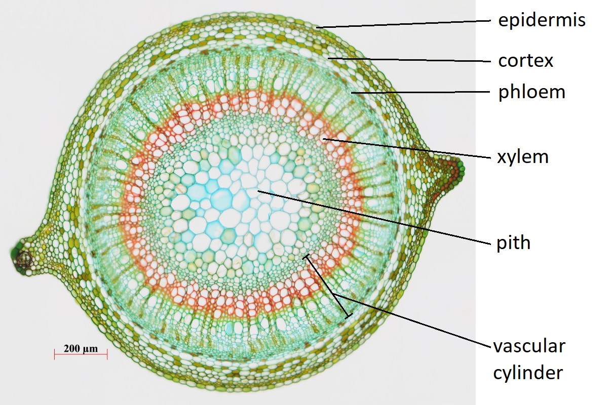
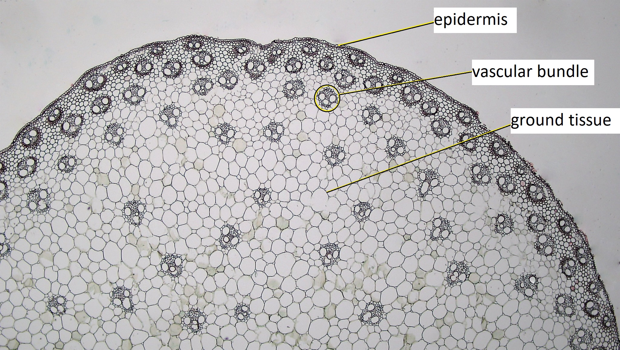
Cross sections reveal three possible arrangements of vascular tissue (steles) in the stem. The first arrangement (solenostele) is present in a few eudicots, such as basswood (Tilia). In the solenostele, the vascular tissue appears as a continuous ring (vascular cylinder; Figure \(\PageIndex{2}\)). The interfascicular regions (pith rays) of parenchyma cells that separate vascular tissue are thus extremely narrow. The second arrangement (eustele) is present in most eudicots such as sunflower (Helianthus) and buttercup (Ranunculus). In the eustele, vascular tissue is clustered into distinct vascular bundles arranged in a ring, allowing for thicker interfascicular regions in between them (Figure \(\PageIndex{3-4}\)). In these solenosteles and eusteles, the vascular tissue separates ground tissue into an outer cortex and central pith. The third arrangement (atactostele) is present in most monocots, such as corn (Zea mays) and a few eudicots. In the atactostele, vascular bundles are scattered throughout the stem (Figure \(\PageIndex{3, 5}\)). While vascular bundles near the outside of the stem are packed more densely in this third arrangement, their distribution is somewhat disorderly. There is no distinction between the cortex and pith in the third arrangement.

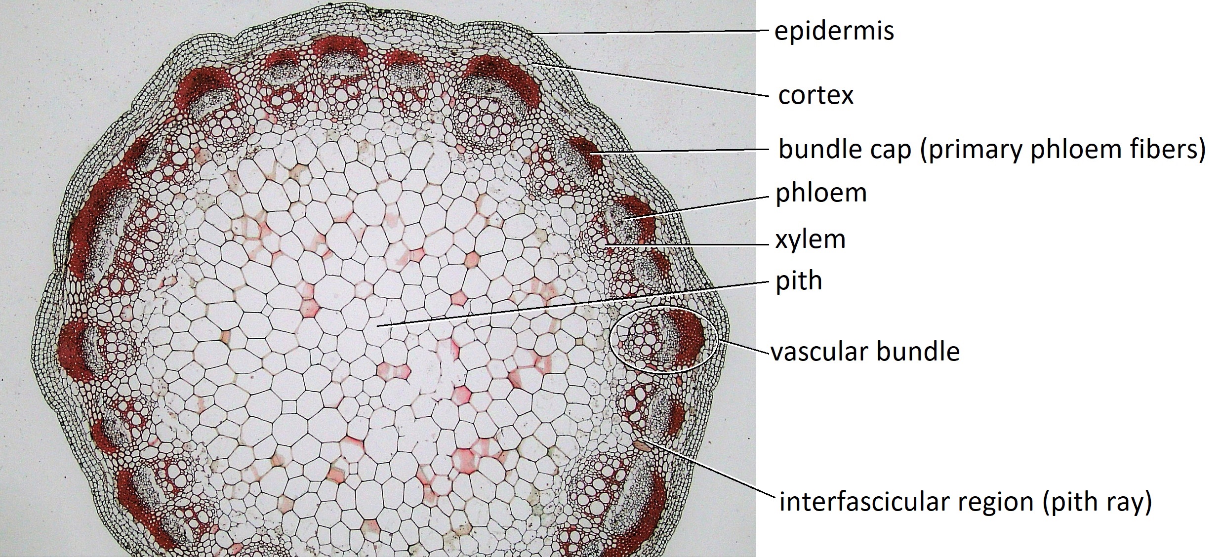
The cells of embryonic tissue called the procambium (see Meristems) divides to produce primary xylem internally and primary phloem externally. In some vascular bundles, some procambial cells remain and form the fascicular cambium in the center of the vascular bundle. Once the stem has finished lengthening, sclerenchyma fibers called primary phloem fibers are produced just outside of the primary phloem. The primary phloem fibers of each vascular bundle are sometimes called phloem caps (bundle caps). If primary phloem fibers surrounded the entire vascular bundle, they form a bundle sheath (Figure \(\PageIndex{6}\)).

Vascular bundles connect leaves and stems. The strands of vascular tissue that connect the leaves to the stem are called leaf traces. Just above leaf traces are portions of stem without vascular tissue called leaf trace gaps. Similarly, branch traces connect axillary shoots to the main stem, leaving branch trace gaps just above them (Figure \(\PageIndex{7-8}\)).


Attributions
Curated and authored by Melissa Ha from the following sources:
- From 5.4: The Stem from Introduction to Botany by Alexey Shipunov (public domain)
- From 30.2 Stems from Biology 2e by OpenStax (licensed CC-BY)