
3.2.3: Internal Root Structure
- Page ID
- 37031

\( \newcommand{\vecs}[1]{\overset { \scriptstyle \rightharpoonup} {\mathbf{#1}} } \)
\( \newcommand{\vecd}[1]{\overset{-\!-\!\rightharpoonup}{\vphantom{a}\smash {#1}}} \)
\( \newcommand{\id}{\mathrm{id}}\) \( \newcommand{\Span}{\mathrm{span}}\)
( \newcommand{\kernel}{\mathrm{null}\,}\) \( \newcommand{\range}{\mathrm{range}\,}\)
\( \newcommand{\RealPart}{\mathrm{Re}}\) \( \newcommand{\ImaginaryPart}{\mathrm{Im}}\)
\( \newcommand{\Argument}{\mathrm{Arg}}\) \( \newcommand{\norm}[1]{\| #1 \|}\)
\( \newcommand{\inner}[2]{\langle #1, #2 \rangle}\)
\( \newcommand{\Span}{\mathrm{span}}\)
\( \newcommand{\id}{\mathrm{id}}\)
\( \newcommand{\Span}{\mathrm{span}}\)
\( \newcommand{\kernel}{\mathrm{null}\,}\)
\( \newcommand{\range}{\mathrm{range}\,}\)
\( \newcommand{\RealPart}{\mathrm{Re}}\)
\( \newcommand{\ImaginaryPart}{\mathrm{Im}}\)
\( \newcommand{\Argument}{\mathrm{Arg}}\)
\( \newcommand{\norm}[1]{\| #1 \|}\)
\( \newcommand{\inner}[2]{\langle #1, #2 \rangle}\)
\( \newcommand{\Span}{\mathrm{span}}\) \( \newcommand{\AA}{\unicode[.8,0]{x212B}}\)
\( \newcommand{\vectorA}[1]{\vec{#1}} % arrow\)
\( \newcommand{\vectorAt}[1]{\vec{\text{#1}}} % arrow\)
\( \newcommand{\vectorB}[1]{\overset { \scriptstyle \rightharpoonup} {\mathbf{#1}} } \)
\( \newcommand{\vectorC}[1]{\textbf{#1}} \)
\( \newcommand{\vectorD}[1]{\overrightarrow{#1}} \)
\( \newcommand{\vectorDt}[1]{\overrightarrow{\text{#1}}} \)
\( \newcommand{\vectE}[1]{\overset{-\!-\!\rightharpoonup}{\vphantom{a}\smash{\mathbf {#1}}}} \)
\( \newcommand{\vecs}[1]{\overset { \scriptstyle \rightharpoonup} {\mathbf{#1}} } \)
\( \newcommand{\vecd}[1]{\overset{-\!-\!\rightharpoonup}{\vphantom{a}\smash {#1}}} \)
\(\newcommand{\avec}{\mathbf a}\) \(\newcommand{\bvec}{\mathbf b}\) \(\newcommand{\cvec}{\mathbf c}\) \(\newcommand{\dvec}{\mathbf d}\) \(\newcommand{\dtil}{\widetilde{\mathbf d}}\) \(\newcommand{\evec}{\mathbf e}\) \(\newcommand{\fvec}{\mathbf f}\) \(\newcommand{\nvec}{\mathbf n}\) \(\newcommand{\pvec}{\mathbf p}\) \(\newcommand{\qvec}{\mathbf q}\) \(\newcommand{\svec}{\mathbf s}\) \(\newcommand{\tvec}{\mathbf t}\) \(\newcommand{\uvec}{\mathbf u}\) \(\newcommand{\vvec}{\mathbf v}\) \(\newcommand{\wvec}{\mathbf w}\) \(\newcommand{\xvec}{\mathbf x}\) \(\newcommand{\yvec}{\mathbf y}\) \(\newcommand{\zvec}{\mathbf z}\) \(\newcommand{\rvec}{\mathbf r}\) \(\newcommand{\mvec}{\mathbf m}\) \(\newcommand{\zerovec}{\mathbf 0}\) \(\newcommand{\onevec}{\mathbf 1}\) \(\newcommand{\real}{\mathbb R}\) \(\newcommand{\twovec}[2]{\left[\begin{array}{r}#1 \\ #2 \end{array}\right]}\) \(\newcommand{\ctwovec}[2]{\left[\begin{array}{c}#1 \\ #2 \end{array}\right]}\) \(\newcommand{\threevec}[3]{\left[\begin{array}{r}#1 \\ #2 \\ #3 \end{array}\right]}\) \(\newcommand{\cthreevec}[3]{\left[\begin{array}{c}#1 \\ #2 \\ #3 \end{array}\right]}\) \(\newcommand{\fourvec}[4]{\left[\begin{array}{r}#1 \\ #2 \\ #3 \\ #4 \end{array}\right]}\) \(\newcommand{\cfourvec}[4]{\left[\begin{array}{c}#1 \\ #2 \\ #3 \\ #4 \end{array}\right]}\) \(\newcommand{\fivevec}[5]{\left[\begin{array}{r}#1 \\ #2 \\ #3 \\ #4 \\ #5 \\ \end{array}\right]}\) \(\newcommand{\cfivevec}[5]{\left[\begin{array}{c}#1 \\ #2 \\ #3 \\ #4 \\ #5 \\ \end{array}\right]}\) \(\newcommand{\mattwo}[4]{\left[\begin{array}{rr}#1 \amp #2 \\ #3 \amp #4 \\ \end{array}\right]}\) \(\newcommand{\laspan}[1]{\text{Span}\{#1\}}\) \(\newcommand{\bcal}{\cal B}\) \(\newcommand{\ccal}{\cal C}\) \(\newcommand{\scal}{\cal S}\) \(\newcommand{\wcal}{\cal W}\) \(\newcommand{\ecal}{\cal E}\) \(\newcommand{\coords}[2]{\left\{#1\right\}_{#2}}\) \(\newcommand{\gray}[1]{\color{gray}{#1}}\) \(\newcommand{\lgray}[1]{\color{lightgray}{#1}}\) \(\newcommand{\rank}{\operatorname{rank}}\) \(\newcommand{\row}{\text{Row}}\) \(\newcommand{\col}{\text{Col}}\) \(\renewcommand{\row}{\text{Row}}\) \(\newcommand{\nul}{\text{Nul}}\) \(\newcommand{\var}{\text{Var}}\) \(\newcommand{\corr}{\text{corr}}\) \(\newcommand{\len}[1]{\left|#1\right|}\) \(\newcommand{\bbar}{\overline{\bvec}}\) \(\newcommand{\bhat}{\widehat{\bvec}}\) \(\newcommand{\bperp}{\bvec^\perp}\) \(\newcommand{\xhat}{\widehat{\xvec}}\) \(\newcommand{\vhat}{\widehat{\vvec}}\) \(\newcommand{\uhat}{\widehat{\uvec}}\) \(\newcommand{\what}{\widehat{\wvec}}\) \(\newcommand{\Sighat}{\widehat{\Sigma}}\) \(\newcommand{\lt}{<}\) \(\newcommand{\gt}{>}\) \(\newcommand{\amp}{&}\) \(\definecolor{fillinmathshade}{gray}{0.9}\)Learning Objectives
- Describe the different structures and zones of a root.
- Compare and contrast a monocot root to a eudicot root.
- Describe secondary root growth and the function of vascular and cork cambium.
Root Anatomy
Root growth begins with seed germination. When the plant embryo emerges from the seed, the radicle of the embryo begins to grow downward and forms the root system. As the root system grows, various structures begin to appear.
Longitudinal Section
If you were to cut a root down longitudinally, you would see the various layers inside. The tip of the root is protected by the root cap, a structure exclusive to roots and unlike any other plant structure. The root cap is continuously replaced because it gets damaged easily as the root pushes through soil. The root tip can be divided into three zones: a zone of cell division, a zone of elongation, and a zone of maturation and differentiation (Figure \(\PageIndex{1}\)). The zone of cell division is a continuation of the root cap; it is made up of the actively dividing cells of the root meristem. The zone of elongation is where the newly formed cells begin to increase in length, thereby lengthening the root. They are older than cells at the zone of cell division. Beginning at the first root hair is the zone of cell maturation where the root cells begin to differentiate into special cell types. The root has an outer layer of cells called the epidermis, which surrounds areas of ground tissue and vascular tissue. The epidermis provides protection and helps in absorption. Root hairs, which are extensions of root epidermal cells, increase the surface area of the root, greatly contributing to the absorption of water and minerals. All three zones are in the first centimeter or so of the root tip.
Figure \(\PageIndex{1}\): A longitudinal view of the root reveals the zones of cell division, elongation, and maturation. Cell division occurs in the apical meristem.
Cross Section
If you were to cut a cross section of the leaf, you could see other features that are not as obvious in the longitudinal section. Inside the root, the ground tissue may form two regions: the cortex and the pith (Figure \(\PageIndex{2}\)). When comparing roots to stems, roots have much more cortex and very little pith. Whereas eudicot roots have no central pith, monocots have a small pith. Both cortex and pith include cells that store photosynthetic products. The cortex is between the epidermis and the vascular tissue, whereas the pith lies between the vascular tissue and the center of the root.
The inner portion of the root contains the vascular tissue (xylem and phloem). This area is called the stele. A layer of cells known as the endodermis borders the stele (Figure \(\PageIndex{2}\)) and is considered the innermost layer of the cortex. The endodermis is exclusive to roots, and serves as a checkpoint for materials entering the root’s vascular system. A waxy substance called suberin is present on the walls of the endodermal cells. This waxy region, known as the Casparian strip, forces water and solutes to cross the plasma membranes of endodermal cells instead of slipping between the cells. This ensures that only materials required by the root pass through the endodermis, while toxic substances and pathogens are generally excluded. The outermost cell layer of the root’s vascular tissue is the pericycle, an area that can give rise to lateral roots.
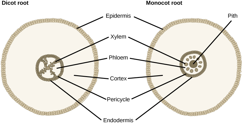
Monocots
Note that the size of the stele in the monocot cross section is large (everything inside the green ring (Figure \(\PageIndex{3}\)). The vascular tissue is arranged in a ring around the pith. This arrangement is called a siphonostele. The cortex surrounds the stele. The endodermis is the innermost layer of the cortex, and the exodermis is the outermost layer of the cortex. The exodermis controls the flow of water, ions, and nutrients. The outermost layer of the root (external to the cortex) is the epidermis, which covers the root and aids in absorption.
Eudicots
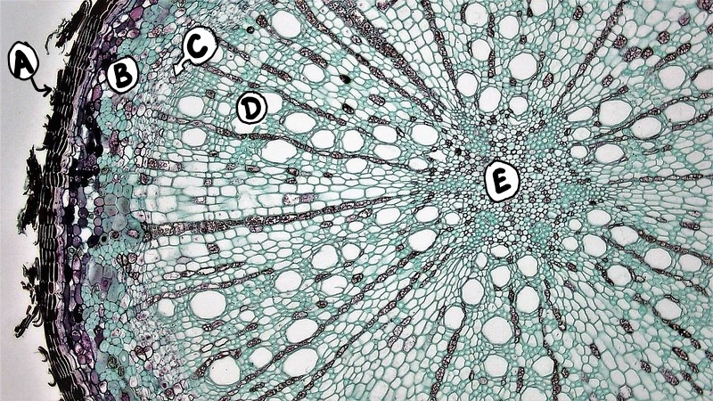
In eudicot roots, the vascular tissue fills the center of the root, and there is no pith. This arrangment is called a protostele. The xylem and phloem of the stele are arranged alternately in an X shape (Figure \(\PageIndex{4}\)). Most of the root is composed of cortex tissue, and the endodermis, the innermost layer of the cortex, borders the stele. The outer layer of the root (external to the cortex) is the epidermis.
.jpg?revision=1&size=bestfit&width=550&height=310)
Secondary Root Growth
Many roots have secondary growth as well as primary growth (figures \(\PageIndex{5-6}\)). This occurs by the production of two types of meristemic tissue, the vascular cambium and the cork cambium. The cork cambium is responsible for the girth or growth in the diameter of the root. This occurs by the cork cambium adding vascular tissue to the root. Cells of the pericycle and procambium (the meristematic tissue between the primary phloem and xylem) begin division, and form a vascular cambium around the primary xylem. The vascular cambium then divides to form secondary xylem on the inside and secondary phloem on the outside.
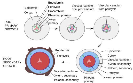
Some roots with secondary growth may form a periderm (a protective layer, replacing the epidermis). This occurs by the formation of a cork cambium which originate from the pericycle. The cork cambium produces parenchyma tissues called phelloderm to the inside of the root and the cork on the outside of the root. Cork cells (phellem) are dead at maturity. They are hollow and the addition of air space in the tissue functions as a protective layer. They also produce a waxy substance called suberin. This wax functions to aid in water loss. It also makes the root more resistant to bacterial and fungal infections. The three layers 1. phelloderm 2. cork cambium and 3. cork cells are collectively known as the periderm.
Attributions
Curated and authored by Kammy Algiers and Melissa Ha from the following sources
- 11.2 Secondary Growth from A Photographic Atlas for Botany by Maria Morrow (CC-BY-NC).
- 30.3 Roots from General Biology by OpenStax (CC-BY).