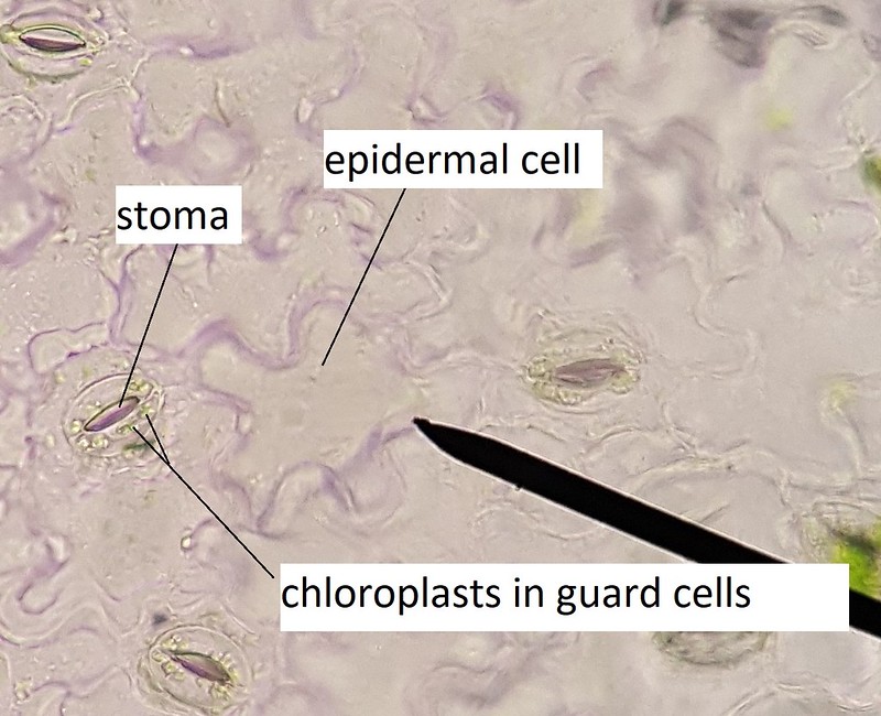
Stomata are pores (holes) in the epidermis of plants. Guard cells are the pairs of cells, shaped a bit like parentheses or two sides of a donut, that flank the stoma. The guard cells regulate when the stoma is open or closed, which in turn regulates gas exchange with the environment and the rate of transpiration.
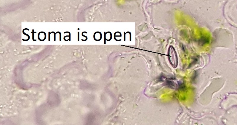
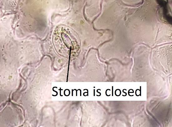
Figure 10.2.2.1: This image is of an epidermal peel. The epidermal cells look like transparent, interlocking puzzle pieces. Interspersed between these cells, there are stomata with guard cells. Each stoma looks a bit like a mouth, with one guard cell acting as the upper lip and another guard cell acting as the lower. Chloroplasts are visible within the guard cells. Photo by Melissa Ha, CC BY-NC .Figure 10.2.2.2: There are two images above. The one on the left shows an open stoma, while the one on the right shows a closed stoma. Photo by Melissa Ha, CC BY-NC.
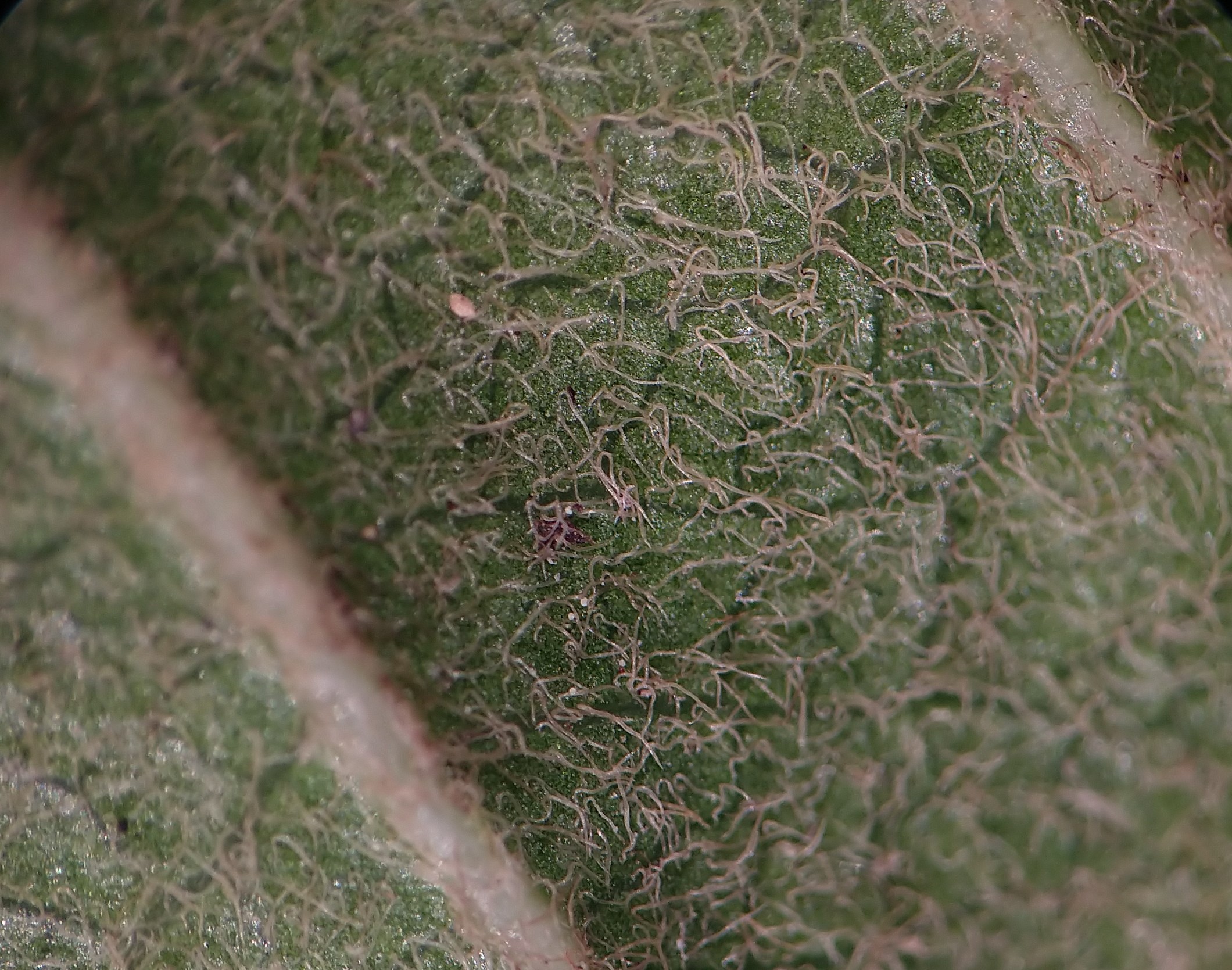
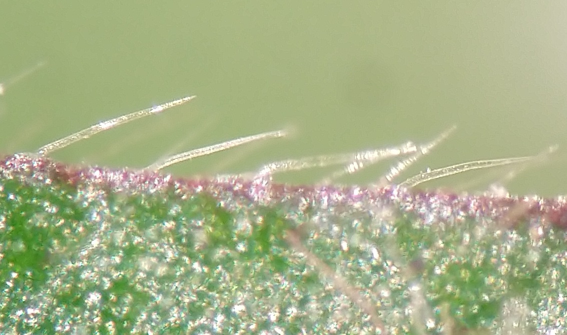
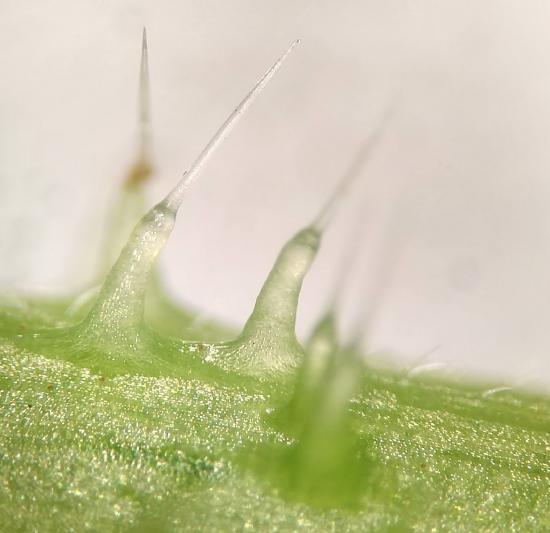
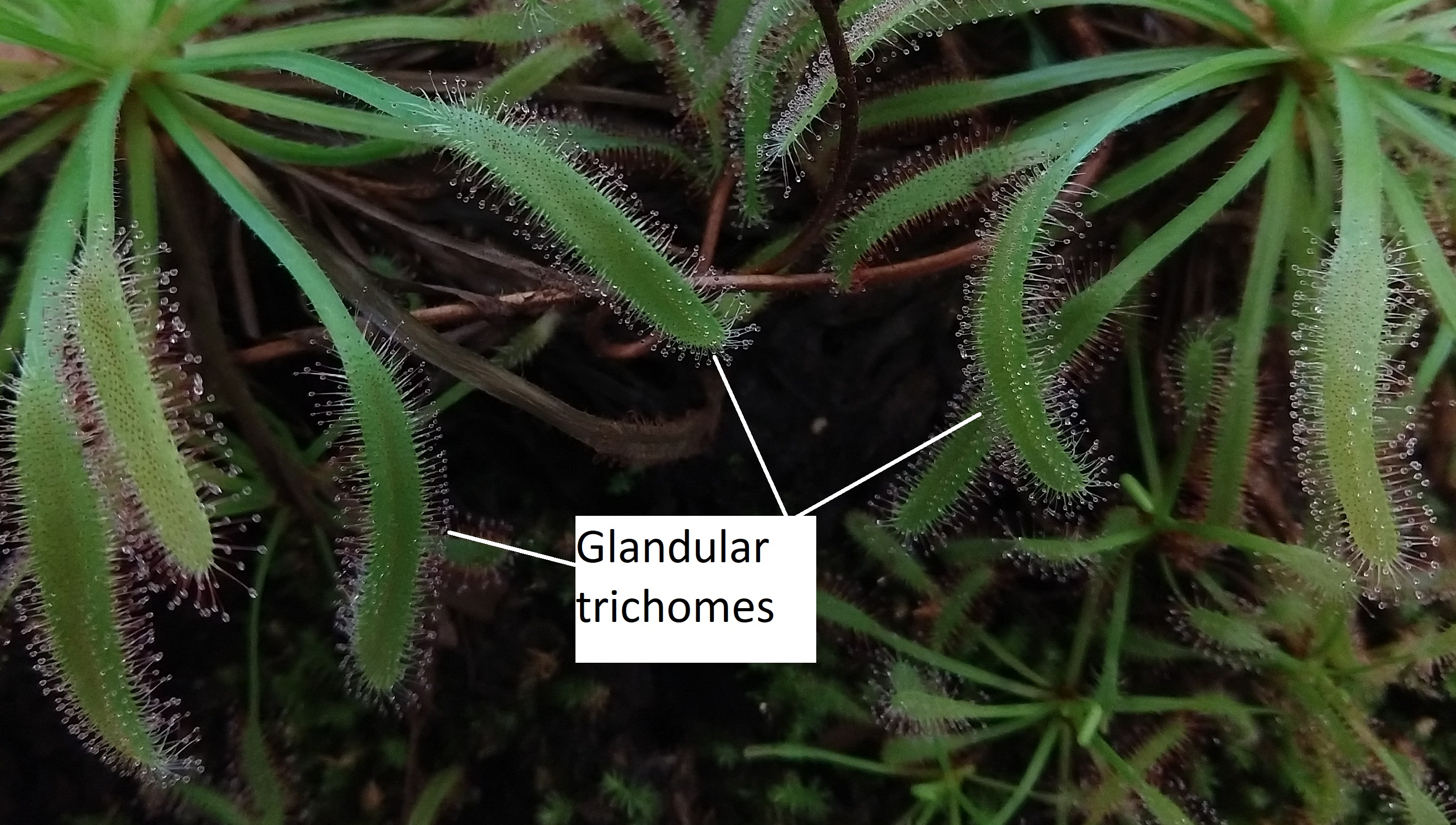
Trichomes
Trichomes are hairs composed of cells on the epidermis of a plant.
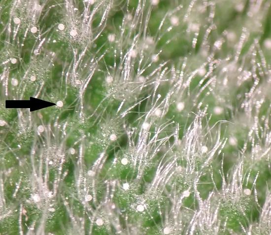
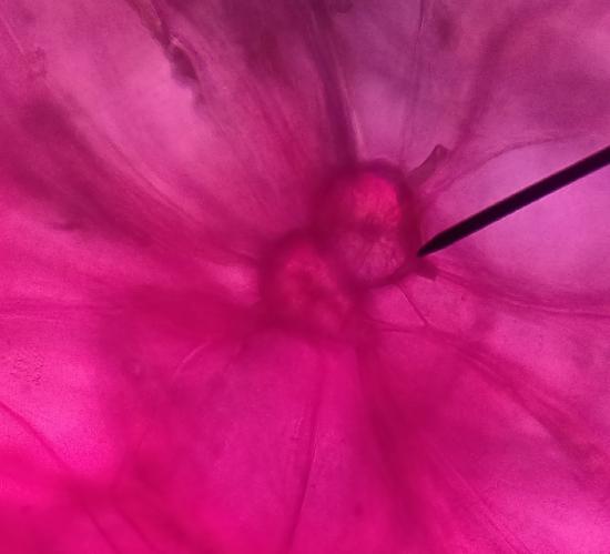
Figure 10.2.2.2: Trichomes on the underside (abaxial) of a leaf (left) and on a leaf margin (right). The trichomes in the photo on the left are long, fuzzy, and intertwining. The trichomes in the photo on the right are straight, separate from each other, and rigid-looking. The different appearance and texture of trichomes is one of the characteristics used to identify plants. Photos by Maria Morrow, CC BY-NC.Figure 10.2.2.3: These nettle trichomes are structured much like a hypodermic needle. The long silica tip breaks off to inject a cocktail of stinging chemicals into any animal that brushes by it.Photo by Maria Morrow, CC BY-NC.Figure 10.2.2.4: The ends of sundew (Drosera sp.) leaves are covered with glandular trichomes. Each trichome has a blob of sticky fluid at the tip. Insects are attracted to the fluid, becoming trapped in it, and the plant can slowly digest their bodies for the mineral nutrients that are lacking in their environment.Photo by Maria Morrow, CC BY-NC.Figure 10.2.2.5: The underside (abaxial surface) of this sage leaf has two types of trichomes: glandular trichomes and peltate trichomes. The glandular trichomes are in tufts on raised bumps of the epidermis. Scattered amongst them are small white pearl-like structures (one is indicated by a black arrow). These are the peltate trichomes, which are disc-like instead of hair-like.Photo by Maria Morrow, CC BY-NC.
Specialized Cells From the Ground Meristem
Two types of specialized sclerenchyma cells that can be produced by the ground meristem are sclereids and fibers.
Sclereids
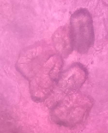
Figure 10.2.2.6: Sclereids are a type of sclerenchyma cell. These sclereids, called stone cells, are produced in the fruits of pears. In the image on the left, two stone cells are surrounded by many large parenchyma cells. There are plasmodesmata (not distinguishable) connecting the sclereids to these parenchyma cells. In the image on the right, there are 6 or 7 stone cells. They have a thick secondary wall with channels running through it (pits). These pits are the former locations of the plasmodesmata. Photos by Maria Morrow, CC BY-NC.
Fibers
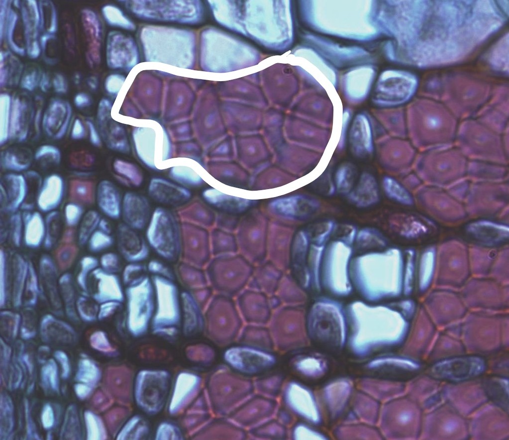
Figure 10.2.2.7: Clusters of fibers in the secondary phloem of a Tilia stem. Phloem cells are parenchyma, so they are not as rigid as xylem cells. A way for plants to strengthen and protect phloem tissue is to include bundles of fibers. One of these bundles has been circled in the image.Photo by Maria Morrow, CC BY-NC.
Specialized Cells From the Procambium
Tracheids and Vessel Elements
Tracheids and vessel elements are cells in the xylem that transport water. They have secondary walls with lignin and are dead at functional maturity.
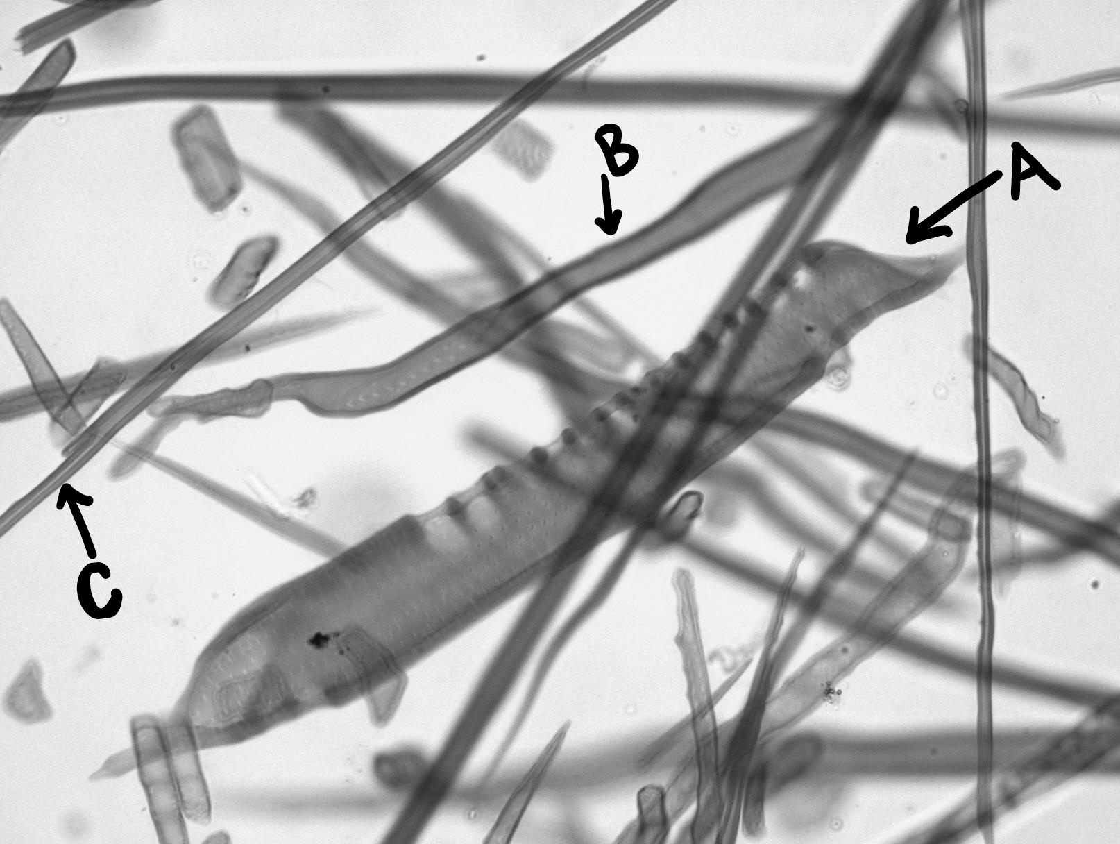
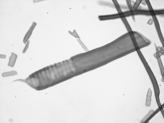
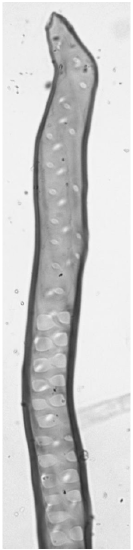
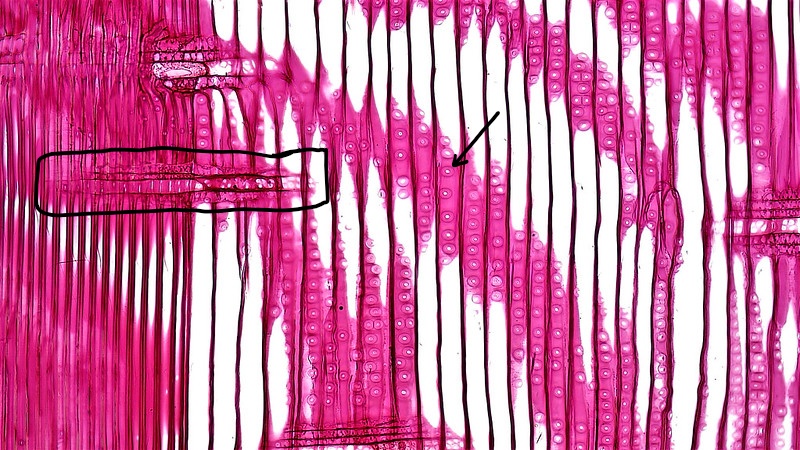
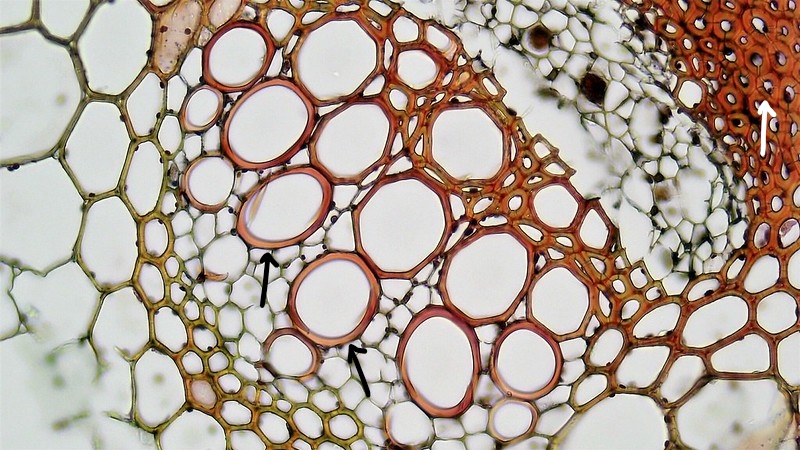
Figure 10.2.2.8: All of these cells are dead and have secondary walls with lignin. The cell labeled A is a vessel element, with larger perforation plates on its ends and strange wall thickenings. B is a tracheid, thinner than a vessel element with rows of holes lining its sides (no perforation plates). C is a fiber, long and thin. Photo by Maria Morrow, CC BY-NC.Figure 10.2.2.9: Conducting cells from the xylem. The first photo shows a vessel element with large openings at the ends. The second photo shows a tracheid, with tapered ends and sets of bordered pits along the length of it. Photos by Maria Morrow, CC BY-NC.Figure 10.2.2.10: A longitudinal section through pine wood. "Gymnosperms like Pinus rely entirely on tracheids for water flow and mechanical support. True xylem vessels and wood parenchyma are lacking. While fibers are generally absent older stems may contain some fiber tracheids. The uniform wood of Pinus consists almost entirely of longitudinally oriented bands of narrow, pale staining tracheids interrupted by short bands of horizontally oriented xylem rays (indicated by box). Xylem rays, usually one cell thick and a few cells high, are composed of clear, thin walled parenchyma cells. The lumen of many tracheids is crossed by branched trabeculae. Tracheid side walls contain large circular bordered pits (indicated by arrow) that function to move water from tracheid to tracheid while preventing air flow from embolized tracheids. The membrane of each bordered pit is marked by a pale, thin, porous outer margo and a thicker, darker, inner torus. Large horizontally oriented resin ducts are lined with living secretory parenchyma that produce resins and many toxic terpenes including turpentines." Image and caption text from Berkshire Community College Bioscience Image Library, CC0, via Wikimedia Commons. Labels added by Maria Morrow.Figure 10.2.2.11: A cross section through a vascular bundle of Helianthus, 400x. "The highly lignified cells walls of xylem (black arrows indicate vessel elements) and mature sclerenchyma (white arrow indicates fibers) are stained red orange. These cells are dead at maturity and can also be distinguished by a heavy cell wall and absence of cytoplasm." Image and caption text from Berkshire Community College Bioscience Image Library, CC0, via Wikimedia Commons. Labels added by Maria Morrow.
Sieve cells, Sieve Tube Elements, and Companion Cells
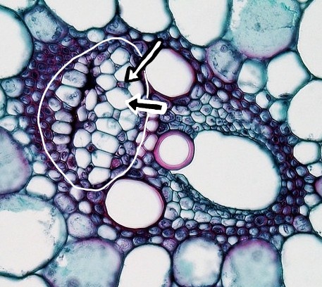
Figure 10.2.2.12: Longitudinal section through phloem. Most of the cells are sieve tube elements. The cells have sieve plates at the ends (marked by arrows). In the event of an injury, P-protein rushes to form a slime plug (B) and close the wound. Sieve tubes are alive but contain very little cell contents, they do not even have a nucleus. Instead, they are controlled by smaller companion cells (A). Photo by Berkshire Community College Bioscience Image Library, CC0, via Wikimedia Commons. Labels added by Maria Morrow.Figure 10.2.2.13: A cross section through a vascular bundle with the phloem circled (white). Within the phloem, there are two types of specialized parenchyma cells: the larger sieve tube elements (black arrow with white border) and the much smaller companion cells (white arrow with black border). Photo by Berkshire Community College Bioscience Image Library, CC0, via Wikimedia Commons. Labels added by Maria Morrow.


_LI.jpg?revision=1&size=bestfit&width=916&height=516)