
22: ONPG Assay
- Page ID
- 146882
\( \newcommand{\vecs}[1]{\overset { \scriptstyle \rightharpoonup} {\mathbf{#1}} } \)
\( \newcommand{\vecd}[1]{\overset{-\!-\!\rightharpoonup}{\vphantom{a}\smash {#1}}} \)
\( \newcommand{\dsum}{\displaystyle\sum\limits} \)
\( \newcommand{\dint}{\displaystyle\int\limits} \)
\( \newcommand{\dlim}{\displaystyle\lim\limits} \)
\( \newcommand{\id}{\mathrm{id}}\) \( \newcommand{\Span}{\mathrm{span}}\)
( \newcommand{\kernel}{\mathrm{null}\,}\) \( \newcommand{\range}{\mathrm{range}\,}\)
\( \newcommand{\RealPart}{\mathrm{Re}}\) \( \newcommand{\ImaginaryPart}{\mathrm{Im}}\)
\( \newcommand{\Argument}{\mathrm{Arg}}\) \( \newcommand{\norm}[1]{\| #1 \|}\)
\( \newcommand{\inner}[2]{\langle #1, #2 \rangle}\)
\( \newcommand{\Span}{\mathrm{span}}\)
\( \newcommand{\id}{\mathrm{id}}\)
\( \newcommand{\Span}{\mathrm{span}}\)
\( \newcommand{\kernel}{\mathrm{null}\,}\)
\( \newcommand{\range}{\mathrm{range}\,}\)
\( \newcommand{\RealPart}{\mathrm{Re}}\)
\( \newcommand{\ImaginaryPart}{\mathrm{Im}}\)
\( \newcommand{\Argument}{\mathrm{Arg}}\)
\( \newcommand{\norm}[1]{\| #1 \|}\)
\( \newcommand{\inner}[2]{\langle #1, #2 \rangle}\)
\( \newcommand{\Span}{\mathrm{span}}\) \( \newcommand{\AA}{\unicode[.8,0]{x212B}}\)
\( \newcommand{\vectorA}[1]{\vec{#1}} % arrow\)
\( \newcommand{\vectorAt}[1]{\vec{\text{#1}}} % arrow\)
\( \newcommand{\vectorB}[1]{\overset { \scriptstyle \rightharpoonup} {\mathbf{#1}} } \)
\( \newcommand{\vectorC}[1]{\textbf{#1}} \)
\( \newcommand{\vectorD}[1]{\overrightarrow{#1}} \)
\( \newcommand{\vectorDt}[1]{\overrightarrow{\text{#1}}} \)
\( \newcommand{\vectE}[1]{\overset{-\!-\!\rightharpoonup}{\vphantom{a}\smash{\mathbf {#1}}}} \)
\( \newcommand{\vecs}[1]{\overset { \scriptstyle \rightharpoonup} {\mathbf{#1}} } \)
\(\newcommand{\longvect}{\overrightarrow}\)
\( \newcommand{\vecd}[1]{\overset{-\!-\!\rightharpoonup}{\vphantom{a}\smash {#1}}} \)
\(\newcommand{\avec}{\mathbf a}\) \(\newcommand{\bvec}{\mathbf b}\) \(\newcommand{\cvec}{\mathbf c}\) \(\newcommand{\dvec}{\mathbf d}\) \(\newcommand{\dtil}{\widetilde{\mathbf d}}\) \(\newcommand{\evec}{\mathbf e}\) \(\newcommand{\fvec}{\mathbf f}\) \(\newcommand{\nvec}{\mathbf n}\) \(\newcommand{\pvec}{\mathbf p}\) \(\newcommand{\qvec}{\mathbf q}\) \(\newcommand{\svec}{\mathbf s}\) \(\newcommand{\tvec}{\mathbf t}\) \(\newcommand{\uvec}{\mathbf u}\) \(\newcommand{\vvec}{\mathbf v}\) \(\newcommand{\wvec}{\mathbf w}\) \(\newcommand{\xvec}{\mathbf x}\) \(\newcommand{\yvec}{\mathbf y}\) \(\newcommand{\zvec}{\mathbf z}\) \(\newcommand{\rvec}{\mathbf r}\) \(\newcommand{\mvec}{\mathbf m}\) \(\newcommand{\zerovec}{\mathbf 0}\) \(\newcommand{\onevec}{\mathbf 1}\) \(\newcommand{\real}{\mathbb R}\) \(\newcommand{\twovec}[2]{\left[\begin{array}{r}#1 \\ #2 \end{array}\right]}\) \(\newcommand{\ctwovec}[2]{\left[\begin{array}{c}#1 \\ #2 \end{array}\right]}\) \(\newcommand{\threevec}[3]{\left[\begin{array}{r}#1 \\ #2 \\ #3 \end{array}\right]}\) \(\newcommand{\cthreevec}[3]{\left[\begin{array}{c}#1 \\ #2 \\ #3 \end{array}\right]}\) \(\newcommand{\fourvec}[4]{\left[\begin{array}{r}#1 \\ #2 \\ #3 \\ #4 \end{array}\right]}\) \(\newcommand{\cfourvec}[4]{\left[\begin{array}{c}#1 \\ #2 \\ #3 \\ #4 \end{array}\right]}\) \(\newcommand{\fivevec}[5]{\left[\begin{array}{r}#1 \\ #2 \\ #3 \\ #4 \\ #5 \\ \end{array}\right]}\) \(\newcommand{\cfivevec}[5]{\left[\begin{array}{c}#1 \\ #2 \\ #3 \\ #4 \\ #5 \\ \end{array}\right]}\) \(\newcommand{\mattwo}[4]{\left[\begin{array}{rr}#1 \amp #2 \\ #3 \amp #4 \\ \end{array}\right]}\) \(\newcommand{\laspan}[1]{\text{Span}\{#1\}}\) \(\newcommand{\bcal}{\cal B}\) \(\newcommand{\ccal}{\cal C}\) \(\newcommand{\scal}{\cal S}\) \(\newcommand{\wcal}{\cal W}\) \(\newcommand{\ecal}{\cal E}\) \(\newcommand{\coords}[2]{\left\{#1\right\}_{#2}}\) \(\newcommand{\gray}[1]{\color{gray}{#1}}\) \(\newcommand{\lgray}[1]{\color{lightgray}{#1}}\) \(\newcommand{\rank}{\operatorname{rank}}\) \(\newcommand{\row}{\text{Row}}\) \(\newcommand{\col}{\text{Col}}\) \(\renewcommand{\row}{\text{Row}}\) \(\newcommand{\nul}{\text{Nul}}\) \(\newcommand{\var}{\text{Var}}\) \(\newcommand{\corr}{\text{corr}}\) \(\newcommand{\len}[1]{\left|#1\right|}\) \(\newcommand{\bbar}{\overline{\bvec}}\) \(\newcommand{\bhat}{\widehat{\bvec}}\) \(\newcommand{\bperp}{\bvec^\perp}\) \(\newcommand{\xhat}{\widehat{\xvec}}\) \(\newcommand{\vhat}{\widehat{\vvec}}\) \(\newcommand{\uhat}{\widehat{\uvec}}\) \(\newcommand{\what}{\widehat{\wvec}}\) \(\newcommand{\Sighat}{\widehat{\Sigma}}\) \(\newcommand{\lt}{<}\) \(\newcommand{\gt}{>}\) \(\newcommand{\amp}{&}\) \(\definecolor{fillinmathshade}{gray}{0.9}\)↵
The ONPG assay is an enzyme activity assay used to measure the activity of the enzyme β-galactosidase (β-gal). The breakdown of ONPG by β-gal results in a color change that can be measured and equated to the activity of β-gal.
Note
The ONPG assay is one of many enzyme activity assays. Enzyme activity assays can be used to measure the activity of a variety of different enzymes and activity measurements can be made using a variety of techniques including colorimetric assays, fluorescence assays, and radioactive assays.
Also known as
O-nitrophenyl β-d-galactopyranoside (ONPG) assay, β-gal activity assay, enzyme activity assay
Samples needed
Cell lysate containing β-gal or purified β-gal enzyme
Method
O-nitrophenyl β-d-galactopyranoside (ONPG) is a synthetic compound that is similar in structure to lactose, the natural substrate of β-gal. β-gal can use ONPG as a substrate and will cleave ONPG to produce galactose and o-nitrophenol. O-nitrophenol has a yellow color and results in a yellow color change.
In general, cell lysate or purified β-gal enzyme is added to a buffer containing ONPG and incubated to allow for ONPG to be able to interact with the β-gal enzyme. Following incubation for the desired time, the reaction is stopped by the addition of a basic solution (typically 1M Na2CO3) to change the pH and halt enzyme activity. Any visible color change can then be observed by eye. This color change can then be measured by a plate reader or spectrophotometer by measuring the absorbance at a wavelength of 405-420 nm.
Controls
Negative control lacking the enzyme or lacking the necessary enzyme substrate can be included. When assaying the activity of β-gal expressed by the lacZ gene on a plasmid, there is the possibility that endogenous lacZ in the chromosome is also producing β-gal. In this case an endogenous control (cells not containing the plasmid expressing lacZ) should also be included to control for the level of endogenous β-gal activity. A positive control, often purified β-gal enzyme, should also be included.
Interpretation
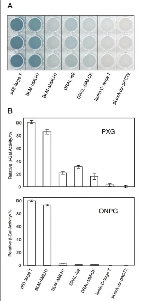
Figure 1. Measurement of protein interactions by pellet X-gal (PXG) and ONPG activity assays. Relevant section of caption for published figure reads “(A) Scanned image of a PXG assay in a 96-well microtiter plate after 30 min of incubation. Three independent transformants were assayed per protein interaction pair. (B) Relative β-gal activity in the PXG and ONPG assays. The p53-large T interaction was set arbitrarily to 100% for both assays. Error bars represent the standard deviation from three independent experiments in both assays.” “Figure 1” by Möckli and Auerbach[1]. [Image description]
The authors wanted to assess if different proteins (p53, large T antigen, hMLH1, truncated hMLH1 (ΔhMLH1), DRAL, MM-CK, and is2) interacted with each other, to do this they utilized a yeast two-hybrid assay. In a yeast two-hybrid assay a yeast transcription factor is split into two domains, and these domains are then fused to protein pairs. Only if the two proteins interact will the full transcription factor be reconstituted and be able to promote a transcription of a reporter gene. In this assay, the lacZ gene encoding for the β-gal enzyme is used as the reporter gene, meaning that the higher β-gal enzyme activity, the stronger the interaction between the two proteins tested.
Panel A shows an image of a 96-well plate where cell lysate containing the two proteins of interest have been intubated with X-gal, which is structurally similar to lactose and can serve as a substrate for β-gal. When X-gal is cleaved by the β-gal enzyme it results in a blue color change. Thus, the stronger the blue color change, the higher the β-gal activity. The authors also performed an ONPG assay to measure β-gal activity by measuring the yellow color change that results from cleavage of the ONPG substrate (not shown). Panel B shows the graphs of the β-gal activity measured by the PXG and ONPG assays. β-gal activity is shown relative to the activity from the p53/large T condition, as these proteins were previously shown to interact and should therefore result in β-gal activity.
Based on the measurements of β-gal activity from the PXG and ONPG assays the authors found the p53/large T interaction was the strongest and the DRAL/MM-CK interaction was the weakest. No β-gal activity was observed in the negative control co-expressing large T antigen and lamin C (that are not known to interact) or empty vectors only (pLexA-dir/ pACT2).
Image Descriptions
Figure 1 image description:
Panel A is an image of a 3 row x 7 column section of a 96-well plate. Each column is three replicates for a protein interaction in a PXG assay. From left to right the columns show: bright blue wells for the p53/large T condition, bright blue wells for the hMLH1/BLM condition, faint blue wells for the BLM-ΔhMLH1 condition, mild blue wells for the DRAL/FHL-2-is2 condition, faint blue wells for the DRAL/FHL-2-MM-CK condition, and clear wells for both the large T antigen/lamin C condition and the empty vector condition.
Panel B is two bar graphs of the relative β-gal activity. The top bar graph is the result of the PXG assay with p53/large T condition at 100% relative activity, the hMLH1/BLM condition at 85%, the BLM-ΔhMLH1 condition at 20%, the DRAL/is2 condition at 35%, the DRAL/MM-CK condition at 18%, and both the large T antigen/lamin C condition and the empty vector condition at around 0%. The bottom bar graph is the results of the ONPG assay with p53/large T condition at 100% relative activity, the hMLH1/BLM condition at 90%, and the rest of the conditions at 2-0%. ↵
Thumbnail
Reaksi_ONPG.jpg↗ by BP63Vincent is licensed under CC BY-SA 3.0↗.
Description: Chemical reaction showing ONPG broken down into galactose and o-nitrophenol (yellow).
Author
Rachel Ende, Tufts University
- Möckli, N. and D. Auerbach. 2004. Quantitative β-Galactosidase Assay Suitable for High-Throughput Applications in the Yeast Two-Hybrid System. BioTechniques 36 (5): 872–76. doi:10.2144/04365PT03. ↵