
9.3: Sex-linked Genes
- Page ID
- 24793

\( \newcommand{\vecs}[1]{\overset { \scriptstyle \rightharpoonup} {\mathbf{#1}} } \)
\( \newcommand{\vecd}[1]{\overset{-\!-\!\rightharpoonup}{\vphantom{a}\smash {#1}}} \)
\( \newcommand{\dsum}{\displaystyle\sum\limits} \)
\( \newcommand{\dint}{\displaystyle\int\limits} \)
\( \newcommand{\dlim}{\displaystyle\lim\limits} \)
\( \newcommand{\id}{\mathrm{id}}\) \( \newcommand{\Span}{\mathrm{span}}\)
( \newcommand{\kernel}{\mathrm{null}\,}\) \( \newcommand{\range}{\mathrm{range}\,}\)
\( \newcommand{\RealPart}{\mathrm{Re}}\) \( \newcommand{\ImaginaryPart}{\mathrm{Im}}\)
\( \newcommand{\Argument}{\mathrm{Arg}}\) \( \newcommand{\norm}[1]{\| #1 \|}\)
\( \newcommand{\inner}[2]{\langle #1, #2 \rangle}\)
\( \newcommand{\Span}{\mathrm{span}}\)
\( \newcommand{\id}{\mathrm{id}}\)
\( \newcommand{\Span}{\mathrm{span}}\)
\( \newcommand{\kernel}{\mathrm{null}\,}\)
\( \newcommand{\range}{\mathrm{range}\,}\)
\( \newcommand{\RealPart}{\mathrm{Re}}\)
\( \newcommand{\ImaginaryPart}{\mathrm{Im}}\)
\( \newcommand{\Argument}{\mathrm{Arg}}\)
\( \newcommand{\norm}[1]{\| #1 \|}\)
\( \newcommand{\inner}[2]{\langle #1, #2 \rangle}\)
\( \newcommand{\Span}{\mathrm{span}}\) \( \newcommand{\AA}{\unicode[.8,0]{x212B}}\)
\( \newcommand{\vectorA}[1]{\vec{#1}} % arrow\)
\( \newcommand{\vectorAt}[1]{\vec{\text{#1}}} % arrow\)
\( \newcommand{\vectorB}[1]{\overset { \scriptstyle \rightharpoonup} {\mathbf{#1}} } \)
\( \newcommand{\vectorC}[1]{\textbf{#1}} \)
\( \newcommand{\vectorD}[1]{\overrightarrow{#1}} \)
\( \newcommand{\vectorDt}[1]{\overrightarrow{\text{#1}}} \)
\( \newcommand{\vectE}[1]{\overset{-\!-\!\rightharpoonup}{\vphantom{a}\smash{\mathbf {#1}}}} \)
\( \newcommand{\vecs}[1]{\overset { \scriptstyle \rightharpoonup} {\mathbf{#1}} } \)
\(\newcommand{\longvect}{\overrightarrow}\)
\( \newcommand{\vecd}[1]{\overset{-\!-\!\rightharpoonup}{\vphantom{a}\smash {#1}}} \)
\(\newcommand{\avec}{\mathbf a}\) \(\newcommand{\bvec}{\mathbf b}\) \(\newcommand{\cvec}{\mathbf c}\) \(\newcommand{\dvec}{\mathbf d}\) \(\newcommand{\dtil}{\widetilde{\mathbf d}}\) \(\newcommand{\evec}{\mathbf e}\) \(\newcommand{\fvec}{\mathbf f}\) \(\newcommand{\nvec}{\mathbf n}\) \(\newcommand{\pvec}{\mathbf p}\) \(\newcommand{\qvec}{\mathbf q}\) \(\newcommand{\svec}{\mathbf s}\) \(\newcommand{\tvec}{\mathbf t}\) \(\newcommand{\uvec}{\mathbf u}\) \(\newcommand{\vvec}{\mathbf v}\) \(\newcommand{\wvec}{\mathbf w}\) \(\newcommand{\xvec}{\mathbf x}\) \(\newcommand{\yvec}{\mathbf y}\) \(\newcommand{\zvec}{\mathbf z}\) \(\newcommand{\rvec}{\mathbf r}\) \(\newcommand{\mvec}{\mathbf m}\) \(\newcommand{\zerovec}{\mathbf 0}\) \(\newcommand{\onevec}{\mathbf 1}\) \(\newcommand{\real}{\mathbb R}\) \(\newcommand{\twovec}[2]{\left[\begin{array}{r}#1 \\ #2 \end{array}\right]}\) \(\newcommand{\ctwovec}[2]{\left[\begin{array}{c}#1 \\ #2 \end{array}\right]}\) \(\newcommand{\threevec}[3]{\left[\begin{array}{r}#1 \\ #2 \\ #3 \end{array}\right]}\) \(\newcommand{\cthreevec}[3]{\left[\begin{array}{c}#1 \\ #2 \\ #3 \end{array}\right]}\) \(\newcommand{\fourvec}[4]{\left[\begin{array}{r}#1 \\ #2 \\ #3 \\ #4 \end{array}\right]}\) \(\newcommand{\cfourvec}[4]{\left[\begin{array}{c}#1 \\ #2 \\ #3 \\ #4 \end{array}\right]}\) \(\newcommand{\fivevec}[5]{\left[\begin{array}{r}#1 \\ #2 \\ #3 \\ #4 \\ #5 \\ \end{array}\right]}\) \(\newcommand{\cfivevec}[5]{\left[\begin{array}{c}#1 \\ #2 \\ #3 \\ #4 \\ #5 \\ \end{array}\right]}\) \(\newcommand{\mattwo}[4]{\left[\begin{array}{rr}#1 \amp #2 \\ #3 \amp #4 \\ \end{array}\right]}\) \(\newcommand{\laspan}[1]{\text{Span}\{#1\}}\) \(\newcommand{\bcal}{\cal B}\) \(\newcommand{\ccal}{\cal C}\) \(\newcommand{\scal}{\cal S}\) \(\newcommand{\wcal}{\cal W}\) \(\newcommand{\ecal}{\cal E}\) \(\newcommand{\coords}[2]{\left\{#1\right\}_{#2}}\) \(\newcommand{\gray}[1]{\color{gray}{#1}}\) \(\newcommand{\lgray}[1]{\color{lightgray}{#1}}\) \(\newcommand{\rank}{\operatorname{rank}}\) \(\newcommand{\row}{\text{Row}}\) \(\newcommand{\col}{\text{Col}}\) \(\renewcommand{\row}{\text{Row}}\) \(\newcommand{\nul}{\text{Nul}}\) \(\newcommand{\var}{\text{Var}}\) \(\newcommand{\corr}{\text{corr}}\) \(\newcommand{\len}[1]{\left|#1\right|}\) \(\newcommand{\bbar}{\overline{\bvec}}\) \(\newcommand{\bhat}{\widehat{\bvec}}\) \(\newcommand{\bperp}{\bvec^\perp}\) \(\newcommand{\xhat}{\widehat{\xvec}}\) \(\newcommand{\vhat}{\widehat{\vvec}}\) \(\newcommand{\uhat}{\widehat{\uvec}}\) \(\newcommand{\what}{\widehat{\wvec}}\) \(\newcommand{\Sighat}{\widehat{\Sigma}}\) \(\newcommand{\lt}{<}\) \(\newcommand{\gt}{>}\) \(\newcommand{\amp}{&}\) \(\definecolor{fillinmathshade}{gray}{0.9}\)Sex Chromosomes
For the most part, mammals have gender determined by the presence of the Y chromosome. This chromosome is gene-poor and a specific area called sex-determining region on Y (SRY) is responsible for the initiation of the male sex determination.

The X-chromosome is rich in genes while the Y-chromosome is a gene desert. The presence of an X-chromosome is absolutely necessary to produce a viable life form and the default gender of mammals is traditionally female.

Chromosomal painting techniques can reveal the gender origin of mammalian cells. By using fluorescent marker sequences that can hybridize specifically to X or Y chromosomes through Fluorescence In Situ Hybridization (FISH), gender can be identified in cells.

The male cells have an X and a Y while the female cells have X and X combination. Credit: Janice Y Ahn, Jeannie T Lee [CC BY 2.0]
Ishihara Tests (Activity)
The genes encoding photoreceptor proteins for the long wave-length (reds) and middle wavelengths (greens) reside on the X chromosome at Xq25. Since the Y-chromosome is not homologous, any mutation to either of these genes that render them non-functional results in an inability to perceive either of those colors. Men are more susceptible to the condition of red-green colorblindness since they are hemizygous. This means that there is no corresponding gene that could complement a deficient red or green photoreceptor gene.
Monochromatic representation of Ishihara test to a colorblind individual as it emerges to something visible to a color-sighted individual.
Dr. Shinobu Ishihara published his test for color perception in 1917 and this test is widely used to detect deficits in color perception. Below are examples of the Ishihara plates. Record the number that you perceive in each plate and discuss with the rest of the class.
Ishihara Test
1. As you go through the plates above, note the number that you see (if any).
2. The genes for the Red and Green receptors are on the X-chromosome, who are most affected by mutation? Create a Punnett Square to illustrate how this works.
3. Can women be color-blind for red/green?
4. Humans have 3 color light receptors and have trichromatic vision. Some women are described as possibly having tetrachromatic vision (seeing 4 colors) and being able to discriminate colors invisible to the rest of us. Describe a mechanism for why this could happen. Why is there a possible gender bias?
Sequence Links:
- Opsin 1 Long Wave sensitive gene (OPN1LW) – red photoreceptor (human)
- Opsin 1 Medium Wave sensitive gene (OPN1MW) – green photoreceptor (human)
The Case of Queen Victoria
Hemophilia literally translates to blood-loving. This is a description of a series of disorders where an individual has an inability to clot blood after a cut. In modern times, clotting factors may be administered to an afflicted individual, but a prior treatment involved blood transfusions. A very famous family had a genetic predisposition to hemophilia and due to the proliferative nature of this family, we have some statistical power to verify predictions on the probabilities of passing the disease state. Below is a partial pedigree for Victoria, Queen of the United Kingdom of Great Britain and Ireland and Empress of India. The filled-in shapes represent individuals who suffered from hemophilia.
Credit: Jeremy Seto (CC-BY-SA)
Questions:
1. From the pedigree above, what can you say about this form of hemophilia with respect to dominance?
2. From this pedigree, can you comment on the probable chromosome where the deficiency occurs?
3. Assign genotypes for Prince Albert and Queen Victoria and perform a Punnett Square to illustrate if their offspring reflect your statements on dominance and chromosome location.
4. Albert and Victoria were 1st cousins. Do you believe this had anything to do with the propagation of this disease? What does your Punnett Square tell you?
5. Highlight the definitive carriers of the disease gene in the pedigree above.
Additional Resources:
- The full case study can be acquired at the National Center for Case Study Teaching in Science.
- Human Factor IX mRNA sequence
X-Inactivation
The mammalian X-chromosome contains significantly more genetic information than the Y-chromosome. This gene dosage is controlled for in females through a process called X-inactivation where one of the X-chromosomes is shut down and highly condensed into a Barr body. Inactivation of the X-chromosome occurs in a stochastic manner that results in females being cellular mosaics where a group of cells has inactivated the paternal X-chromosome and other patches of cells have inactivated the maternal X-chromosome. The most striking example of mosaicism is the calico cat. A calico cat (tortoiseshell cat) is always a female. One of the genes that encodes coat color in cats resides on the X-chromosome and exist as either orange or black alleles. Due to the stochastic inactivation, the patterning of orange and black fur is a distinctive quality of calicos.

Credit: Howcheng [CC-BY-SA-3.0]
While the genetic information for the orange or black coat color exists in all cells, they are not equally expressed. This type of heritable trait in spite of the presence of the genetic material (DNA) is called epigenetic to imply that it is “above” (epi) genetics.
Drosophila: Thomas Hunt Morgan
Around 1908, Thomas Hunt Morgan began to explore the genetics of what was to become a model organism, Drosophila melanogaster (Fruit fly). This small organism had a relatively short life cycle, great fecundity and was easily managed. From these flies that normally have red eye coloring, he and his students found white-eyed mutants. The lab noted that white-eyed flies were almost exclusively male. This gender imbalance led Morgan to believe that the trait was sex-linked.

In 1911, Morgan published a paper that described the inheritance patterns of 5 eye-colors in Drosophila (Morgan, 1911).
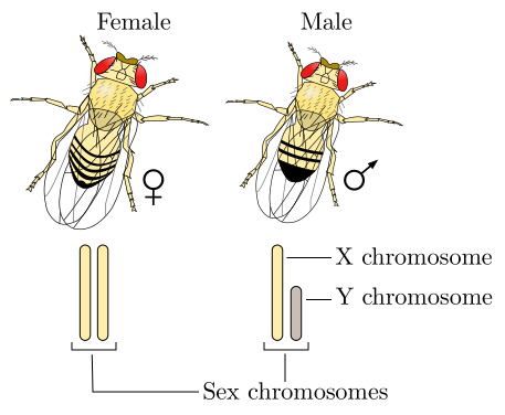
Drosophila follows a sex determination based on the ratio of X:A chromosomes and not by the presence of a Y as in mammals. A 1:1 ratio results in a female and a 1:2 ratio results in a male where the Y is ignored. The standard 2N = 8. Credit: GYassineMrabet [CC-BY-SA 4.0]
While DNA was not yet known as the source of genetic information, Morgan’s studies revealed that the location of genes most likely resided on the chromosomes. By cataloging many mutations in the lab, he was able to construct a map of gene locations. His 1922 paper specifically stated that some traits were sex-linked and therefore residing on the sex chromosome. When performing crosses of white-eyed males to wild-type females, he continued to find white-eyed trait only in males. However, in the subsequent cross of females from that generation with white-eyed males, the presence of white-eyed males and females were revealed. This indicated that the white-eyed trait was recessive and resided on the X chromosome.
Analysis of the transmission of “White-Eyed” color in Drosophila. Credit: Jeremy Seto (CC-BY-SA)
Morgan received the Nobel Prize in Physiology or Medicine in 1933 for his inference of chromosomes being a physical mechanism for packaging genetic information in the cells.
- Morgan TH. THE ORIGIN OF FIVE MUTATIONS IN EYE COLOR IN DROSOPHILA AND THEIR MODES OF INHERITANCE. Science. 1911 Apr 7;33(849):534-7