
5.2: Acellular Entities - Viruses, Prions, and Viroids
- Page ID
- 104937

Acellular Entities: Viruses, Prions, and Viroids
- Please read and watch the following Mandatory Resources
- Reading the material for understanding, and taking notes during videos, will take approximately 2 hours.
- Optional Activities are embedded.
- To navigate to the next section, use the Contents menu at the top of the page OR the right arrow on the side of the page.
- If on a mobile device, use the Contents menu at the top of the page OR the links at the bottom of the page.
Learning Objectives
- Describe how viruses were first discovered and how they are detected
- Identify three hypotheses about how viruses evolved
- Explain the Baltimore classification systems for viruses
- Describe prions and their basic properties
- Define viroids and their targets of infection
Introduction - Viruses
No one knows exactly when viruses emerged or from where they came, since viruses do not leave physical footprints such as fossils. Modern viruses are thought to be a mosaic of bits and pieces of nucleic acids picked up from past hosts along their evolutionary paths.
Viruses exist in a netherworld between a living organism and a nonliving entity. Living things grow, metabolize, reproduce, and evolve. Viruses exhibit some of these abilities but cannot do any of them on their own. They must infect a host cell and hijack the host’s replication machinery to produce (nearly) identical progeny virus particles. Since their reproduction is not perfect, they can evolve through natural selection. They do not have their own metabolism and are entirely dependent on their host cells to replicate. They do not metabolize or grow but rather are assembled into their mature form by the host cell.
Viruses are acellular parasitic entities that are not classified within any domain or kingdom. Viruses are diverse entities. They vary in their structure, their replication methods, and in their target hosts. Nearly all forms of life—from bacteria and archaea to eukaryotes such as plants, animals, and fungi—have viruses that infect them. While most biological diversity can be understood through evolutionary history such as how species have adapted to conditions and environments, much about virus origins and evolution remains unknown.
In this 4-minute video, viruses are described as the most common biological unit on Earth, outnumbering all other types combined.
Question after watching: Why do scientists talk about "types" of viruses rather than "species" of viruses?
This 11-minute video points out, viruses are all around us, they evolve, grow, and can be killed.
Question after watching: What hypotheses are offered to reconsider viruses as being alive? Are you convinced? Why or why not?
Virus Discovery and Detection
Viruses were first discovered after the development of a porcelain filter, called the Chamberland-Pasteur filter, which could remove all bacteria visible in the microscope from any liquid sample. In 1886, Adolph Meyer demonstrated that a disease of tobacco plants, tobacco mosaic disease, could be transferred from a diseased plant to a healthy one via liquid plant extracts. In 1892, Dmitri Ivanowski showed that this disease could be transmitted in this way even after the Chamberland-Pasteur filter had removed all viable bacteria from the extract. Still, it was many years before it was proven that these “filterable” infectious agents were not simply very small bacteria but were a new type of very small, disease-causing particle.
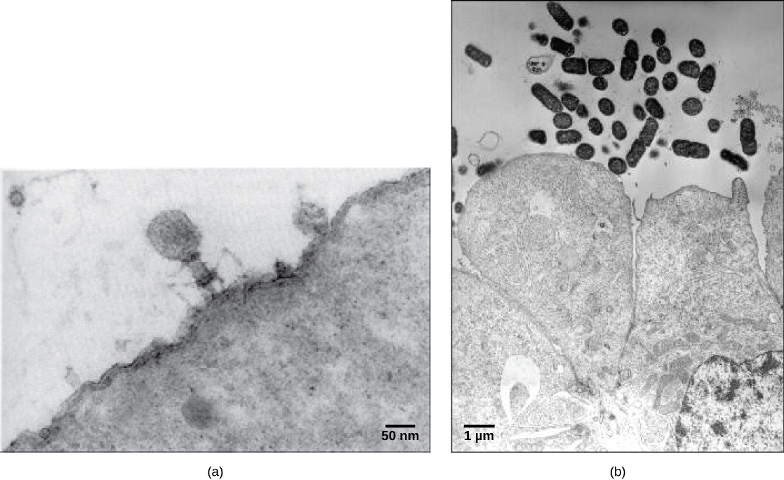
Virions, single virus particles outside of the cell, are very small, about 20–250 nanometers in diameter. These individual virus particles are the infectious form of a virus outside the host cell. Unlike bacteria (which are about 100x larger), we cannot see viruses with a light microscope (except for some large virions of the poxvirus family). It was not until the development of the electron microscope in the late 1930s that scientists got their first look of the structure of the tobacco mosaic virus (TMV) (Figure \(\PageIndex{1}\)) and other viruses (Figure \(\PageIndex{2}\)). The surface structure of virions can be observed by both scanning and transmission electron microscopy, whereas the internal structures of the virus can only be observed in images from a transmission electron microscope. The use of these technologies has allowed for the discovery of many viruses from all types of living organisms. They were initially grouped by shared morphology. Later, groups of viruses were classified by the type of nucleic acid they contained, DNA or RNA, and whether their nucleic acid was single- or double-stranded. More recently, molecular analysis of viral replicative cycles has further refined their classification.

Evolution of Viruses
Although biologists have accumulated a significant amount of knowledge about how present-day viruses evolve, much less is known about how viruses originated in the first place. When exploring the evolutionary history of most organisms, scientists can look at fossil records and similar physical evidence. However, viruses do not fossilize, so researchers must conjecture by investigating how today’s viruses evolve and by using biochemical and genetic information to create speculative virus histories.
While most findings agree that viruses do not have a single common ancestor, scholars have yet to agree on a hypothesis about the origins of viruses. One such hypothesis, called devolution or the regressive hypothesis, proposes that viruses evolved from free-living cells that progressively lost the ability to reproduce and metabolize on their own. A second hypothesis (called escapist or the progressive hypothesis) suggests that viruses originated from RNA and DNA molecules that escaped from a host cell. This theory accounts for viruses having either an RNA or a DNA genome. A third hypothesis posits a system of self-replication similar to that of other self-replicating molecules, likely evolving alongside the cells they rely on as hosts. Studies of some plant pathogens support this hypothesis.
As technology advances, scientists may develop and refine further hypotheses to explain the origin of viruses. The emerging field called virus molecular systematics attempts to do that through comparisons of sequenced genetic material. Researchers in this field hope their work could someday lead to advances in the treatments for the ailments of viral diseases.
This 2-minute video describes the speed at which viruses evolve through mutations.
Questions after watching: Why do some evolutionary scientists focus on viruses? Why is it important to humans?
The SARS-CoV-2 (Covid-19) pandemic has altered life throughout the world. This 1.5-minute video describes how SARS-CoV-2 infects humans and reproduces.
Question after watching: What are three things you learned from this video that you did not know prior to watching?
Viral Structure and Function
Viruses are acellular, meaning they are biological entities that do not have a cellular structure. Therefore, they lack most of the components of cells, such as organelles, ribosomes, and the plasma membrane. A virion consists of a nucleic acid core, an outer protein coating or capsid, and sometimes (notably among viruses that infect animal cells) an outer envelope made of protein and phospholipid membranes derived from the host cell. Viruses may also contain additional proteins, such as enzymes. The most obvious difference between members of viral families is their morphology, which is quite diverse. An interesting feature of viral complexity is that the complexity of the host does not correlate with the complexity of the virion. Some of the most complex virion structures are observed in bacteriophages, viruses that infect the simplest living organisms, bacteria.
Viruses come in many shapes and sizes, but these are consistent and distinct for each viral family. All virions have a nucleic acid genome covered by a protective layer of proteins, called a capsid. The capsid is made up of protein subunits called capsomeres. Some viral capsids are simple polyhedral “spheres,” whereas others are quite complex in structure.
Unlike nearly all living organisms that use DNA as their genetic material, viruses may use either DNA or RNA to encode their information. Some viruses are composed of single-stranded RNA, double-stranded RNA, single-stranded DNA, or double-stranded DNA. It may be linear or circular. While most viruses contain a single nucleic acid, others have genomes that have several, which are called segments (analogous to chromosomes). The virus core contains the genome or total genetic content of the virus. Viral genomes tend to be small, containing only those genes that encode proteins that the virus cannot get from the host cell.
In this 7-minute video, discover the wide variety of viral shapes and types.
Question after watching: Why are viruses dependent on other organisms for their reproduction?
Virus Classification
To understand the features shared among different groups of viruses, a classification scheme is necessary. Biologists have used several classification systems in the past, based on the morphology and genetics of different viruses. However, these earlier classification methods grouped viruses differently, based on which features of the virus they were using to classify them. The most commonly used classification method today is called the Baltimore classification scheme and is based on how messenger RNA (mRNA) is generated in each type of virus.
Baltimore Classification
The most commonly used system of virus classification was developed by Nobel Prize-winning biologist David Baltimore in the early 1970s. In addition to the differences in morphology and genetics mentioned above, the Baltimore classification scheme groups viruses according to how the mRNA is produced during the replicative cycle of the virus. The characteristics of each group in the Baltimore classification are summarized in Table \(\PageIndex{1}\) with examples of each group.
| Group | Characteristics | Mode of mRNA Production | Example |
|---|---|---|---|
| I | Double-stranded DNA | mRNA is transcribed directly from the DNA template | Herpes simplex (herpesvirus) |
| II | Single-stranded DNA | DNA is converted to double-stranded form before RNA is transcribed | Canine parvovirus (parvovirus) |
| III | Double-stranded RNA | mRNA is transcribed from the RNA genome | Childhood gastroenteritis (rotavirus) |
| IV | Single stranded RNA (+) | Genome functions as mRNA | Common cold (pircornavirus) |
| V | Single stranded RNA (-) | mRNA is transcribed from the RNA genome | Rabies (rhabdovirus) |
| VI | Single stranded RNA viruses with reverse transcriptase | Reverse transcriptase makes DNA from the RNA genome; DNA is then incorporated in the host genome; mRNA is transcribed from the incorporated DNA | Human immunodeficiency virus (HIV) |
| VII | Double stranded DNA viruses with reverse transcriptase | The viral genome is double-stranded DNA, but viral DNA is replicated through an RNA intermediate; the RNA may serve directly as mRNA or as a template to make mRNA | Hepatitis B virus (hepadnavirus) |
Virus Replication
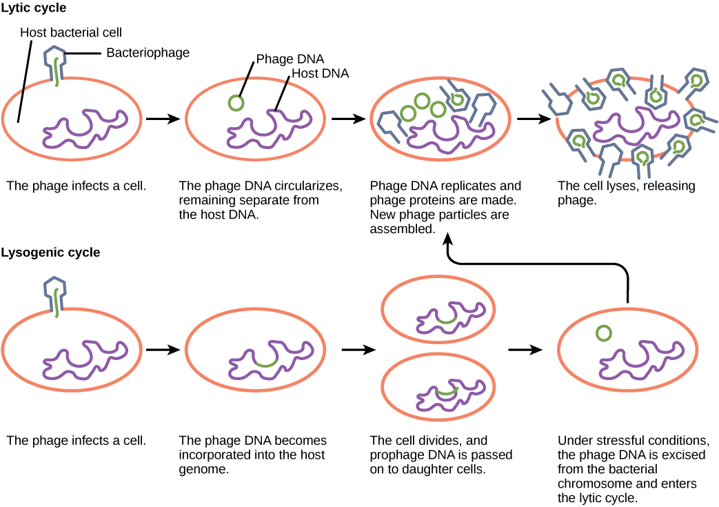
Viral replication within a living cell always produces changes in the cell, sometimes resulting in cell death and sometimes slowly killing the infected cells. There are six basic stages in the virus replication cycle: attachment, penetration, uncoating, replication, assembly, and release. A viral infection may be productive, resulting in new virions, or nonproductive, which means that the virus remains inside the cell without producing new virions. Bacteriophages are viruses that infect bacteria. They have two different modes of replication: the lytic cycle, where the virus replicates and bursts out of the bacteria, and the lysogenic cycle, which involves the incorporation of the viral genome into the bacterial host genome (Figure \(\PageIndex{3}\)).
Note that the narrator misspeaks when she says that viruses are 'microorgansms'. Viruses are often very specific as to which hosts and which cells within the host they will infect.
Viruses can be seen as obligate, intracellular parasites. A virus must attach to a living cell's surface, be taken inside, manufacture its proteins and copy its genome, and find a way to escape the cell so that the virus can infect other cells. Viruses can infect only certain species and only certain cells within that host. Cells that a virus may use to replicate are called permissive. For most viruses, the molecular basis for this specificity is that a particular surface molecule known as the viral receptor must be found on the host cell surface for the virus to attach. Also, metabolic and host cell immune response differences seen in different cell types based on differential gene expression are a likely factor in the type of cells a virus may target. The permissive cell must make the substances that the virus needs or the virus will not be able to replicate there.
A virus must use cell processes to replicate. The viral replication cycle can produce dramatic biochemical and structural changes in the host cell, which may cause cell damage. These changes, called cytopathic (causing cell damage) effects, can change cell functions or even destroy the cell. Some infected cells, such as those infected by the common cold virus known as rhinovirus, die through lysis (bursting) or apoptosis (programmed cell death or “cell suicide”), releasing all progeny virions at once. The symptoms of viral diseases result from the immune response to the virus, which attempts to control and eliminate the virus from the body, and from cell damage caused by the virus. Many animal viruses, such as HIV (human immunodeficiency virus), leave the infected cells of the immune system by a process known as budding, where virions leave the cell individually. During the budding process, the cell does not undergo lysis and is not immediately killed. However, the damage to the cells that the virus infects may make it impossible for the cells to function normally, even though the cells remain alive for a period of time.
Influenza virus is packaged in a viral envelope that fuses with the plasma membrane. This way, the virus can exit the host cell without killing it. What advantage does the virus gain by keeping the host cell alive?
- Answer
-
The host cell can continue to make new virus particles.
Career Connection: Virologist
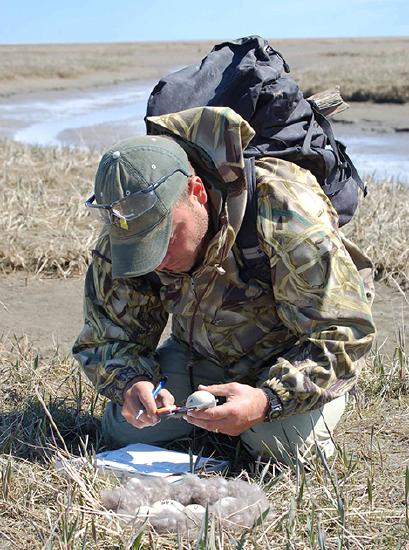
Virology is the study of viruses, and a virologist is an individual trained in this discipline. Training in virology can lead to many different career paths. Virologists are actively involved in academic research and teaching in colleges and medical schools. Some virologists treat patients or are involved in the development and/or production of vaccines. They might participate in epidemiologic studies (Figure \(\PageIndex{3}\)) or become science writers, to name just a few possible careers.
If you think you may be interested in a career in virology, find a mentor in the field. Many large medical centers have departments of virology, and smaller hospitals usually have virology labs within their microbiology departments. Volunteer in a virology lab for a semester or work in one over the summer. Discussing the profession and getting a first-hand look at the work will help you decide whether a career in virology is right for you.
Viral Infections
Most human disease-causing pathogens (60%+) come from animals. This transference of a disease from an animal to a human being is called zoonoses, and all such diseases collectively are called zoonotic diseases.
Question after watching: What are the different ways that zoonotics are transmitted are transmitted from animals to humans and back to animals?
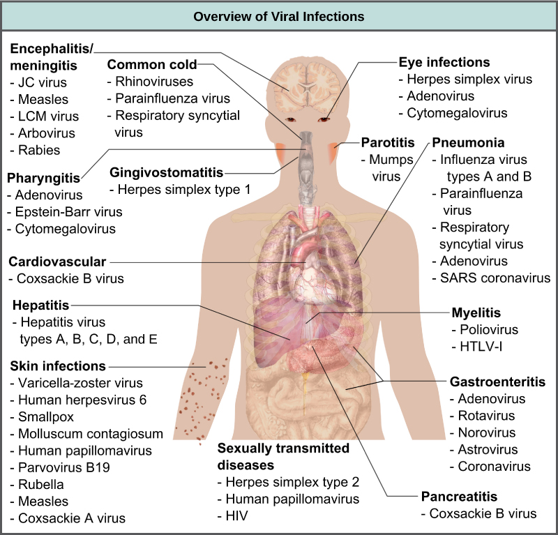
Viruses cause a variety of diseases in animals, including humans, ranging from the common cold to potentially fatal illnesses like meningitis (Figure \(\PageIndex{5}\)). These diseases can be treated by antiviral drugs or by vaccines, but some viruses, such as HIV, are capable of both avoiding the immune response and mutating to become resistant to antiviral drugs. On the other hand, viruses may have also had large influences in evolution, leading to new species, and even help by keeping bacterial populations in check.
Vaccines for Prevention
While we do have limited numbers of effective antiviral drugs, such as those used to treat HIV and influenza, the primary method of controlling viral disease is by vaccination, which is intended to prevent outbreaks by building immunity to a virus or virus family. Vaccines may be prepared using live viruses, killed viruses, or molecular subunits of the virus. The killed viral vaccines and subunit viruses are both incapable of causing disease.
Live viral vaccines are designed in the laboratory to cause few symptoms in recipients while giving them protective immunity against future infections. Polio was one disease that represented a milestone in the use of vaccines. Mass immunization campaigns in the 1950s (killed vaccine) and 1960s (live vaccine) significantly reduced the incidence of the disease, which caused muscle paralysis in children and generated a great amount of fear in the general population when regional epidemics occurred. The success of the polio vaccine paved the way for the routine dispensation of childhood vaccines against measles, mumps, rubella, chickenpox, and other diseases.
Some vaccines are in continuous development because certain viruses, such as influenza and HIV, have a high mutation rate compared to other viruses and normal host cells. With influenza, mutations in the surface molecules of the virus help the organism evade the protective immunity that may have been obtained in a previous influenza season, making it necessary for individuals to get vaccinated every year. Other viruses, such as those that cause the childhood diseases measles, mumps, and rubella, mutate so infrequently that the same vaccine is used year after year.
Learn the science behind how vaccines trigger an immune response and teach our bodies to recognize dangerous pathogens.
Question after watching: Why is it that vaccines not only protect those who get vaccinated, but others as well.
Vaccines and Anti-viral Drugs for Treatment
In some cases, vaccines can be used to treat an active viral infection. The concept behind this is that by giving the vaccine, immunity is boosted without adding more disease-causing virus. In the case of rabies, a fatal neurological disease transmitted via the saliva of rabies virus-infected animals, the progression of the disease from the time of the animal bite to the time it enters the central nervous system may be 2 weeks or longer. This is enough time to vaccinate an individual who suspects that they have been bitten by a rabid animal, and their boosted immune response is sufficient to prevent the virus from entering nervous tissue.
Another way of treating viral infections is the use of antiviral drugs. These drugs often have limited success in curing viral disease, but in many cases, they have been used to control and reduce symptoms for a wide variety of viral diseases. For most viruses, these drugs can inhibit the virus by blocking the actions of one or more of its proteins. It is important that the targeted proteins be encoded by viral genes and that these molecules are not present in a healthy host cell. Antiviral drugs like Paxlovid have been critical in treating Covid-19, especially in vulnerable populations.
Other Acellular Entities: Prions and Viroids
Prions and viroids are pathogens (agents with the ability to cause disease) that have simpler structures than viruses but, in the case of prions, still can produce deadly diseases.
Prions
Prions, so-called because they are made of a protein, are infectious particles—smaller than viruses—that contain no nucleic acids (neither DNA nor RNA). Historically, the idea of an infectious agent that did not use nucleic acids was considered impossible, but pioneering work by Nobel Prize-winning biologist Stanley Prusiner has convinced most biologists that such agents exist.
Fatal neurodegenerative diseases, such as kuru in humans and bovine spongiform encephalopathy (BSE) in cattle (commonly called “mad cow disease”) were shown to be transmitted by prions. The disease was spread by the consumption of meat, nervous tissue, or internal organs. Individuals with kuru and BSE show symptoms of loss of motor control and unusual behaviors, such as uncontrolled bursts of laughter with kuru, followed by death.
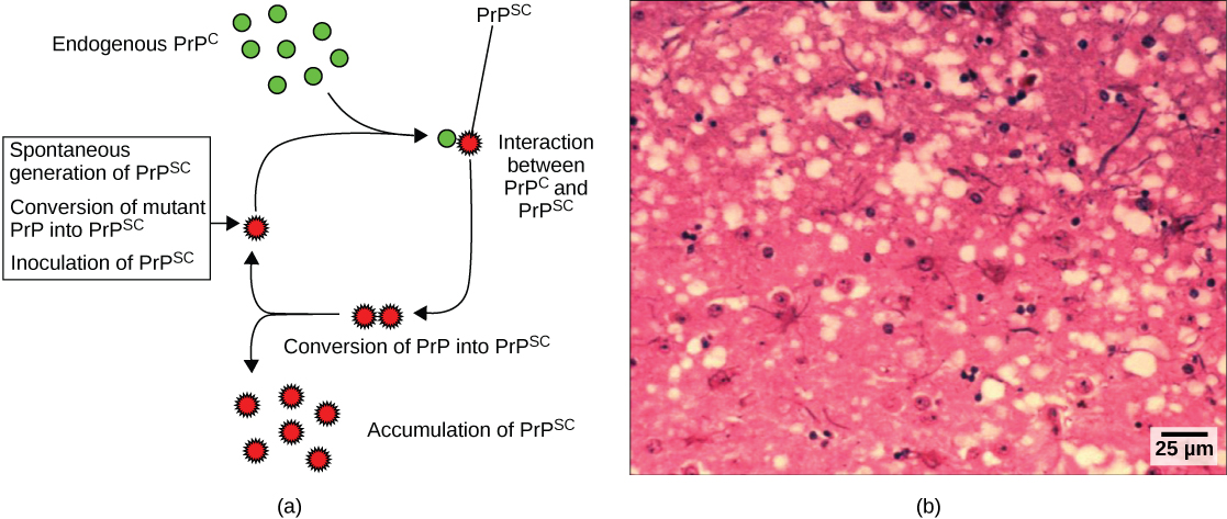
BSE was initially thought to only affect cattle. Cattle dying of the disease were shown to have developed lesions or “holes” in the brain, causing the brain tissue to resemble a sponge. Later, however, it was shown that a similar encephalopathy in humans known as Creutzfeldt-Jakob disease (CJD) could be acquired from eating meet from animals with BSE (Figure \(\PageIndex{4}\)). This led to bans by several countries on the importation of British beef and causing considerable economic damage to the British cattle industry. BSE still exists in some areas of the world, and although a rare disease, CJD is difficult to detect and to treat. The disease can be spread from human to human by blood, so many countries have banned blood donation from regions associated with BSE.
The cause of spongiform encephalopathies, such as kuru and BSE, is an infectious structural variant of a normal cellular protein called PrP (prion protein). It is this variant that constitutes the prion particle. PrP exists in two forms, PrPc, the normal form of the protein, and PrPsc, the infectious form. Once introduced into the body, the PrPsc prion binds to normal PrPc protein and converts it to PrPsc. This leads to an exponential increase of PrPsc proteins, which aggregate. PrPsc is folded abnormally, and the resulting conformation (shape) is directly responsible for the lesions seen in the brains of infected cattle with BSE and humans with CJD. Thus, the prion seems to be an entirely new form of infectious agent, the first one found whose transmission is not reliant upon genes made of DNA or RNA.
In this 5-minute video, Hank describes prions - misfolded proteins that are responsible for destroying brain cells and infect new hosts by eating an abnormal prion protein.
Question after watching: Why were some women and children more at risk to Kuru than men? Why were some resistant to Kuru?
Which of the following is not associated with prions?
- replicating shapes
- mad cow disease
- DNA
- mutations in the gene for a protein
- Answer
-
C
Viroids
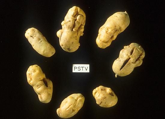
Viroids are plant pathogens: small, single-stranded, circular RNA particles that are much simpler than a virus. They do not have a capsid or outer envelope, but like viruses can reproduce only within a host cell. Viroids do not, however, manufacture any proteins, and they only produce a single, specific RNA molecule. Human diseases caused by viroids have yet to be identified.
Viroids are known to infect plants (Figure \(\PageIndex{5}\)) and are responsible for crop failures and the loss of millions of dollars in agricultural revenue each year. Some of the plants they infect include potatoes, cucumbers, tomatoes, chrysanthemums, avocados, and coconut palms.
This 4-minute video describes the discovery of viroids: infectious, replicating bits of RNA.
Question after watching: How are viroids helping biologists investigate the origin of life?
Which statement is true of viroids?
- They are single-stranded RNA particles.
- They reproduce only outside of the cell.
- They produce proteins.
- They affect both plants and animals.
- Answer
-
A. They are single-stranded RNA particles.
Thumbnail: Ebola virus. (Public Domain; CDC).