
4: Obtaining Pure Cultures from a Mixed Population
- Page ID
- 105483
\( \newcommand{\vecs}[1]{\overset { \scriptstyle \rightharpoonup} {\mathbf{#1}} } \)
\( \newcommand{\vecd}[1]{\overset{-\!-\!\rightharpoonup}{\vphantom{a}\smash {#1}}} \)
\( \newcommand{\dsum}{\displaystyle\sum\limits} \)
\( \newcommand{\dint}{\displaystyle\int\limits} \)
\( \newcommand{\dlim}{\displaystyle\lim\limits} \)
\( \newcommand{\id}{\mathrm{id}}\) \( \newcommand{\Span}{\mathrm{span}}\)
( \newcommand{\kernel}{\mathrm{null}\,}\) \( \newcommand{\range}{\mathrm{range}\,}\)
\( \newcommand{\RealPart}{\mathrm{Re}}\) \( \newcommand{\ImaginaryPart}{\mathrm{Im}}\)
\( \newcommand{\Argument}{\mathrm{Arg}}\) \( \newcommand{\norm}[1]{\| #1 \|}\)
\( \newcommand{\inner}[2]{\langle #1, #2 \rangle}\)
\( \newcommand{\Span}{\mathrm{span}}\)
\( \newcommand{\id}{\mathrm{id}}\)
\( \newcommand{\Span}{\mathrm{span}}\)
\( \newcommand{\kernel}{\mathrm{null}\,}\)
\( \newcommand{\range}{\mathrm{range}\,}\)
\( \newcommand{\RealPart}{\mathrm{Re}}\)
\( \newcommand{\ImaginaryPart}{\mathrm{Im}}\)
\( \newcommand{\Argument}{\mathrm{Arg}}\)
\( \newcommand{\norm}[1]{\| #1 \|}\)
\( \newcommand{\inner}[2]{\langle #1, #2 \rangle}\)
\( \newcommand{\Span}{\mathrm{span}}\) \( \newcommand{\AA}{\unicode[.8,0]{x212B}}\)
\( \newcommand{\vectorA}[1]{\vec{#1}} % arrow\)
\( \newcommand{\vectorAt}[1]{\vec{\text{#1}}} % arrow\)
\( \newcommand{\vectorB}[1]{\overset { \scriptstyle \rightharpoonup} {\mathbf{#1}} } \)
\( \newcommand{\vectorC}[1]{\textbf{#1}} \)
\( \newcommand{\vectorD}[1]{\overrightarrow{#1}} \)
\( \newcommand{\vectorDt}[1]{\overrightarrow{\text{#1}}} \)
\( \newcommand{\vectE}[1]{\overset{-\!-\!\rightharpoonup}{\vphantom{a}\smash{\mathbf {#1}}}} \)
\( \newcommand{\vecs}[1]{\overset { \scriptstyle \rightharpoonup} {\mathbf{#1}} } \)
\(\newcommand{\longvect}{\overrightarrow}\)
\( \newcommand{\vecd}[1]{\overset{-\!-\!\rightharpoonup}{\vphantom{a}\smash {#1}}} \)
\(\newcommand{\avec}{\mathbf a}\) \(\newcommand{\bvec}{\mathbf b}\) \(\newcommand{\cvec}{\mathbf c}\) \(\newcommand{\dvec}{\mathbf d}\) \(\newcommand{\dtil}{\widetilde{\mathbf d}}\) \(\newcommand{\evec}{\mathbf e}\) \(\newcommand{\fvec}{\mathbf f}\) \(\newcommand{\nvec}{\mathbf n}\) \(\newcommand{\pvec}{\mathbf p}\) \(\newcommand{\qvec}{\mathbf q}\) \(\newcommand{\svec}{\mathbf s}\) \(\newcommand{\tvec}{\mathbf t}\) \(\newcommand{\uvec}{\mathbf u}\) \(\newcommand{\vvec}{\mathbf v}\) \(\newcommand{\wvec}{\mathbf w}\) \(\newcommand{\xvec}{\mathbf x}\) \(\newcommand{\yvec}{\mathbf y}\) \(\newcommand{\zvec}{\mathbf z}\) \(\newcommand{\rvec}{\mathbf r}\) \(\newcommand{\mvec}{\mathbf m}\) \(\newcommand{\zerovec}{\mathbf 0}\) \(\newcommand{\onevec}{\mathbf 1}\) \(\newcommand{\real}{\mathbb R}\) \(\newcommand{\twovec}[2]{\left[\begin{array}{r}#1 \\ #2 \end{array}\right]}\) \(\newcommand{\ctwovec}[2]{\left[\begin{array}{c}#1 \\ #2 \end{array}\right]}\) \(\newcommand{\threevec}[3]{\left[\begin{array}{r}#1 \\ #2 \\ #3 \end{array}\right]}\) \(\newcommand{\cthreevec}[3]{\left[\begin{array}{c}#1 \\ #2 \\ #3 \end{array}\right]}\) \(\newcommand{\fourvec}[4]{\left[\begin{array}{r}#1 \\ #2 \\ #3 \\ #4 \end{array}\right]}\) \(\newcommand{\cfourvec}[4]{\left[\begin{array}{c}#1 \\ #2 \\ #3 \\ #4 \end{array}\right]}\) \(\newcommand{\fivevec}[5]{\left[\begin{array}{r}#1 \\ #2 \\ #3 \\ #4 \\ #5 \\ \end{array}\right]}\) \(\newcommand{\cfivevec}[5]{\left[\begin{array}{c}#1 \\ #2 \\ #3 \\ #4 \\ #5 \\ \end{array}\right]}\) \(\newcommand{\mattwo}[4]{\left[\begin{array}{rr}#1 \amp #2 \\ #3 \amp #4 \\ \end{array}\right]}\) \(\newcommand{\laspan}[1]{\text{Span}\{#1\}}\) \(\newcommand{\bcal}{\cal B}\) \(\newcommand{\ccal}{\cal C}\) \(\newcommand{\scal}{\cal S}\) \(\newcommand{\wcal}{\cal W}\) \(\newcommand{\ecal}{\cal E}\) \(\newcommand{\coords}[2]{\left\{#1\right\}_{#2}}\) \(\newcommand{\gray}[1]{\color{gray}{#1}}\) \(\newcommand{\lgray}[1]{\color{lightgray}{#1}}\) \(\newcommand{\rank}{\operatorname{rank}}\) \(\newcommand{\row}{\text{Row}}\) \(\newcommand{\col}{\text{Col}}\) \(\renewcommand{\row}{\text{Row}}\) \(\newcommand{\nul}{\text{Nul}}\) \(\newcommand{\var}{\text{Var}}\) \(\newcommand{\corr}{\text{corr}}\) \(\newcommand{\len}[1]{\left|#1\right|}\) \(\newcommand{\bbar}{\overline{\bvec}}\) \(\newcommand{\bhat}{\widehat{\bvec}}\) \(\newcommand{\bperp}{\bvec^\perp}\) \(\newcommand{\xhat}{\widehat{\xvec}}\) \(\newcommand{\vhat}{\widehat{\vvec}}\) \(\newcommand{\uhat}{\widehat{\uvec}}\) \(\newcommand{\what}{\widehat{\wvec}}\) \(\newcommand{\Sighat}{\widehat{\Sigma}}\) \(\newcommand{\lt}{<}\) \(\newcommand{\gt}{>}\) \(\newcommand{\amp}{&}\) \(\definecolor{fillinmathshade}{gray}{0.9}\)- Identify and perform different methods for dilution of bacteria.
- Define and give examples of special media: differential, selective, enrichment.
- Interpret results of growth on special media.
- Successfully isolate and identify monocultures from a mixed culture of bacteria.
- Describe and identify colony morphology.
A. Introduction: Dilution Techniques and Special Media
As you now know, microorganisms exist in nature as mixed populations. However, to study microorganisms in the laboratory we must have them in the form of a pure culture, that is, one in which all organisms are descendants of the same organism. Two major steps are involved in obtaining pure cultures from a mixed population:
- First, the mixture must be diluted until the various individual microorganisms become separated far enough apart on an agar surface that after incubation they form visible colonies isolated from the colonies of other microorganisms. This plate is called an isolation plate.
- Then, an isolated colony can be aseptically "picked off" the isolation plate and transferred to new sterile medium. After incubation, all organisms in the new culture will be descendants of the same organism, that is, a pure culture.

The most common way of separating bacterial cells on the agar surface to obtain isolated colonies is the streak plate method we used in the previous lab to inoculate a petri plate. It provides a simple and rapid method of diluting the sample by mechanical means. As the loop is streaked across the agar surface, more and more bacteria are rubbed off until individual separated organisms are deposited on the agar. After incubation, the area at the beginning of the streak pattern will show confluent growth while the area near the end of the pattern should show discrete colonies.
THE POUR PLATE AND SPIN PLATE METHODS OF ISOLATION
Another method of separating bacteria is the pour plate method. With the pour plate method, the bacteria are mixed with melted agar until evenly distributed and separated throughout the liquid. The melted agar is then poured into an empty plate and allowed to solidify. After incubation, discrete bacterial colonies can then be found growing both on the agar and in the agar.
The spin plate method involves diluting the bacterial sample in tubes of sterile water, saline, or broth. Small samples of the diluted bacteria are then pipetted onto the surface of agar plates. A sterile, bent-glass rod is then used to spread the bacteria evenly over the entire agar surface in order to see isolated colonies. We will use this technique as part of the plate count method of enumerating bacteria.
USE OF SPECIALIZED MEDIA
To supplement mechanical techniques of isolation such as the streak plate method, many special-purpose media are available to the microbiologist to aid in the isolation and identification of specific microorganisms. These special purpose media fall into four groups: selective media, differential media, enrichment media, and combination selective and differential media.
- Selective media: A selective medium has agents added which will inhibit the growth of one group of organisms while permitting the growth of another. For example, Columbia CNA agar has the antibiotics colistin and nalidixic acid added which inhibit the growth of Gram-negative bacteria but not the growth of Gram-positives. It is, therefore, said to be selective for Gram-positive organisms, and would be useful in separating a mixture of Gram-positive and Gram-negative bacteria.
- Differential media: A differential medium contains additives that cause an observable color change in the medium when a particular chemical reaction occurs. They are useful in differentiating bacteria according to some biochemical characteristic. In other words, they indicate whether or not a certain organism can carry out a specific biochemical reaction during its normal metabolism. Many such media will be used in future labs to aid in the identification of microorganisms.
- Enrichment media: An enrichment medium contains additives that enhance the growth of certain organisms. This is useful when the organism you wish to culture is present in relatively small numbers compared to the other organisms growing in the mixture.
- Combination selective and differential media: A combination selective and differential medium permits the growth of one group of organisms while inhibiting the growth of another. In addition, it differentiates those organisms that grow based on whether they can carry out particular chemical reactions.
For Example: MacConkey Agar
MacConkey agar is a selective medium used for the isolation of non-fastidious Gram-negative rods, particularly members of the family Enterobacteriaceae and the genus Pseudomonas, and the differentiation of lactose fermenting from lactose non-fermenting Gram-negative bacilli. MacConkey agar contains the dye crystal violet well as bile salts that inhibit the growth of most Gram-positive bacteria but do not affect the growth of most Gram-negatives. If the Gram-negative bacterium ferments the sugar lactose in the medium, the acid end products lower the pH of the medium. The neutral red in the agar turns red in color once the pH drops below 6.8. As the pH drops, the neutral red is absorbed by the bacteria, causing the colonies to appear bright pink to red.
Results are interpreted as follows:
- Strong fermentation of lactose with high levels of acid production by the bacteria causes the colonies and confluent growth to appear bright pink to red. The resulting acid, at high enough concentrations, can also causes the bile salts in the medium to precipitate out of solution causing a pink precipitate (cloudiness) to appear in the agar surrounding the growth.
- Weak fermentation of lactose by the bacteria causes the colonies and confluent growth to appear pink to red, but without the precipitation of bile salts there is no pink precipitate (cloudiness) in the agar surrounding the growth.
- If the bacteria do not ferment lactose, the colonies and confluent growth appear colorless and the agar surrounding the bacteria remains relatively transparent.
Typical colony morphology on MacConkey agar is as follows:
- Escherichia coli: colonies and confluent growth appear bright pink to red and surrounded by a pink precipitate (cloudiness) in the agar surrounding the growth.
- Enterobacter and Klebsiella: colonies and confluent growth appear bright pink to red but are not surrounded by a pink precipitate (cloudiness) in the agar surrounding the growth.
- Salmonella, Serratia, Proteus, and Shigella: colorless colonies; agar relatively transparent.
There are literally hundreds of special-purpose media available to the microbiologist. Today we will combine both a mechanical isolation technique (the streak plate) with selective and selective-differential media to obtain pure cultures from a mixture of bacteria. In future labs, you will work to isolate and identify pathogenic bacteria using many additional special-purpose media.
Lab Procedure A:
MEDIA
One plate of each of the following media: Trypticase Soy agar, Columbia CNA agar, and MacConkey agar.
ORGANISMS
During this lab you will be given a broth culture containing a mixture of Gram-positive and Gram-negative bacteria. Over the next three labs you will attempt to obtain pure cultures of each organism in your mixture and determine which two bacteria you have. Today you will try to separate the bacteria in the mixture in order to obtain isolated colonies; next lab you will identify the three bacteria in your mixture and pick off single isolated colonies of each of the three bacteria in order to get a pure culture of each. The following lab you will prepare microscopy slides of each of the three pure cultures to determine if they are indeed pure.
Procedure:
- On the bottom of each of the three petri plate you are using today, divide the plate into thirds with your wax marker and label as shown below. This will guide your streaking.
- Although Trypticase Soy agar (TSA), which grows both Gram-positive and Gram-negative bacteria, is not normally used as an isolation medium, we will attempt to obtain isolated colonies of the two organisms in your mixture by using strictly mechanical methods. Often, however, one bacterium overgrows another in a mixture and by the time you spread out the more abundant organism enough to get isolated colonies, the one in smaller numbers is no longer on the loop so you may not see single colonies of each on the TSA next time.
- Streak your mixture on a plate of Trypticase Soy agar.
- Streak the same mixture for isolation on a plate of Columbia CNA agar (selective for Gram-positive bacteria).
- Streak the same mixture for isolation on a plate of MacConkey agar (selective for Gram-negative bacteria and differential for certain members of the bacterial family Enterobacteriaceae).
- Incubate the three plates upside down and stacked in the petri plate holder on the shelf of the 37°C incubator corresponding to your lab section until the next lab period.
B. Introduction: Describing your Observations
Bacterial Colony Morphology
Bacteria grow on solid media as colonies. A colony is defined as a visible mass of microorganisms all originating from a single mother cell, therefore a colony constitutes a clone of bacteria all genetically alike.
In the identification of bacteria and fungi much weight is placed on how the organism grows in or on media. This exercise will help you identify the cultural characteristics of a bacterium on an agar plate - called colony morphology. Although one might not necessarily see the importance of colonial morphology at first, it really can be important when identifying the bacterium. Features of the colonies may help to pinpoint the identity of the bacterium. Different species of bacteria can produce very different colonies.

In the above picture of a mixed culture, an agar plate that has been exposed to the air and many different colony morphologies can be identified. Nine obviously different colonies are numbered: some colony types recur in various areas of the plate (note # 3 and # 4). Not only are pigment differences seen, but also size, edge, pattern, opacity, and shine. Two circles have been drawn around merging colonies, where the species of the 2 colonies are different. Trying to pick a bit of one of those adjacent colonies increases the chances of picking up another mixed culture, consisting of the 2 species that were merged together. ALWAYS pick a well-isolated colony when subculturing.
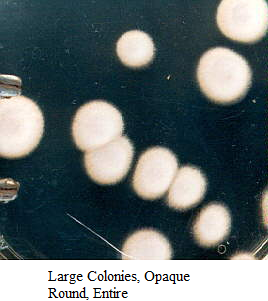
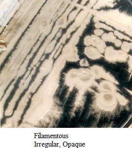
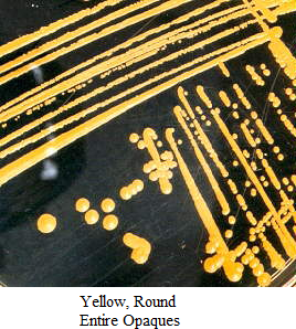
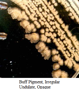
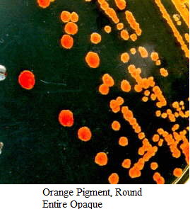
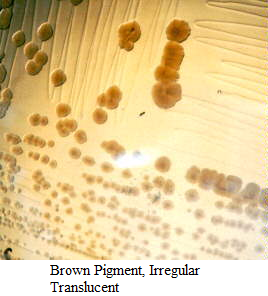
WHOLE SHAPE OF COLONY
Varies from round to irregular to filamentous and rhizoid (root-like)
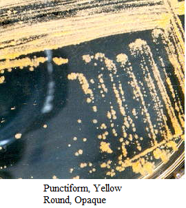
SIZE OF COLONY
Can vary from large colonies to tiny colonies less than 1 millimeter (mm) = punctiform (pin-point). Measure with a mm ruler.
EDGE/MARGIN OF COLONY
Magnified edge shape (use a dissecting microscope to see the margin edge well)
CHROMOGENESIS
Color of colonies, pigmentation: white, buff, red, purple, etc.
Some pigments are water-soluble, others are not.
If you take a large inoculum and place it in a tube of water or saline, do you see color?
Do you see any pigment if the organism is growing in a broth medium?
Does incubation temperature affect the color?
Does the entire colony have the color, or is it more like a bull’s eye?
OPACITY OF COLONY
Is the colony transparent (clear), opaque (not transparent or clear), translucent (almost clear, but distorted vision–like looking through frosted glass), iridescent (changing colors in reflected light)?
ELEVATION OF COLONY
How much does the colony rise above the agar (turn the plate on end to determine height)?
SURFACE OF COLONY
Smooth, glistening, rough, dull (opposite of glistening), rugose (wrinkled)
CONSISTENCY or TEXTURE
Butyrous (buttery), viscid (sticks to loop, hard to get off), brittle/friable (dry, breaks apart), mucoid (sticky, mucus-like)
ORGANISMS
- Agar plates of various bacteria (examples = Pseudomonas, Chromobacterium, Micrococcus, Bacillus, Streptomyces, Streptococcus, and Neisseria)
Procedure:
- Use a plate which has well-isolated colonies. Look at the largest colonies with the naked eye to determine general shape and chromogenesis.
- Use a dissecting/stereoscopic microscope for more detail. Place the plate RIGHTSIDE UP on the stage, leaving the petri dish cover ON (Otherwise, your culture will become contaminated.) There are 2 lenses on our scopes—10X and 20X - the black lens knob is on the right side of the head of the microscope. The magnification is especially helpful for the study of elevation, surface, opacity, size, and edge. There are 2 lights on these microscopes that you might find helpful, either using one at a time, or both, or even sometimes without them. Two small black rotating knobs on either side of the base control the 2 lights, one light from above and one light from below the stage.
- Or you may want to use the Quebec colony counter since it has a magnifying glass, and a light behind the plate stage. Make sure that the dish is right-side up.
- If you see water condensation on the lid cover, take a KimWipe and carefully remove the water from the cover, then quickly replacing the cover on the dish.
- In order to determine CONSISTENCY, you need to use your inoculating loop or needle to pick up the colony and determine the consistency of the inoculum material as the loop leaves the agar medium.


Images of Bacterial Colonies
RESULTS
1. Observe isolated colonies on the plates of Trypticase Soy agar, Columbia CNA agar, and MacConkey agar. Record your observations and conclusions.
2. Using any of the three plates on which they are growing:
a. Aseptically pick off a single isolated colony of each of the two bacteria from your original mixture that you have just identified and aseptically transfer them to separate plates of Trypticase Soy agar. Remember to streak the plate for isolation.
b. When picking off single colonies, remove the top portion of the colony without touching the agar surface itself to avoid picking up any inhibited bacteria from the surface of the agar. Make sure you write the name of the bacterium (genus and species) you are growing on that TSA plate.
c. Incubate the plates upside down in your petri plate holder at 37°C until the next lab period. These will be your pure cultures for future staining experiments.
Contributors and Attributions
Dr. Gary Kaiser (COMMUNITY COLLEGE OF BALTIMORE COUNTY, CATONSVILLE CAMPUS)
Jackie Reynolds, Professor of Biology (Richland College)