
Reading: Cnidarians
- Page ID
- 7693

\( \newcommand{\vecs}[1]{\overset { \scriptstyle \rightharpoonup} {\mathbf{#1}} } \)
\( \newcommand{\vecd}[1]{\overset{-\!-\!\rightharpoonup}{\vphantom{a}\smash {#1}}} \)
\( \newcommand{\dsum}{\displaystyle\sum\limits} \)
\( \newcommand{\dint}{\displaystyle\int\limits} \)
\( \newcommand{\dlim}{\displaystyle\lim\limits} \)
\( \newcommand{\id}{\mathrm{id}}\) \( \newcommand{\Span}{\mathrm{span}}\)
( \newcommand{\kernel}{\mathrm{null}\,}\) \( \newcommand{\range}{\mathrm{range}\,}\)
\( \newcommand{\RealPart}{\mathrm{Re}}\) \( \newcommand{\ImaginaryPart}{\mathrm{Im}}\)
\( \newcommand{\Argument}{\mathrm{Arg}}\) \( \newcommand{\norm}[1]{\| #1 \|}\)
\( \newcommand{\inner}[2]{\langle #1, #2 \rangle}\)
\( \newcommand{\Span}{\mathrm{span}}\)
\( \newcommand{\id}{\mathrm{id}}\)
\( \newcommand{\Span}{\mathrm{span}}\)
\( \newcommand{\kernel}{\mathrm{null}\,}\)
\( \newcommand{\range}{\mathrm{range}\,}\)
\( \newcommand{\RealPart}{\mathrm{Re}}\)
\( \newcommand{\ImaginaryPart}{\mathrm{Im}}\)
\( \newcommand{\Argument}{\mathrm{Arg}}\)
\( \newcommand{\norm}[1]{\| #1 \|}\)
\( \newcommand{\inner}[2]{\langle #1, #2 \rangle}\)
\( \newcommand{\Span}{\mathrm{span}}\) \( \newcommand{\AA}{\unicode[.8,0]{x212B}}\)
\( \newcommand{\vectorA}[1]{\vec{#1}} % arrow\)
\( \newcommand{\vectorAt}[1]{\vec{\text{#1}}} % arrow\)
\( \newcommand{\vectorB}[1]{\overset { \scriptstyle \rightharpoonup} {\mathbf{#1}} } \)
\( \newcommand{\vectorC}[1]{\textbf{#1}} \)
\( \newcommand{\vectorD}[1]{\overrightarrow{#1}} \)
\( \newcommand{\vectorDt}[1]{\overrightarrow{\text{#1}}} \)
\( \newcommand{\vectE}[1]{\overset{-\!-\!\rightharpoonup}{\vphantom{a}\smash{\mathbf {#1}}}} \)
\( \newcommand{\vecs}[1]{\overset { \scriptstyle \rightharpoonup} {\mathbf{#1}} } \)
\(\newcommand{\longvect}{\overrightarrow}\)
\( \newcommand{\vecd}[1]{\overset{-\!-\!\rightharpoonup}{\vphantom{a}\smash {#1}}} \)
\(\newcommand{\avec}{\mathbf a}\) \(\newcommand{\bvec}{\mathbf b}\) \(\newcommand{\cvec}{\mathbf c}\) \(\newcommand{\dvec}{\mathbf d}\) \(\newcommand{\dtil}{\widetilde{\mathbf d}}\) \(\newcommand{\evec}{\mathbf e}\) \(\newcommand{\fvec}{\mathbf f}\) \(\newcommand{\nvec}{\mathbf n}\) \(\newcommand{\pvec}{\mathbf p}\) \(\newcommand{\qvec}{\mathbf q}\) \(\newcommand{\svec}{\mathbf s}\) \(\newcommand{\tvec}{\mathbf t}\) \(\newcommand{\uvec}{\mathbf u}\) \(\newcommand{\vvec}{\mathbf v}\) \(\newcommand{\wvec}{\mathbf w}\) \(\newcommand{\xvec}{\mathbf x}\) \(\newcommand{\yvec}{\mathbf y}\) \(\newcommand{\zvec}{\mathbf z}\) \(\newcommand{\rvec}{\mathbf r}\) \(\newcommand{\mvec}{\mathbf m}\) \(\newcommand{\zerovec}{\mathbf 0}\) \(\newcommand{\onevec}{\mathbf 1}\) \(\newcommand{\real}{\mathbb R}\) \(\newcommand{\twovec}[2]{\left[\begin{array}{r}#1 \\ #2 \end{array}\right]}\) \(\newcommand{\ctwovec}[2]{\left[\begin{array}{c}#1 \\ #2 \end{array}\right]}\) \(\newcommand{\threevec}[3]{\left[\begin{array}{r}#1 \\ #2 \\ #3 \end{array}\right]}\) \(\newcommand{\cthreevec}[3]{\left[\begin{array}{c}#1 \\ #2 \\ #3 \end{array}\right]}\) \(\newcommand{\fourvec}[4]{\left[\begin{array}{r}#1 \\ #2 \\ #3 \\ #4 \end{array}\right]}\) \(\newcommand{\cfourvec}[4]{\left[\begin{array}{c}#1 \\ #2 \\ #3 \\ #4 \end{array}\right]}\) \(\newcommand{\fivevec}[5]{\left[\begin{array}{r}#1 \\ #2 \\ #3 \\ #4 \\ #5 \\ \end{array}\right]}\) \(\newcommand{\cfivevec}[5]{\left[\begin{array}{c}#1 \\ #2 \\ #3 \\ #4 \\ #5 \\ \end{array}\right]}\) \(\newcommand{\mattwo}[4]{\left[\begin{array}{rr}#1 \amp #2 \\ #3 \amp #4 \\ \end{array}\right]}\) \(\newcommand{\laspan}[1]{\text{Span}\{#1\}}\) \(\newcommand{\bcal}{\cal B}\) \(\newcommand{\ccal}{\cal C}\) \(\newcommand{\scal}{\cal S}\) \(\newcommand{\wcal}{\cal W}\) \(\newcommand{\ecal}{\cal E}\) \(\newcommand{\coords}[2]{\left\{#1\right\}_{#2}}\) \(\newcommand{\gray}[1]{\color{gray}{#1}}\) \(\newcommand{\lgray}[1]{\color{lightgray}{#1}}\) \(\newcommand{\rank}{\operatorname{rank}}\) \(\newcommand{\row}{\text{Row}}\) \(\newcommand{\col}{\text{Col}}\) \(\renewcommand{\row}{\text{Row}}\) \(\newcommand{\nul}{\text{Nul}}\) \(\newcommand{\var}{\text{Var}}\) \(\newcommand{\corr}{\text{corr}}\) \(\newcommand{\len}[1]{\left|#1\right|}\) \(\newcommand{\bbar}{\overline{\bvec}}\) \(\newcommand{\bhat}{\widehat{\bvec}}\) \(\newcommand{\bperp}{\bvec^\perp}\) \(\newcommand{\xhat}{\widehat{\xvec}}\) \(\newcommand{\vhat}{\widehat{\vvec}}\) \(\newcommand{\uhat}{\widehat{\uvec}}\) \(\newcommand{\what}{\widehat{\wvec}}\) \(\newcommand{\Sighat}{\widehat{\Sigma}}\) \(\newcommand{\lt}{<}\) \(\newcommand{\gt}{>}\) \(\newcommand{\amp}{&}\) \(\definecolor{fillinmathshade}{gray}{0.9}\)This laboratory exercise covers the following animals. You should learn this classification scheme and be able to classify the animals into these categories.
- Phylum: Cnidaria
- Class: Hydrozoa (Hydra and relatives)
- Class: Anthozoa (Sea Anemones and Corals)
- Class: Scyphozoa (Jellyfishes)
Some examples of Cnidarians are hydra, jellyfishes, corals, sea anemones, and Portuguese man-of-wars.
Characteristics
Radial Symmetry
The body parts of a radially symmetrical animal are arranged around a central axis so that each part extends from the center. The animal can be cut along the axis in more than one plane to produce identical halves. Animals that exhibit radial symmetry tend to be sessile (immobile). Radial symmetry allows them to reach out in all directions.
Cnidarians have two tissue layers. The outer layer is the epidermis. It is formed from ectoderm. The inner layer, the gastrodermis, secretes digestive juices into the inner space called the gastrovascular cavity. The gastrodermis is formed from endoderm.

Cnidarians do not have mesoderm and therefore do not have organs.
A nonliving gelatinous material called mesoglea separates the two tissue layers. A nerve net is located between the epidermis and mesoglea. The body contains long structures called tentacles that can be moved to capture prey. The tentacles contain stinging cells called cnidocytes and within each one is a capsule called a nematocyst, which discharges to either trap or sting the prey. Contractile (muscle-like) fibers are found in both the epidermis and the gastrodermis. Their movements are not complex because they do not have a brain.
Cnidarians have a hydrostatic skeleton. The contractile fibers act against the fluid-filled gastrovascular cavity. The movements are like a balloon; the animal can be short and thick or long and thin. Cnidarians have a saclike gut and extracellular digestion.
Two body forms are found among the Cnidarians, a polyp and a medusa. A polyp is attached and has the tentacles and mouth directed upward. A medusa is free-floating and has the mouth and tentacles on the ventral surface. It resembles an upside-down polyp. Some species have both a polyp and a medusa in their life cycle, others have one or the other form dominant.
Hydrozoa
Hydra
- Use a dropper to place a live Hydra on a slide. Examine the Hydra using a dissection microscope.
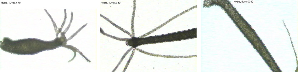
- Hydra reproduce both sexually and asexually by budding. Try to find a live Hydra with buds. If you cannot find a live Hydra budding, look for budding in a prepared slide of Hydra.
- Add a drop of vinegar to the slide containing Hydra. Describe what happened to the cnidocytes.

- Examine microscope slides of hydra l.s. and hydra c.s. Look for the presence of two tissue layers. Identify stinging cells (Cnidocytes) in a slide of the whole animal.


Figure 5. Portion of a Hydra tentacle showing cnidocytes

Figure 5. Left: Hydra l.s. X 40. Right: Hydra l.s. with ingested food X 40


Other Hydrozoans
Examine preserved specimens of Gonionemius, Polyorchis, and Physalia.

Figure 6. Left: Gonionemus, preserved. Middle: Polyorchis, preserved. Right: Portuguese Man-Of-War
Sea Anemones and Coral (Class Anthozoa)
Examine a sea anemone and coral.

Figure 7. Sea anemone, preserved

Figure 8. Left: Astrangia (Northern Coral) Skeleton. Middle: Coral Skeleton. Right: Coral Skeleton.
Jellyfish (Class Schyphozoa)
Examine preserved jellyfish on display.

Figure 9. Aurelia
LICENSES AND ATTRIBUTIONS
CC LICENSED CONTENT, SHARED PREVIOUSLY
- Cnidarians, Biology 102. Authored by: Michael J. Gregory, Ph.D.. Provided by: LibreTexts. Located at: http://bio.libretexts.org/Under_Construction/BioStuff/BIO_102/Laboratory_Exercises/Cnidarians. Project: The Biology Web. License: CC BY-NC-SA: Attribution-NonCommercial-ShareAlike