22.4: Male Reproductive System
- Page ID
- 53847
Male Reproductive System
The testes produce spermatozoa (sperm), the male reproductive cells. Spermatozoa are produced in the seminiferous tubules and collected by the rete testis (rete = net). Spermatozoa travels from the rete testis to the head of the epididymis where they mature and are stored until ejaculation. The vas deferens transports spermatozoa from the tail of the epididymis to the prostate gland. Seminal glands, the prostate gland, and the bulbourethral glands add fluid secretions to spermatozoa to form semen. These additional fluids support the spermatozoa and their survival as well as facilitate their transfer to the female reproductive tract.
Above: Male reproductive system, lateral view of the left side with structures shown with sagittal sections.
The epididymis, vas deferens, ejaculatory duct and urethra form a system of tubules for the transport of spermatozoa from testes to the pelvic cavity. There they will be combined with the secretions of the accessory glands to form semen.
Above: Structures of the male genitalia.
In order for proper and efficiently development of spermatozoa, the testes must be cooler than core body temperature (95°F is optimal for spermatogenesis and 98.6°F is core body temperature). The testes cool some just from being suspended away from the body, but the core temperature arterial blood from the gonadal artery must also be cooled before reaching the testis capillary beds. As blood is cooled in the peripheral capillary beds of the scrotal walls, it collects into a network of gonadal veins called the pampiniform plexus (pampiniform = shaped like a vine or tendril). As it ascends to the abdomen, this venous plexus wraps around the gonadal artery, cooling the arterial blood to sufficient temperature.
Above: Internal structure of the testis and epididymis.
Sperm cells are produced in the seminiferous tubules in the testes. A cross section through the process of spermatogenesis (a type of meiosis). Close examination of a cross section of a seminiferous tubule shows that sperm cells develop beginning close to the inner lining of the seminiferous tubule, beginning with specialized stem cells called spermatogonia, and develop as they move closer to the lumen of the seminiferous tubules. Primary spermatocytes (cells that undergo the first meiotic division) and secondary spermatocytes (cells that undergo the second meiotic division) are intermediate cells produced as spermatozoa develop. Spermatogenesis produces immature spermatids that ultimately mature into spermatozoa.
Above: The testis is both an exocrine (producing spermatozoa) and an endocrine (producing androgens) gland. This immature testis, cut in cross section, provides an overview of the testis proper, its enclosing tunics, and its location in the scrotum. Also seen are the epididymis and the ductus deferens lying posterior to the testis. Tissue is magnified by 10x.
Above: The exocrine function of the testis is performed by the epithelium lining the convoluted portions of seminiferous tubules. Each convoluted tubule is lined by a stratified epithelium composed of two cell types. Germ cells divide and cytodifferentiate to form haploid spermatozoa. Sertoli cells nourish and protect germ cells during their formation before releasing them into the lumen of the tubule. Tissue is magnified by 1000x.
Above: Structures of the spermatic cord and structures enveloping the testis.
|
Structure |
Location |
Function |
|---|---|---|
|
bulbourethral glands (Cowper's glands) |
pair (right and left) of glands posterior to the bulb of the penis and membranous urethra |
produce ~1% of the total volume of semen; produce a thick mucus that cleans and neutralizes the urethra of residual urine and increases the mobility of sperm in the vagina |
|
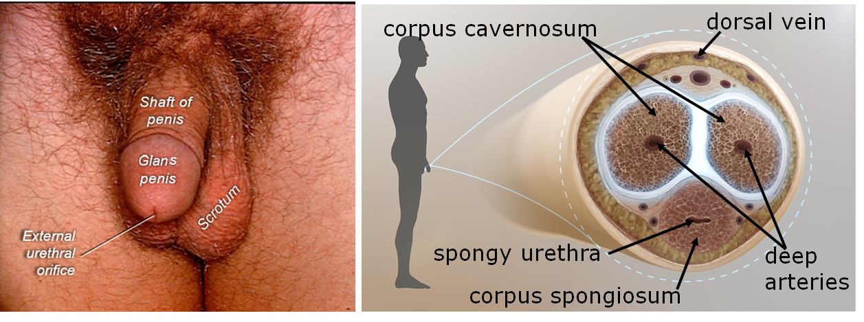
corpus cavernosum |
two of the three bodies of erectile tissue bodies in the penis |
become engorged with blood, causing an erection |
|
corpus spongiosum |
one of the three bodies of erectile tissue in the penis; the spongy urethra passes through the corpus spongiosum |
become engorged with blood, causing an erection |
|
cremasteric muscle |
very thin muscle that surrounds the spermatic cord and testes |
elevate the testes to warm them when the testes become too cold for optimal sperm production and development |
|
dartos muscle |
a very thin muscle in the walls of the scrotum |
elevate the scrotum to warm it when the testes are too cold for optimal sperm production and development |
|
ejaculatory ducts |
channels in the prostate gland formed when vas deferens joins with duct from the seminal vesicle |
spermatozoa and semen fluids flow through toward the prostatic urethra |
|
epididymis |
paired organ (right and left) located on the posterior aspect of the testes; consists of head, body and tail regions |
location where spermatids mature and become spermatozoa; store spermatozoa until ejaculation; spermatozoa account for about 3% of semen volume; an average of 200 – 500 million sperm released from an ejaculation |
|
external urethral orifice |
urethral opening on the penis |
location where urine and semen leave the body |
|
glans penis |
the highly sensitive bulbous structure at the end of the corpus spongiosum and tip of the penis |
contains the external urethral orifice and is covered by the prepuce in uncircumcised males |
|
membranous urethra |
short segment of the urethra that passes through the floor of the pelvis |
transports urine and semen |
|
penis |
external genitalia composed of shaft and glans penis |
male copulatory organ |
|
prepuce (foreskin) |
loose skin of the penis which covers the glans and is often removed by a process called circumcision |
protects glans penis |
|
prostate gland |
gland inferior to the urinary bladder; prostatic urethra and ejaculatory ducts pass through |
contributes ~26% to the volume of semen; secretes fluid that stabilizes sperm and protects them from the acidic environment of the vagina |
|
prostatic urethra |
region of the urethra that passes through the prostate; occurs from the bladder to the membranous urethra |
transports urine and semen |
|
scrotum |
skin surrounding the testis |
protection of the testis and associated tissues |
|
seminal vesicles |
pair (right and left) of tubular glands; posterior to the urinary bladder |
produce fluid that mixes with spermatozoa in the vas deferens just before the prostate gland; fluid from the seminal vesicles contributes the majority (~70%) of the volume of semen (semen = the mixture of sperm and supporting fluids); fluid from the seminal vesicles contains a lot of fructose, a sugar that is used by spermatozoa as their main energy source |
|
seminiferous tubules |
tubules within the testes |
site of spermatogenesis (production of spermatozoa); produce millions of spermatozoa per day |
|
spermatic cord |
bundle of vessels (blood and lymphatic vessels) and nerves traveling from the trunk to the testes |
a connective tissue sheath consisting of blood vessels, nerves and the cremaster muscle |
|
spongy urethra |
region of the urethra between the membranous urethra and the external urethral orifice; passes through the corpus spongiosum |
transports urine and semen |
|
testis (pl. testes) |
pair of organs within the scrotum; located outside of the abdominopelvic cavity; covered with tunica albuginea ("white tunic") |
produces spermatozoa and testosterone (a hormone) |
|
tunica albuginea |
a dense connective tissue capsule or "white tunic" that covers the testes; inward extensions of the tunica albuginea divides the seminiferous tubules into lobes |
|
|
vas deferens (ductus deferens) |
Passes from the epididymis, through the spermatic cord, through the inguinal canal, into the pelvic cavity and superiorly over the bladder |
upon ejaculation, sperm is received here from the epididymis by peristalsis |

Above: (Left) External male genitalia. (Right) Cross section of the penis.
Attributions
- "Anatomy 204L: Laboratory Manual (Second Edition)" by Ethan Snow, University of North Dakota is licensed under CC BY-NC 4.0
- "Anatomy and Physiology Lab Reference" by Laird C Sheldahl, OpenOregonEducational Resources, Mt. Hood Community College is licensed under CC BY-SA 4.0
- "Digital Histology" by Department of Anatomy and Neurobiology and the Office of Faculty Affairs, Virginia Commonwealth University School of Medicine and the ALT Lab at Virginia Commonwealth University is licensed under CC BY 4.0
- "Male Reproductive System" by Dongho Kim is licensed under CC BY-NC-SA 4.0
- "Rete testis.jpg" by KDS444 is licensed under CC BY-SA 3.0
- "Wiki Images" by https://www.scientificanimations.com/wiki-images/ is licensed under CC BY-SA 4.0


