
20.6: Duodenum, Pancreas, Liver, and Gallbladder
- Page ID
- 53821

Duodenum, Pancreas, Liver, and Gallbladder
Duodenum
Chyme passing through the pyloric sphincter moves into the duodenum which needs to neutralizes the highly acidic solution from the stomach, continue chemical digestion, and begin absorption of nutrients. In the duodenum there is a papilla (a small rounded protuberance) called the major duodenal papilla where secretions from the pancreas, liver, and gallbladder enter the duodenum to neutralize stomach acid, continue chemical digestion, and aid in absorption of nutrients.

Above: The duodenum, part of the small intestine, receives chyme from the stomach and neutralizes the gastric juices and continues chemical digestion with secretions from the pancreas.
Pancreas
The pancreas has both endocrine and exocrine functions: it produces glucagon and insulin to regulate blood sugar levels (endocrine hormones) and digestive enzymes to aide in digestion (exocrine). There are three main regions of the pancreas: The head, body and tail.
The main pancreatic duct combines with the common bile duct to form the hepatopancreatic ampulla. The hepatopancreatic sphincter controls the flow of bile and pancreatic juices through the ampulla and out the major duodenal papilla. The accessory pancreatic duct leads to the minor duodenal papilla.
Exocrine secretions containing digestive enzymes from the pancreas follow the following path:
- Pancreatic digestive enzymes produced in pancreatic acinar cells
- pancreatic duct
- common bile duct
- through the hepatopancreatic sphincter
- through the duodenal papilla
- into the duodenum

Above: The duodenum, pancreas, and gall bladder including the common bile duct where secretions from the liver and gall bladder combine with pancreatic secretions and are released into the duodenum through the duodenal papilla. Release of secretions is controlled by the hepatopancreatic sphincter.

Above: Cadaver images of pancreas and duodenum.
Liver
The liver cleans and detoxifies blood entering it from the hepatic portal vein, balances the amount of nutrients in the blood including glucose and amino acids, and produces bile. Bile is a greenish-brown alkaline fluid that aids in digestion by emulsifying fats.
Bile produced in the liver follows the following path into the duodenum:
- Bile is produced in the liver
- right/left hepatic ducts
- common hepatic duct
- common bile duct
- through the hepatopancreatic sphincter
- through the duodenal papilla
- into the duodenum

Above: Cadaver images of liver and gallbladder. (Left) Anterior view of the liver and (right) posterior view of the liver.

Above: (Top) Superior view of the liver with the top of the image being anterior and the bottom of the image being posterior. (Bottom) Inferoposterior view of the liver.
The liver is composed of four lobes: right lobe, left lobe, quadrate lobe, and caudate lobe. Between the right and left lobes is the falciform ligament, a fold of the peritoneum (serosa) which also attaches the liver to the anterior body wall and diaphragm. The coronary ligament is a ligament composed of peritoneum along the coronal plane of the superior aspect of the liver, attaching it to the diaphragm and the right kidney.
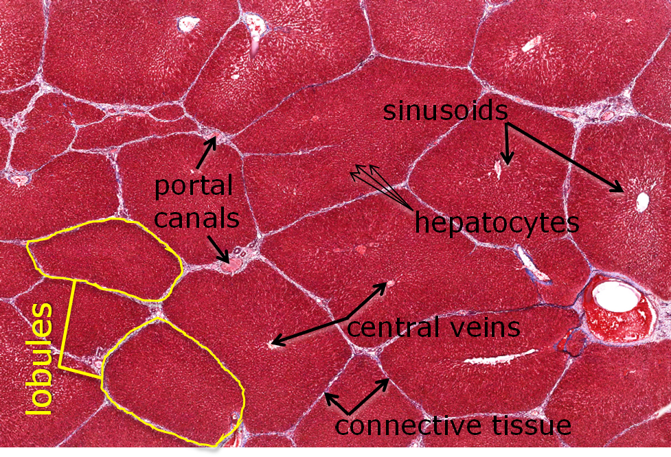
Above: The lobule, the basic structure of the liver, is formed by anastomosing rows of hepatocytes and intervening sinusoids, which extend from the periphery of the lobule toward a central vein. Portal canals, located at the marginal angles around the perimeter of the lobule, contain branches of the hepatic portal vein, hepatic artery and bile duct. Blood enters the liver in the hepatic portal vein or hepatic artery; branches of both vessels are located in a portal canal. From these branches, blood enters hepatic sinusoids between plates of hepatocytes and is carried into the central vein. Bile, the exocrine liver product, is released into tiny tunnel-like passageways called bile canaliculi. Canaliculi are located between the boundaries of adjacent hepatocytes and are continuous with bile ducts in the hepatic portal canals. Bile drains from the canaliculi to the bile duct, passing through an enlarging duct system and finally opening into the duodenum. This image of pig liver tissue shows bands of connective tissue outlining lobules; these distinctions are not so obvious in the human. Tissue is magnified by 40x.
Gallbladder
The gallbladder stores and concentrates bile. Bile is produced in the liver and is transported into the gallbladder for storage through the right and left hepatic ducts, to the common hepatic duct, then through the cystic duct. The gallbladder has different regions called the fundus (dome-shaped), body (main region), and neck (narrow region leading to the cystic duct).
Some of the bile produced by the liver is stored in the gallbladder. Stored bile follows the following path:
- Bile is produced in the liver
- Right/left hepatic ducts
- Common hepatic duct
- Cystic duct
- Gallbladder for storage
Since bile is important for the digestion of fats, individuals who have had their gallbladders removed do not have a lot of bile stored to help with fat digestion. Therefore, these individuals have to carefully monitor their diet since they cannot tolerate consuming a large amount of fat at one time.

Above: The gallbladder.
Clinical Application: Blood Sugar and Diabetes
After you consume a meal, your body digests that food to break down the sugars, nutrients, vitamins, minerals, proteins, etc. into usable and readily-absorbed forms. The sugar (in the form of glucose) is absorbed into the circulatory system and taken first to the liver (via the portal v.) to be processed and then to the rest of the body. When the blood sugar level is high, the pancreas produces insulin, a hormone that stimulates the liver to convert glycose to glycogen (the storage form of glucose) and body tissues to uptake glucose from the blood. Both of these actions will cause the level of sugar in the blood to lower. After some time has passed and your body has a low blood sugar level from using the glucose that was in your blood for energy, the pancreas will produce glucagon, a hormone that stimulates the liver to convert previously made glycogen back into glucose and release it into circulation for transportation. This action will cause the level of sugar in the blood to raise. The figure below represents the communication between the pancreas, liver, and tissue cells to maintain homeostatic blood sugar levels.

Above: Diagram showing how the liver and pancreas work together to maintain blood glucose levels.
Diabetes is a disease that causes abnormal blood sugar levels. There are three main types of diabetes. Type 1 diabetes is a condition where the pancreas produces little to no insulin. Type 2 diabetes is a condition where the liver is unable to adequately convert glucose into glycogen. Prediabetes is a condition where high blood sugar is caused by hindered glucose processing, but it is not severe enough to be Type 2 diabetes because it can be controllable by diet modification.
Attributions
- "Anatomy 204L: Laboratory Manual (Second Edition)" by Ethan Snow, University of North Dakota is licensed under CC BY-NC 4.0
- "Digital Histology" by Department of Anatomy and Neurobiology and the Office of Faculty Affairs, Virginia Commonwealth University School of Medicine and the ALT Lab at Virginia Commonwealth University is licensed under CC BY 4.0
- "Gray's Anatomy plates" by Henry Vandyke Carte is in the Public Domain
- "Medical gallery of Blausen Medical 2014" by Blausen.com staff is licensed under CC BY 3.0