
12.4: Muscles of the Posterior Trunk
- Page ID
- 53687

Muscles of the Posterior Trunk
Posterior Neck & Back

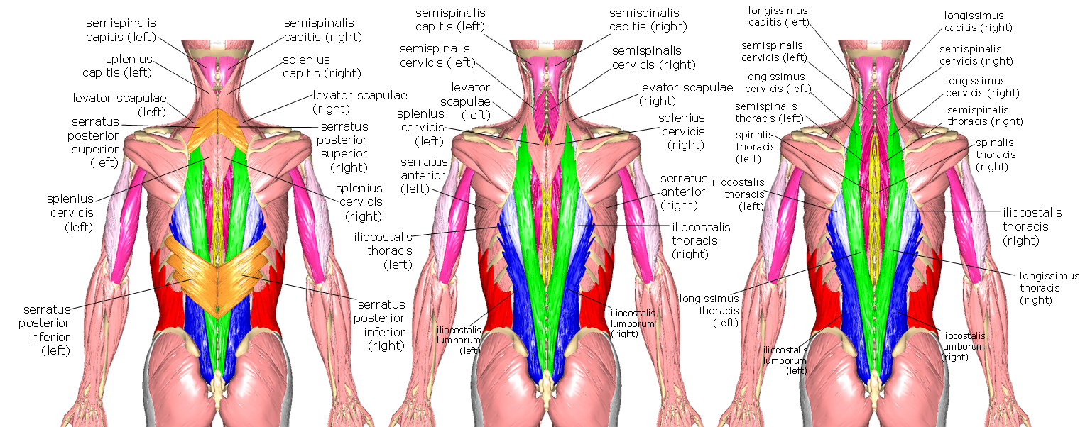
Above: Posterior trunk (left) superficial muscles without the deltoids, (middle) superficial muscles without the trapezius, and (right) posterior trunk without the deltoids, trapezius, and latissimus dorsi.
In an exercise setting, "lats" refers to the latissimus dorsi, a paired muscle that covers most of the lower back with its lateral fibers. The upper back is covered by the large trapezius muscle (aka "traps") that is almost diamond-shaped as it extends from the neck, out to the shoulders, then tapers in midway down the back. There are three sections of trapezius: upper (descending) trapezius, middle (transverse) trapezius, and lower (ascending) trapezius.
Similar to serratus anterior on the anterior trunk, the serratus posterior muscles resemble a serrated knife. There are two serratus posterior muscles - one superior and one inferior.
There are sets of muscles associated with the vertebral column with different regions. Those named with "capitis" attach to the head, "cervicis" relates to the cervical vertebrae, "thoracis" relates to the thoracic vertebrae, and "lumborum" relates to the lumbar vertebrae. These muscles from medial to lateral are called spinalis (shown in yellow), semispinalis (shown in pink shades along the vertebrae), longissimus (shown in green shades), and iliocostalis (shown in blue shades). These muscles act to flex and extend the vertebral column. Even deeper muscles associated with the vertebral column include quadratus lumborum and multifidus.
Above: Deeper muscles of the posterior trunk. (Left) Posterior trunk without trapezius, deltoids, latissimus dorsi, rhomboid major, and rhomboid minor. (Middle) Posterior trunk as shown at left except without serratus posterior superior, serratus posterior inferior, and splenius capitis.

Above: The deepest muscles of the posterior trunk (left) without trapezius, deltoids, latissimus dorsi, rhomboid major, rhomboid minor, serratus posterior superior, serratus posterior inferior, splenius capitis, all iliocostalis muscles, longissimus thoracis, semispinalis thoracis, all spinalis muscles, and abdominal muscles. (Right) The posterior trunk shown at left without intercostal muscles, semispinalis capitis, and longissimus cervicus with multifidus colored in purple.

Above: Cadaver images of the muscles of the posterior trunk and neck.
Rotator Cuff
The rotator cuff is the name given to the group of four muscles that are largely responsible for the ability to rotate the arm: supraspinatus, infraspinatus, subscapularis, and teres minor. Three of the four rotator cuff muscles are deep to the deltoid and trapezius muscles and cannot be seen unless those muscles are first removed and one is on the anterior side of the scapula bone and cannot be seen from the surface. On the anterior side of scapula bone is the subscapularis. It is triangular in shape and covers the entire anterior aspect of the scapula. Its origin is along the subscapular fossa that makes up most of the anterior “wing” of the scapula and it inserts on the lesser tubercle of the humerus bone.
On the posterior side of the scapula bone are the other three muscles of the rotator cuff. All three insert on the greater tubercle of the humerus, allowing them, in combination with the subscapularis, to control rotation of the arm. The supraspinatus muscle is above the spine of the scapula in the supraspinous fossa. The infraspinatus muscle is below the spine of the scapula in the infraspinous fossa. The relatively thin teres minor muscle is the most inferior of the rotator cuff muscles.
The teres major muscle has its origin on the scapula, like the rotator cuff muscles, but is not involved in rotating the arm. It inserts lower on the humerus than the rotator cuff muscles and is involved in adducting the arm (bringing it closer to the midline of the body).

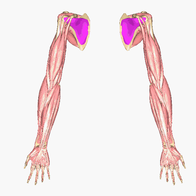
Above: Muscles of the rotator cuff. (Top left) Posterior muscles of the rotator cuff and (top right) anterior view of the scapulae showing the subscapularis muscles on the anterior aspects of the subscapular fossae of the scapulae. (Top right and bottom) The subscapularis muscles are shown without the rest of the body since they are positioned at the posterior of the thoracic cage and are difficult to see.

Above: Cadaver images of the muscles of the rotator cuff.
Attributions
- "Anatomy 204L: Laboratory Manual (Second Edition)" by Ethan Snow, University of North Dakota is licensed under CC BY-NC 4.0
- "Anatomy and Physiology I Lab" by Victoria Vidal is licensed under CC BY 4.0
- "BIOL 250 Human Anatomy Lab Manual SU 19" by Yancy Aquino, Skyline College is licensed under CC BY-NC-SA 4.0
- "BodyParts3D/Anatomography" by The Database Center for Life Science is licensed under CC BY-SA 2.1