12.3: Muscles of the Anterior Trunk
- Page ID
- 53686
Muscles of the Anterior Trunk
Some of the trunk muscles have been given nicknames by gym rats. For instance, the "pecs" are the pectoralis major muscles at each breast. Deep to pectoralis major are the paired pectoralis minor muscles. Between the ribs are three layers of intercostal muscles: external intercostals (most superficial), inner intercostals (intermediate), and innermost intercostals (deepest intercostal muscles).Although the internal intercostal muscles are deep to the external intercostal muscles, paying attention to the direction of muscle fibers for each muscle can help you identify them correctly. From an anterior view, internal intercostal muscle fibers run in an inferolateral direction from superior to inferior, whereas external intercostal muscle fibers run in an inferomedial direction from superior to inferior.
Extending from the back and wrapping around the sides of the rib cage are the paired serratus anterior muscles. This muscle’s anterior edges are serrated like the teeth of a saw because this muscle’s origins are on ribs 1 through 8 and each serration is the attachment point to another rib.
Rectus abdominis, the medial, paired muscles that can create an abdominal "six-pack," is enclosed by the rectus sheath, also known as the rectus fascia. The rectus sheath is formed by aponeuroses (white, fibrous, flat, tendon-like tissue) from the external oblique, internal oblique, and transverse abdominis muscles. The rectus sheath fuses along a medial line called the linea alba, literally meaning the "white line." Linea alba is a fibrous tissue attached to the pubic symphysis and the xiphoid process of the sternum and is a point of insertion for abdominal muscle.
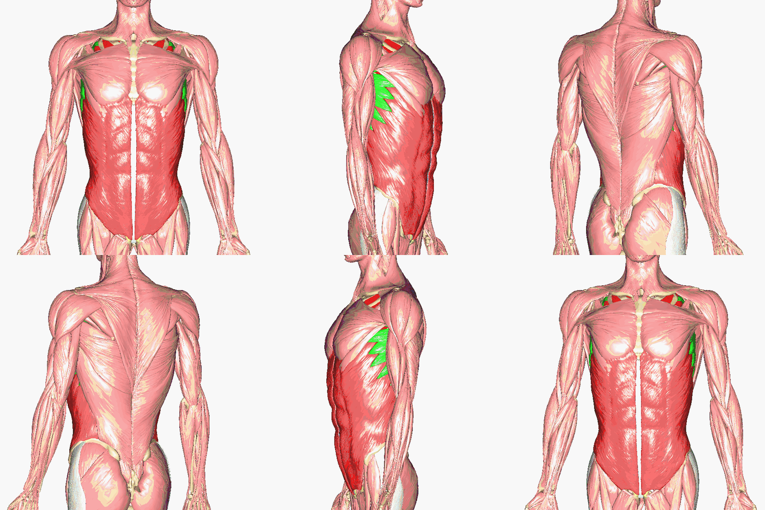
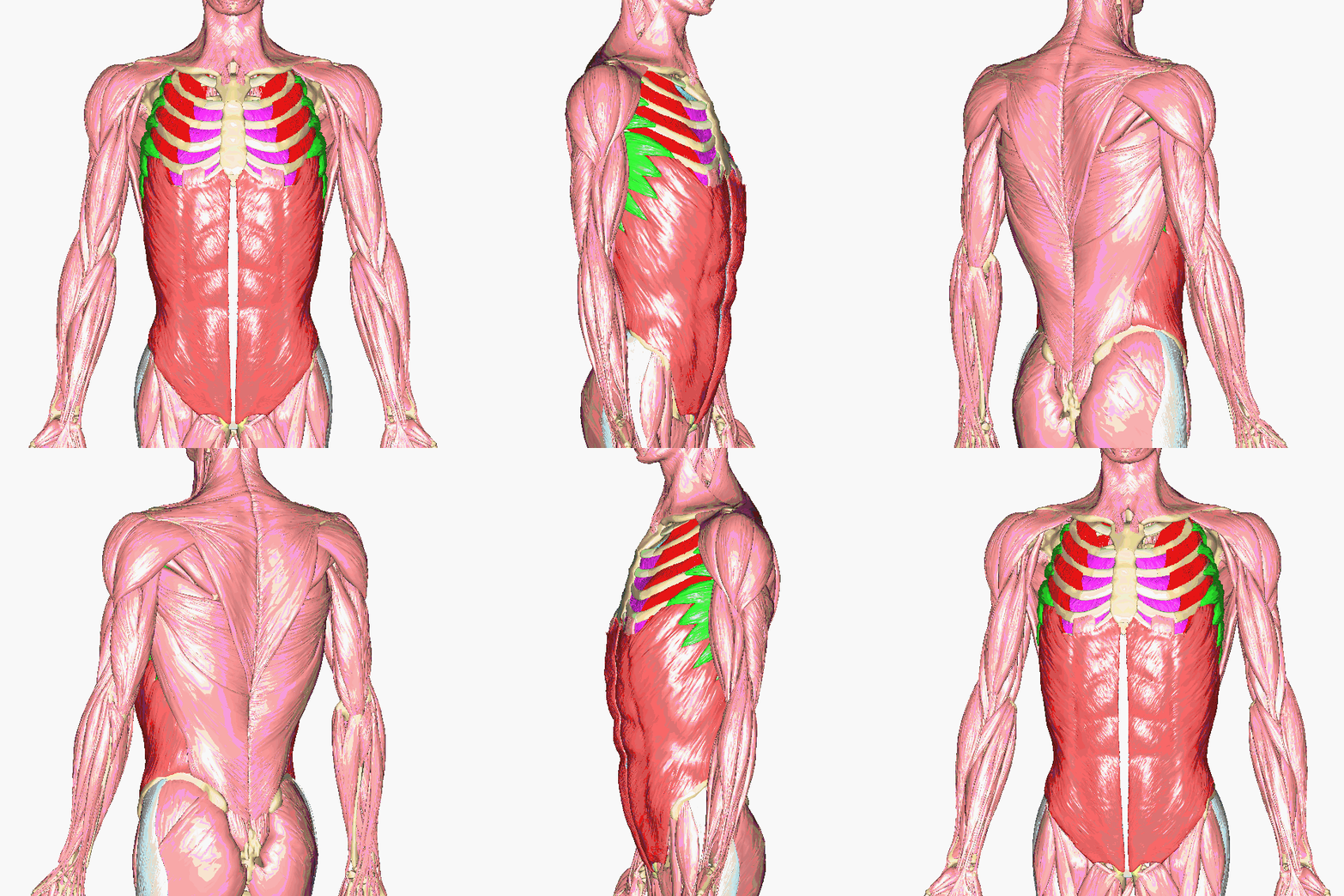
Above: Superficial anterior trunk muscles (top left and middle) with all muscles and (top right and bottom) with pectoralis major removed. External obliques are shown in light red, external intercostals are shown in bright red, serratus anterior is shown in green, and internal intercostals are shown in magenta.
Above: Cross section of abdominal muscles, superior view, with anterior at the top of the image and posterior at the bottom of the image. The rectus sheath, the aponeuroses of the external obliques, internal obliques, and transverse abdominis, and enclosing the rectus abdominis muscles is shown in blue.
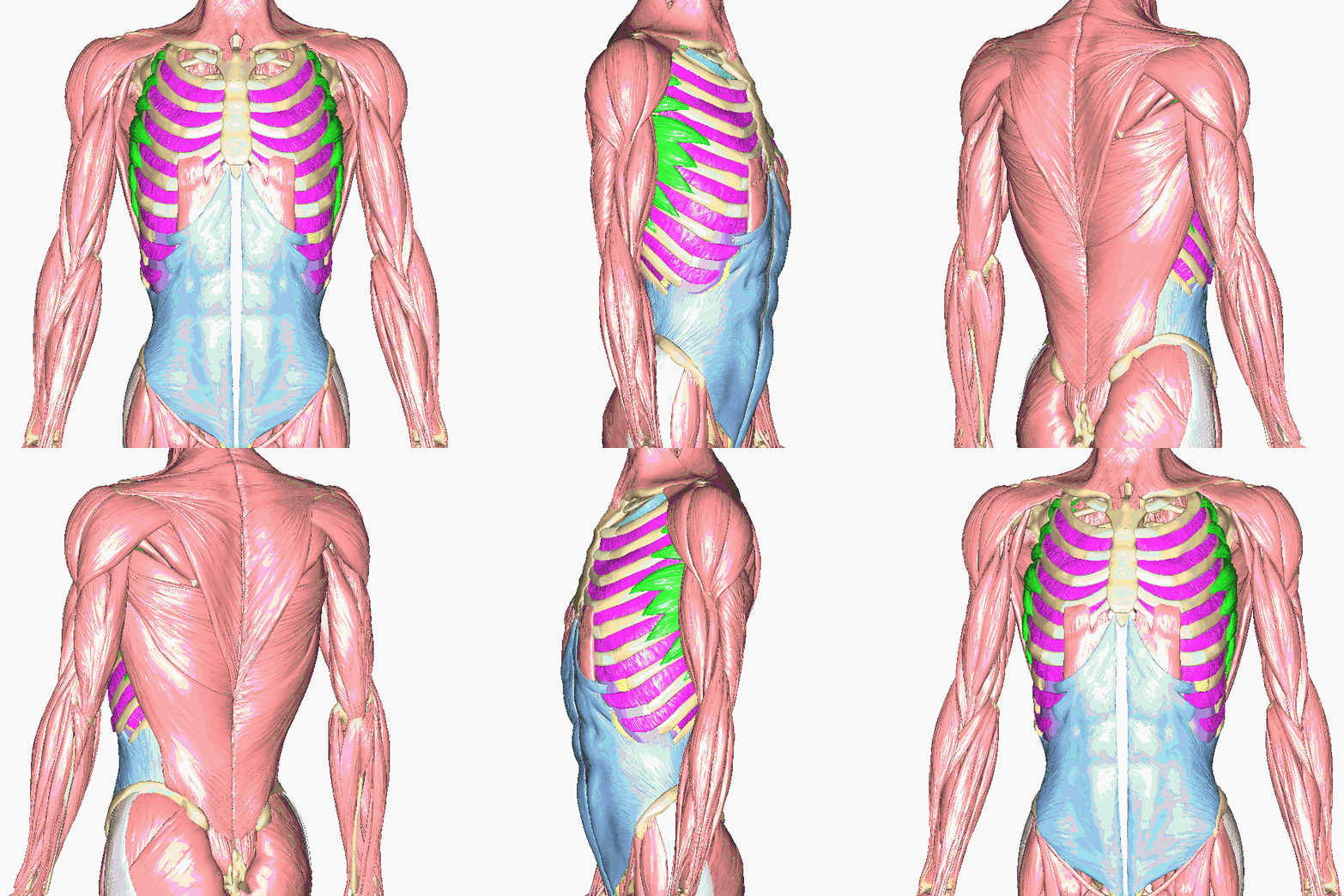
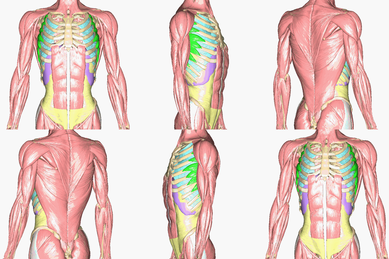
Above: Deeper anterior trunk muscles (top left and middle) without pectoralis major, pectoralis minor, and the external obliques (top middle and bottom) also without internal intercostals and internal obliques, and (right) also without innermost intercostals, serratus anterior, and transversus abdominis. External intercostals are shown in bright red, serratus anterior is shown in green, internal intercostals are shown in magenta, internal obliques are shown in light blue, transversus abdominis in light yellow, innermost intercostals in cyan, and the diaphragm in lilac.
What gym rats call the "core muscles" are three layers of muscle that sit over the abdomen. The outer layer is the external oblique muscle, with its aponeurosis covering the medial abdomen. Under the external oblique are the internal obliques on the sides of the abdomen and the rectus abdominis muscle in-between the internal obliques. The fibers of the internal obliques run up at an angle, opposite in direction to the fiber angle of the external obliques. The rectus abdominis muscle is also known as the abs. The deepest layer has the transversus abdominis muscle, whose fibers run laterally. Its fibers are concentrated at the sides of the abdomen and, like the external oblique, has an aponeurosis covering the medial abdomen under the rectus abdominis.
"Obliques" are the external oblique muscle whose fibers angle down as it covers both sides of the abdominal region. The external oblique muscle has two sets of fibers, which cover the left and right abdomen, that are connected by a wide aponeurosis sheet in the center of the abdomen. In most muscle models that aponeurosis sheet is cut away to reveal the rectus abdominis muscles below it.
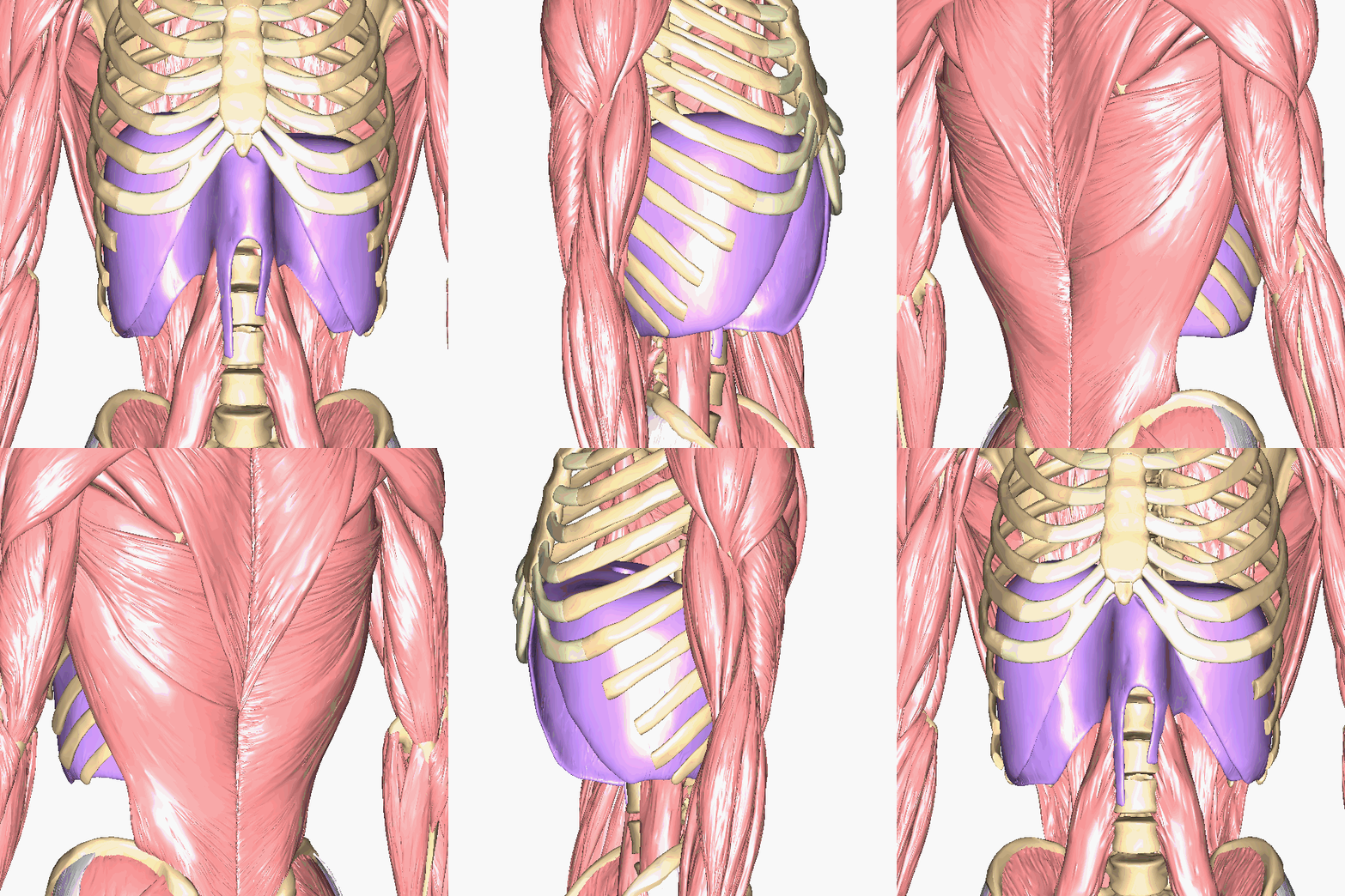
Above: The diaphragm.
Above: Cadaver images of the muscles of the anterior thorax.
Above: Cadaver image of the muscles of the abdominal region.
Attributions
- "Anatomy 204L: Laboratory Manual (Second Edition)" by Ethan Snow, University of North Dakota is licensed under CC BY-NC 4.0
- "Anatomy and Physiology I Lab" by Victoria Vidal is licensed under CC BY 4.0
- "BIOL 250 Human Anatomy Lab Manual SU 19" by Yancy Aquino, Skyline College is licensed under CC BY-NC-SA 4.0
- "BodyParts3D/Anatomography" by The Database Center for Life Science is licensed under CC BY-SA 2.1
- "Gray's Anatomy plates" by Henry Vandyke Carte is in the Public Domain


