
10.3: Additional Reference Figures
- Page ID
- 53671

\( \newcommand{\vecs}[1]{\overset { \scriptstyle \rightharpoonup} {\mathbf{#1}} } \)
\( \newcommand{\vecd}[1]{\overset{-\!-\!\rightharpoonup}{\vphantom{a}\smash {#1}}} \)
\( \newcommand{\id}{\mathrm{id}}\) \( \newcommand{\Span}{\mathrm{span}}\)
( \newcommand{\kernel}{\mathrm{null}\,}\) \( \newcommand{\range}{\mathrm{range}\,}\)
\( \newcommand{\RealPart}{\mathrm{Re}}\) \( \newcommand{\ImaginaryPart}{\mathrm{Im}}\)
\( \newcommand{\Argument}{\mathrm{Arg}}\) \( \newcommand{\norm}[1]{\| #1 \|}\)
\( \newcommand{\inner}[2]{\langle #1, #2 \rangle}\)
\( \newcommand{\Span}{\mathrm{span}}\)
\( \newcommand{\id}{\mathrm{id}}\)
\( \newcommand{\Span}{\mathrm{span}}\)
\( \newcommand{\kernel}{\mathrm{null}\,}\)
\( \newcommand{\range}{\mathrm{range}\,}\)
\( \newcommand{\RealPart}{\mathrm{Re}}\)
\( \newcommand{\ImaginaryPart}{\mathrm{Im}}\)
\( \newcommand{\Argument}{\mathrm{Arg}}\)
\( \newcommand{\norm}[1]{\| #1 \|}\)
\( \newcommand{\inner}[2]{\langle #1, #2 \rangle}\)
\( \newcommand{\Span}{\mathrm{span}}\) \( \newcommand{\AA}{\unicode[.8,0]{x212B}}\)
\( \newcommand{\vectorA}[1]{\vec{#1}} % arrow\)
\( \newcommand{\vectorAt}[1]{\vec{\text{#1}}} % arrow\)
\( \newcommand{\vectorB}[1]{\overset { \scriptstyle \rightharpoonup} {\mathbf{#1}} } \)
\( \newcommand{\vectorC}[1]{\textbf{#1}} \)
\( \newcommand{\vectorD}[1]{\overrightarrow{#1}} \)
\( \newcommand{\vectorDt}[1]{\overrightarrow{\text{#1}}} \)
\( \newcommand{\vectE}[1]{\overset{-\!-\!\rightharpoonup}{\vphantom{a}\smash{\mathbf {#1}}}} \)
\( \newcommand{\vecs}[1]{\overset { \scriptstyle \rightharpoonup} {\mathbf{#1}} } \)
\( \newcommand{\vecd}[1]{\overset{-\!-\!\rightharpoonup}{\vphantom{a}\smash {#1}}} \)
\(\newcommand{\avec}{\mathbf a}\) \(\newcommand{\bvec}{\mathbf b}\) \(\newcommand{\cvec}{\mathbf c}\) \(\newcommand{\dvec}{\mathbf d}\) \(\newcommand{\dtil}{\widetilde{\mathbf d}}\) \(\newcommand{\evec}{\mathbf e}\) \(\newcommand{\fvec}{\mathbf f}\) \(\newcommand{\nvec}{\mathbf n}\) \(\newcommand{\pvec}{\mathbf p}\) \(\newcommand{\qvec}{\mathbf q}\) \(\newcommand{\svec}{\mathbf s}\) \(\newcommand{\tvec}{\mathbf t}\) \(\newcommand{\uvec}{\mathbf u}\) \(\newcommand{\vvec}{\mathbf v}\) \(\newcommand{\wvec}{\mathbf w}\) \(\newcommand{\xvec}{\mathbf x}\) \(\newcommand{\yvec}{\mathbf y}\) \(\newcommand{\zvec}{\mathbf z}\) \(\newcommand{\rvec}{\mathbf r}\) \(\newcommand{\mvec}{\mathbf m}\) \(\newcommand{\zerovec}{\mathbf 0}\) \(\newcommand{\onevec}{\mathbf 1}\) \(\newcommand{\real}{\mathbb R}\) \(\newcommand{\twovec}[2]{\left[\begin{array}{r}#1 \\ #2 \end{array}\right]}\) \(\newcommand{\ctwovec}[2]{\left[\begin{array}{c}#1 \\ #2 \end{array}\right]}\) \(\newcommand{\threevec}[3]{\left[\begin{array}{r}#1 \\ #2 \\ #3 \end{array}\right]}\) \(\newcommand{\cthreevec}[3]{\left[\begin{array}{c}#1 \\ #2 \\ #3 \end{array}\right]}\) \(\newcommand{\fourvec}[4]{\left[\begin{array}{r}#1 \\ #2 \\ #3 \\ #4 \end{array}\right]}\) \(\newcommand{\cfourvec}[4]{\left[\begin{array}{c}#1 \\ #2 \\ #3 \\ #4 \end{array}\right]}\) \(\newcommand{\fivevec}[5]{\left[\begin{array}{r}#1 \\ #2 \\ #3 \\ #4 \\ #5 \\ \end{array}\right]}\) \(\newcommand{\cfivevec}[5]{\left[\begin{array}{c}#1 \\ #2 \\ #3 \\ #4 \\ #5 \\ \end{array}\right]}\) \(\newcommand{\mattwo}[4]{\left[\begin{array}{rr}#1 \amp #2 \\ #3 \amp #4 \\ \end{array}\right]}\) \(\newcommand{\laspan}[1]{\text{Span}\{#1\}}\) \(\newcommand{\bcal}{\cal B}\) \(\newcommand{\ccal}{\cal C}\) \(\newcommand{\scal}{\cal S}\) \(\newcommand{\wcal}{\cal W}\) \(\newcommand{\ecal}{\cal E}\) \(\newcommand{\coords}[2]{\left\{#1\right\}_{#2}}\) \(\newcommand{\gray}[1]{\color{gray}{#1}}\) \(\newcommand{\lgray}[1]{\color{lightgray}{#1}}\) \(\newcommand{\rank}{\operatorname{rank}}\) \(\newcommand{\row}{\text{Row}}\) \(\newcommand{\col}{\text{Col}}\) \(\renewcommand{\row}{\text{Row}}\) \(\newcommand{\nul}{\text{Nul}}\) \(\newcommand{\var}{\text{Var}}\) \(\newcommand{\corr}{\text{corr}}\) \(\newcommand{\len}[1]{\left|#1\right|}\) \(\newcommand{\bbar}{\overline{\bvec}}\) \(\newcommand{\bhat}{\widehat{\bvec}}\) \(\newcommand{\bperp}{\bvec^\perp}\) \(\newcommand{\xhat}{\widehat{\xvec}}\) \(\newcommand{\vhat}{\widehat{\vvec}}\) \(\newcommand{\uhat}{\widehat{\uvec}}\) \(\newcommand{\what}{\widehat{\wvec}}\) \(\newcommand{\Sighat}{\widehat{\Sigma}}\) \(\newcommand{\lt}{<}\) \(\newcommand{\gt}{>}\) \(\newcommand{\amp}{&}\) \(\definecolor{fillinmathshade}{gray}{0.9}\)Additional Reference Figures
Mandible Joints
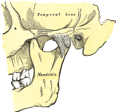
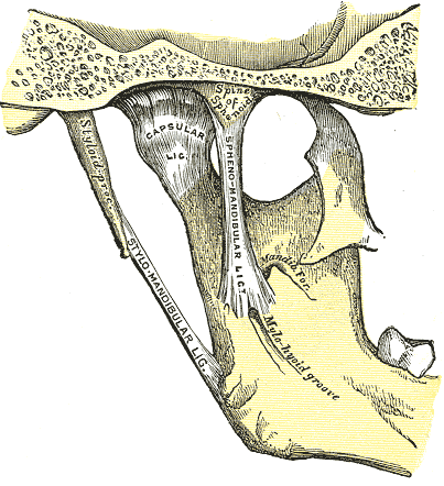
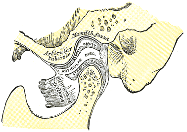
Above: Articulation of the mandible. (Top) Lateral aspect of the left side of the mandible. (Middle) Medial aspect of the left side of the mandible. (Bottom) Sagittal section of the articulation of the mandible.
Vertebral Joints
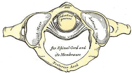
Above: Articulation between odontoid process (dens of C2 or axis) and atlas (C1). Anterior is at the top of the illustration and posterior is at the bottom.
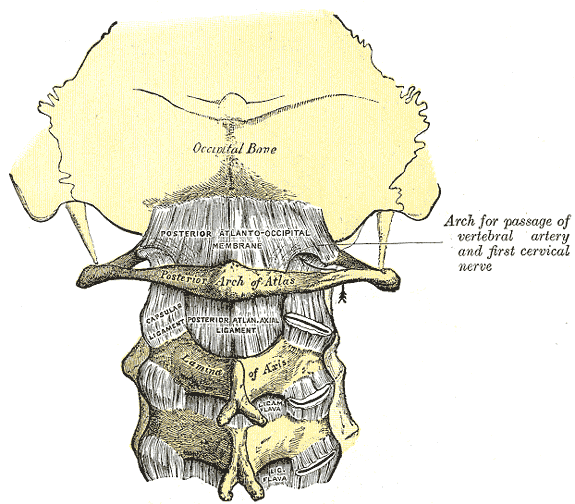
Above: Posterior atlantooccipital membrane and atlantoaxial ligament. Posterior view of the occipital bone of the skull and the vertebral column.
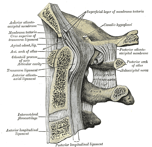
Above: Median sagittal section through the occipital bone and first three cervical vertebrae. The left side of this illustration is anterior and the right side is posterior.

Above: Median sagittal section of two lumbar vertebrae and their ligaments. Anterior is at the left of the illustration and posterior is at the right of the illustration.

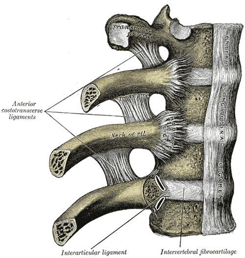
Above: Costotransverse articulation. Seen from above. Anterior is at the top of the illustration and posterior is at the bottom.
Above: Costovertebral articulations. Anterior view.
Sternal Joints

Above: Sternocostal and interchondral articulations. Anterior view.
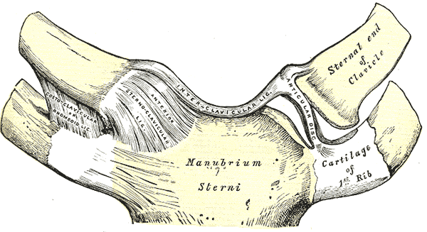
Above: Sternoclavicular articulation. Anterior view.
Pelvic Joints
Above: Anterior articulations of pelvis.

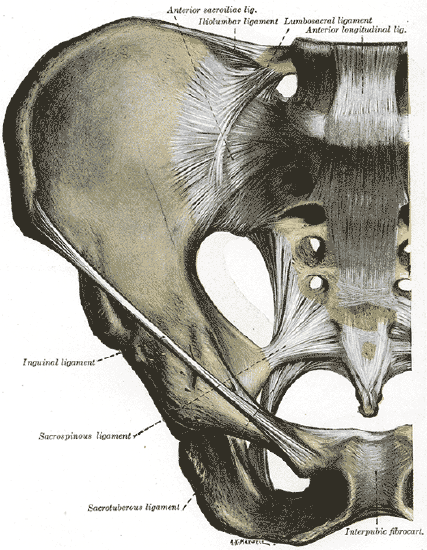
Above: Posterior articulations of pelvis.

Above: Symphysis pubis exposed by a coronal section.
Shoulder Joints
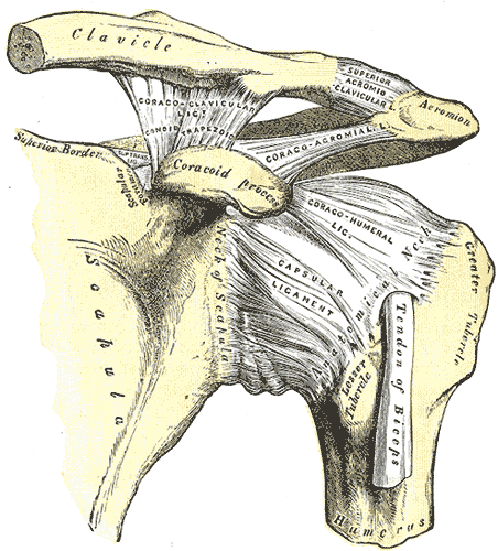
Above: The left shoulder and acromioclavicular joints, and the proper ligaments of the scapula.

Above: Glenoid fossa of the right scapula. Lateral view.
Elbow Joint
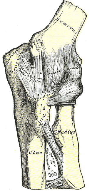
Above: Left elbow joint, showing anterior and internal ligaments

Above: Left elbow joint, showing posterior and external ligaments.
Hand Joints
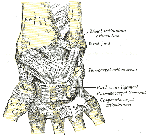
Above: Ligaments of the right wrist. Anterior view.
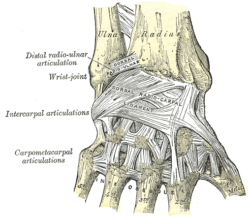
Above: Ligaments of the right wrist. Posterior view.
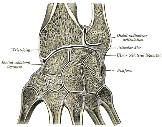
Above: Vertical section through the articulations at the wrist, showing the synovial cavities.
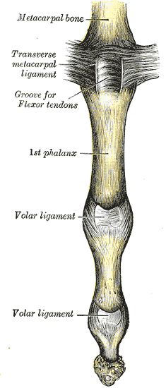
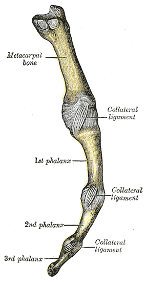
Above: Metacarpophalangeal articulation and articulations of digit. (Left) Volar aspect and (right) ulnar aspect.
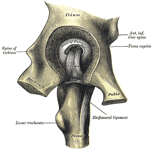
Hip Joint

Above: Right hip-joint from the front.

Above: The hip-joint from behind.
Above: Left hip-joint, opened by removing the floor of the acetabulum from within the pelvis.
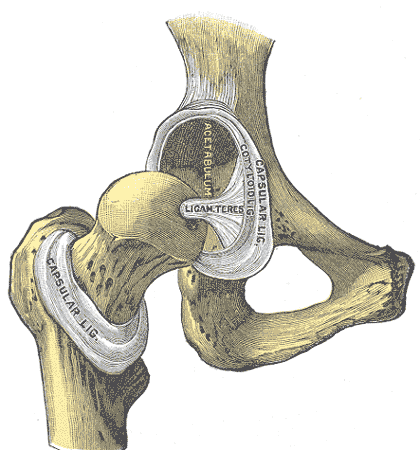
Above: Hip-joint, front view. The capsular ligament has been largely removed.
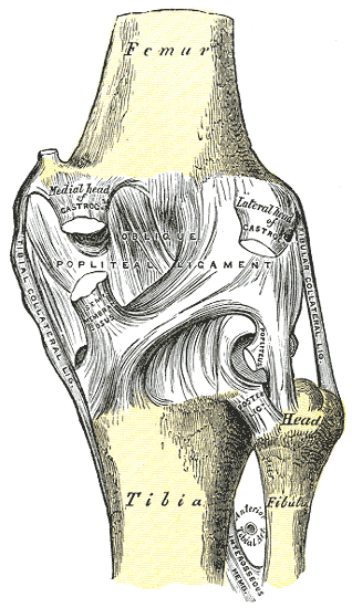
Knee Joint
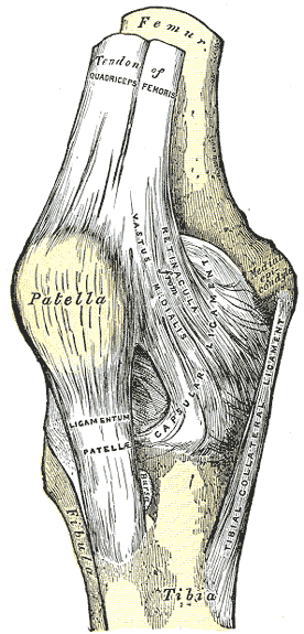

Above: Right knee-joint. (Left) Anterior view, (middle and right) posterior view).

Above: Left knee-joint from behind, showing interior ligaments.
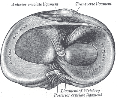
Above: Head of right tibia seen from above, showing menisci and attachments of ligaments.

Above: Sagittal section of right knee-joint.
Foot Joints

Above: Ligaments of the medial aspect of the foot.

Above: The ligaments of the foot from the lateral aspect.

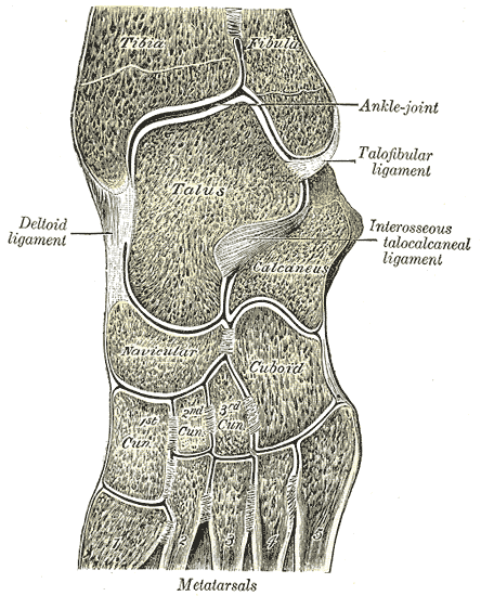
Above: Coronal section through right talocrural and talocalcaneal joints.
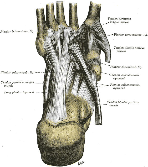
Above: Ligaments of the sole of the foot, with the tendons of the peroneus longus, tibialis posterior and tibialis anterior muscles.
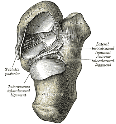
Above: Talocalcaneal and talocalcaneonavicular articulations exposed from above by removing the talus.
Above: Oblique section of left intertarsal and tarsometatarsal articulations, showing the synovial cavities.
Attributions
- "Gray's Anatomy plates" by Henry Vandyke Carte is in the Public Domain