
10.4: Laboratory Activities and Assignment
- Page ID
- 53672

Laboratory Activities and Assignment
Part 1: Review of Articulations & Movements
Articulations
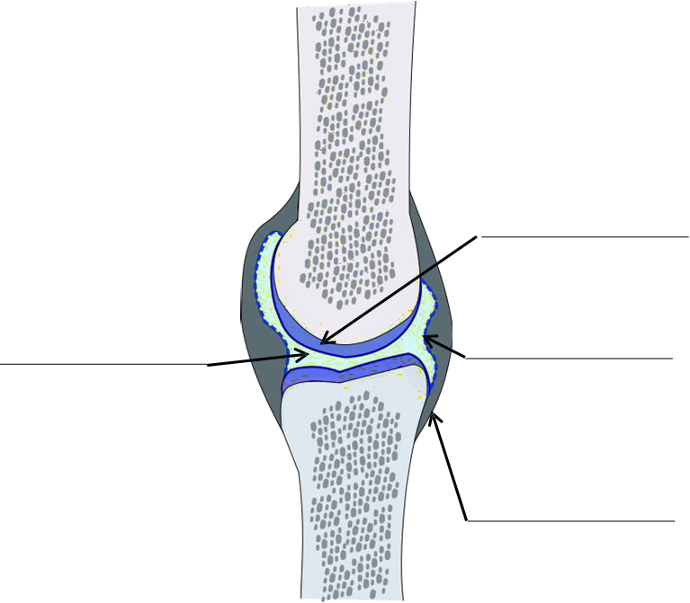
1. On the diagram below of a synovial joint, label the figure with the terms listed below:
|
|
2. Create a diagram showing the relationships (i.e. show in diagram form which types of joints belong to the same categories and which ones do not) between the following types of joints:
|
|
|
3. On the diagram below of the glenohumeral joint (shoulder), label the figure with the terms listed below:
|
|
|

4. On the diagram below of the knee joint (anterior view), label the figure with the terms listed below:
|
|
|

5. On the diagram below of the knee joint (anterior view), label the figure with the terms listed below:
|
|
|

Movements
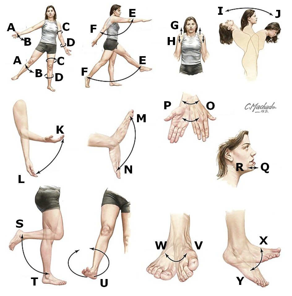
1. For each letter on the figure below, indicate the type of motion shown from the choices below. Some choices are used multiple times.
|
|
|
|
A: _________________ B: _________________ C: _________________ D: _________________ E: _________________ F: _________________ G: _________________ H: _________________ I: _________________ |
J: __________________ K: __________________ L: __________________ M: __________________ N: __________________ O: __________________ P: __________________ Q: __________________ |
R: ________________ S: ________________ T: ________________ U: ________________ V: ________________ W: _______________ X: ________________ Y: ________________ |
Part 2: Articulation Laboratory Activities
1. Classify joints by structure and function. Inspect the anatomical models available to you. Classify the joints by structure and function. Also add the structural subcategory to your answers. Write your answers in the table provided below.
|
Structural Classifications & Subcategories: |
Functional Classifications: |
|---|---|
|
1. fibrous
2. cartilaginous
3. synovial
|
|
|
Joint |
Structural classification & Subcategories |
Functional classification |
|---|---|---|
|
atlantoaxial joint (C1 and C2) |
||
|
atlantooccipital joint (C1 and occipital bone) |
||
|
coronal suture |
||
|
costochondral joint |
||
|
glenohumeral joint (shoulder) |
||
|
intervertebral joint |
||
|
knee joint |
||
|
radioulnar joint |
||
|
sagittal suture |
||
|
tibiofibular joint |
||
|
tooth joint |
2. Examine an anatomical model of a knee joint. Identify the following structures on the model.
|
Structure |
Identified (✓/X) |
|---|---|
|
anterior cruciate ligament (ACL) |
|
|
articular surface of the femur |
|
|
bursae |
|
|
femur |
|
|
joint capsule |
|
|
lateral collateral ligament |
|
|
lateral condyle of femur |
|
|
lateral meniscus |
|
|
medial collateral ligament |
|
|
medial condyle of femur |
|
|
medial meniscus |
|
|
patella |
|
|
patellar ligament |
|
|
posterior cruciate ligament (PCL) |
|
|
quadriceps muscle |
|
|
synovial cavity (with synovial fluid) |
|
|
synovial membrane |
|
|
tendon of rectus femoris |
|
|
tibia |
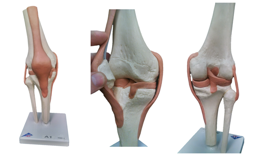
3. Use the anatomical model of a knee joint to label the images below with the structures listed:
|
|
|
Part 3: Movement Laboratory Activities
1. Collaborating with your lab group, choose a body part for each of the movement terminologies below and physically move your body in this way. Describe how you moved for each below:
a. flexion:
b. extension:
c. circumduction:
d. elevation:
e. depression:
f. medial rotation:
g. lateral rotation:
h. dorsiflexion:
i. plantar flexion:
j. pronation:
k. supination:
l. inversion:
m. eversion:
n. protrusion:
o. retrusion:
p. opposition:
q. reposition:
2. With your lab group, move your body in each of the ways described below. Discuss these movements with your lab group and describe these movements using anatomical movement terminologies. You will need to use multiple movement terms for each:
a. Walking:
b. Stand and pivot your entire body to stand facing the direction to your right:
c. Beginning in a normal standing position, move your arms as if you were doing a jumping jack:
d. Simulate the movements you would do to grab a frying pan from a counter and move it to the stove:
Attributions
Part 1: Review of Articulations & Movements
- "Anatomy and Physiology Lab Homework" by Laird C Sheldahl is licensed under CC BY-SA 4.0
- "Anatomy and Physiology Lab Reference" by Laird C Sheldahl, OpenOregonEducational Resources, Mt. Hood Community College is licensed under CC BY-SA 4.0
- "Introduction to Anatomy" by Paul Hudson is licensed under CC BY-NC-SA 4.0
Part 2: Articulation Laboratory Activities
- "Anatomy and Physiology I Lab" by Victoria Vidal is licensed under CC BY 4.0