
9.2: Shoulder Girdle/Pectoral Girdle
- Page ID
- 53650

Shoulder Girdle/Pectoral Girdle
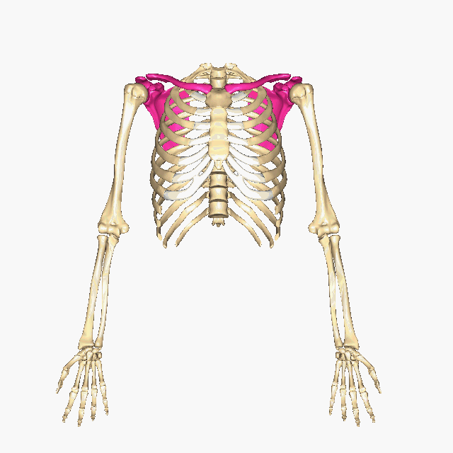
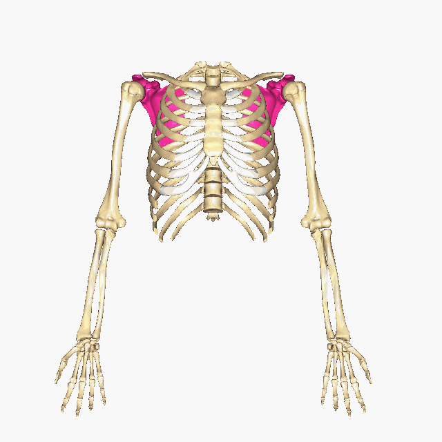
Above: The pectoral/shoulder girdle is shown in pink. Each has a scapula and a clavicle.
There is a pectoral girdle above each arm, consisting of a scapula (shoulder blade) and a clavicle (collar bone). The shoulder girdle, also known as the pectoral girdle, forms a socket and support system for the upper limb. Click here to see the positions of the shoulder/pectoral girdles.

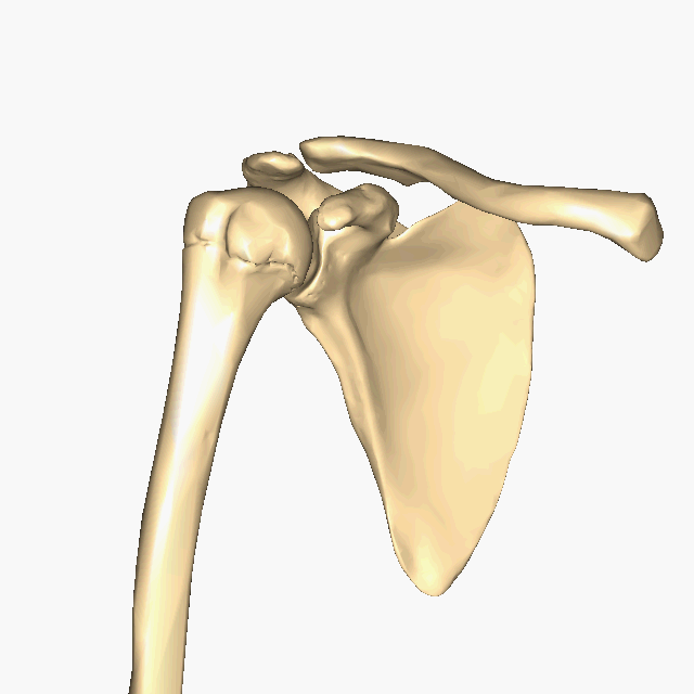
Above: The right shoulder/pectoral girdle show with the humerus (upper limb). (A) Anterior view, (B) lateral view, and (C) posterior view of the shoulder/pectoral girdle with the humerus. The joint between the humerus and the scapula is called the glenohumeral joint since the humerus ("humeral") articulates with the glenoid cavity ("gleno") of the scapula. The joint between the clavicle and the scapula is called the acromioclavicular joint since the clavicle ("clavicular") articulates with the acromial process ("acromio") of the scapula.
Clavicle
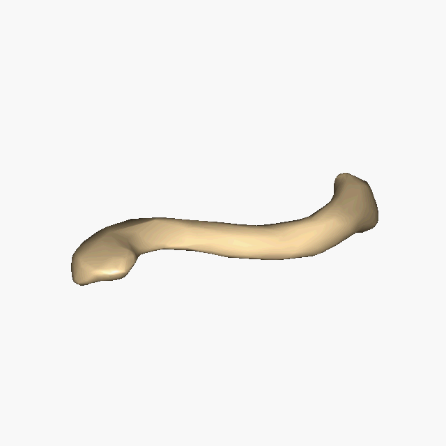
The clavicle is a vaguely S-shaped bone which articulates with the scapula at its lateral end and articulates with the sternum at its medial end. The flatter broader end is the acromial end, which articulates with the acromion process of the scapula. The superior surface of the acromial end is smoother than its inferior surface, which can help you determine whether a particular clavicle bone comes from the left or right. The more circular end is the sternal end, which articulates with one of the sternal notches on the manubrium of the sternum. The conoid tubercle found on the inferior, posterolateral aspect of the clavicle is an attachment point for the conoid ligament connecting the clavicle to the coracoid process of the scapula. Similarly, the trapezoid line of the clavicle also attaches a ligament (trapezoid ligament) joining the clavicle to the coracoid process of the scapula. An acromioclavicular ligament attaches the acromial end of the clavicle to the acromion process of the scapula.

Above: A-C shows the right clavicle and D-F shows the left clavicle. A and D have a superior view, B and E have a superomedial view, and C and F have an inferior view.
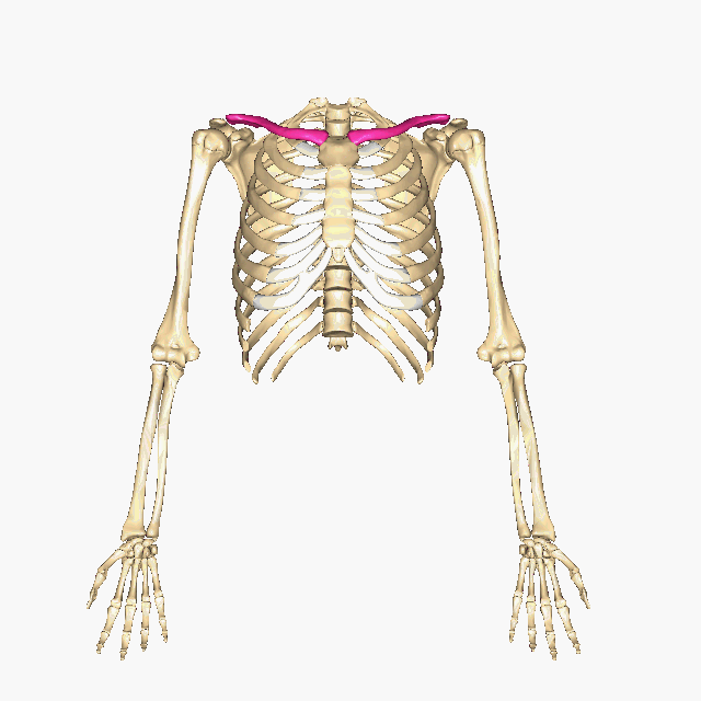
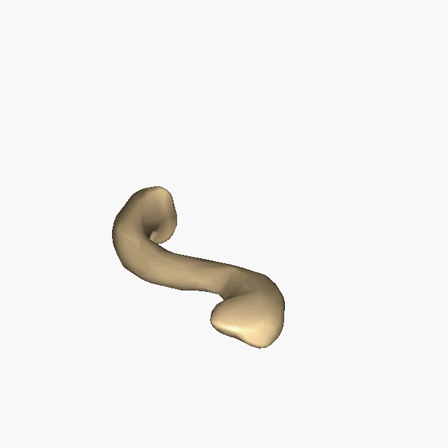
Above: (Top) Position of the clavicles. (Middle) Right clavicle. (Bottom) Left clavicle.
Scapula

Above: A-C shows the right scapula and D-F shows the left scapula. A and D have a anterior views, B and E have a lateral views, and C and F have posterior views.
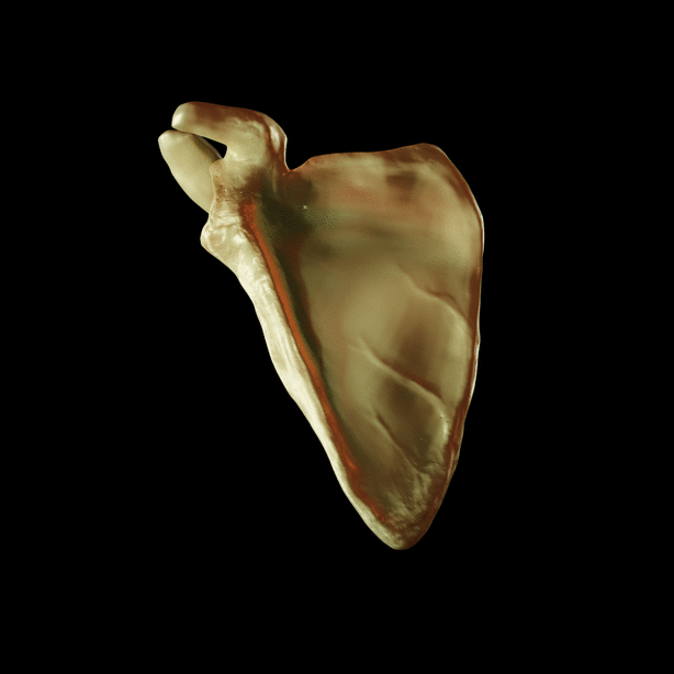
Above: (Top) Position of scapulae. (Bottom) Right scapula.
The smooth anterior surface (subscapular fossa) of the scapula is positioned facing the posterior lateral aspect of the thoracic cage. Rotator cuff muscles, that The scapula, commonly known as the shoulder blade, forms articulations with the clavicle and with the humerus. The scapula articulates with the clavicle at the acromion process and the coracoid process also creates attachments with ligaments attaching to the clavicle. The glenoid cavity of the scapula articulates with the head of the humerus of the upper limb forming the glenohumeral joint. stabilize the shoulder joint, attach scapula including supraspinatus (located in the supraspinous fossa), infraspinatus (located in the infraspinous fossa), subscapularis (located in the subscapular fossa), and teres minor (attached to the lateral border of the scapula).
Attributions
- "3d Scapula Rendered.gif" by DrJanaOfficial is licensed under CC BY-SA 4.0
- "BIOL 250 Human Anatomy Lab Manual SU 19" by Yancy Aquino, Skyline College is licensed under CC BY-NC-SA 4.0
- "BodyParts3D/Anatomography" by The Database Center for Life Science is licensed under CC BY-SA 2.1
- "Clavicle 3d Model.gif" by DrJanaOfficial is licensed under CC BY-SA 4.0