
8.2.2: Interior of the Cranium
- Page ID
- 53618

\( \newcommand{\vecs}[1]{\overset { \scriptstyle \rightharpoonup} {\mathbf{#1}} } \)
\( \newcommand{\vecd}[1]{\overset{-\!-\!\rightharpoonup}{\vphantom{a}\smash {#1}}} \)
\( \newcommand{\id}{\mathrm{id}}\) \( \newcommand{\Span}{\mathrm{span}}\)
( \newcommand{\kernel}{\mathrm{null}\,}\) \( \newcommand{\range}{\mathrm{range}\,}\)
\( \newcommand{\RealPart}{\mathrm{Re}}\) \( \newcommand{\ImaginaryPart}{\mathrm{Im}}\)
\( \newcommand{\Argument}{\mathrm{Arg}}\) \( \newcommand{\norm}[1]{\| #1 \|}\)
\( \newcommand{\inner}[2]{\langle #1, #2 \rangle}\)
\( \newcommand{\Span}{\mathrm{span}}\)
\( \newcommand{\id}{\mathrm{id}}\)
\( \newcommand{\Span}{\mathrm{span}}\)
\( \newcommand{\kernel}{\mathrm{null}\,}\)
\( \newcommand{\range}{\mathrm{range}\,}\)
\( \newcommand{\RealPart}{\mathrm{Re}}\)
\( \newcommand{\ImaginaryPart}{\mathrm{Im}}\)
\( \newcommand{\Argument}{\mathrm{Arg}}\)
\( \newcommand{\norm}[1]{\| #1 \|}\)
\( \newcommand{\inner}[2]{\langle #1, #2 \rangle}\)
\( \newcommand{\Span}{\mathrm{span}}\) \( \newcommand{\AA}{\unicode[.8,0]{x212B}}\)
\( \newcommand{\vectorA}[1]{\vec{#1}} % arrow\)
\( \newcommand{\vectorAt}[1]{\vec{\text{#1}}} % arrow\)
\( \newcommand{\vectorB}[1]{\overset { \scriptstyle \rightharpoonup} {\mathbf{#1}} } \)
\( \newcommand{\vectorC}[1]{\textbf{#1}} \)
\( \newcommand{\vectorD}[1]{\overrightarrow{#1}} \)
\( \newcommand{\vectorDt}[1]{\overrightarrow{\text{#1}}} \)
\( \newcommand{\vectE}[1]{\overset{-\!-\!\rightharpoonup}{\vphantom{a}\smash{\mathbf {#1}}}} \)
\( \newcommand{\vecs}[1]{\overset { \scriptstyle \rightharpoonup} {\mathbf{#1}} } \)
\( \newcommand{\vecd}[1]{\overset{-\!-\!\rightharpoonup}{\vphantom{a}\smash {#1}}} \)
Interior of the Cranium
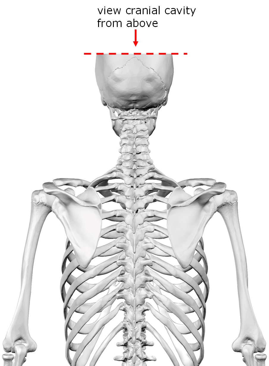
Above: Figure shows the view of the cranial cavity below.
If the skull were sliced in the transverse plane (separating superior and inferior) to expose the inferior aspect of the cranial cavity, where the underside of the brain is supported by the skull, and it was viewed from above (superior view), the skull would appear as the two images below. The base of the cranial cavity is composed of three main fossae (basin-like structures): the anterior cranial fossa, the middle cranial fossa, and the posterior cranial fossa.
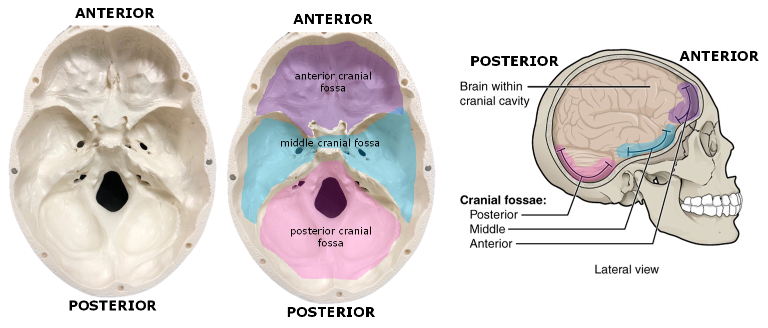
Above: The fossae of the cranial cavity. (Left) Superior view of the cranial cavity of the skull unaltered. (Middle) Superior view of the cranial cavity of the skull colored to show the anterior cranial fossa, the middle cranial fossa, and the posterior cranial fossa. (Right) Lateral view of the skull showing that the anterior cranial fossa is more anterior and is more superior than the other cranial fossae. The middle cranial fossa is inferior to the anterior cranial fossa but more superior to the posterior cranial fossa.
The base of the cranial cavity is created by the frontal bone, ethmoid bone, sphenoid bone, two temporal bones, and occipital bone.
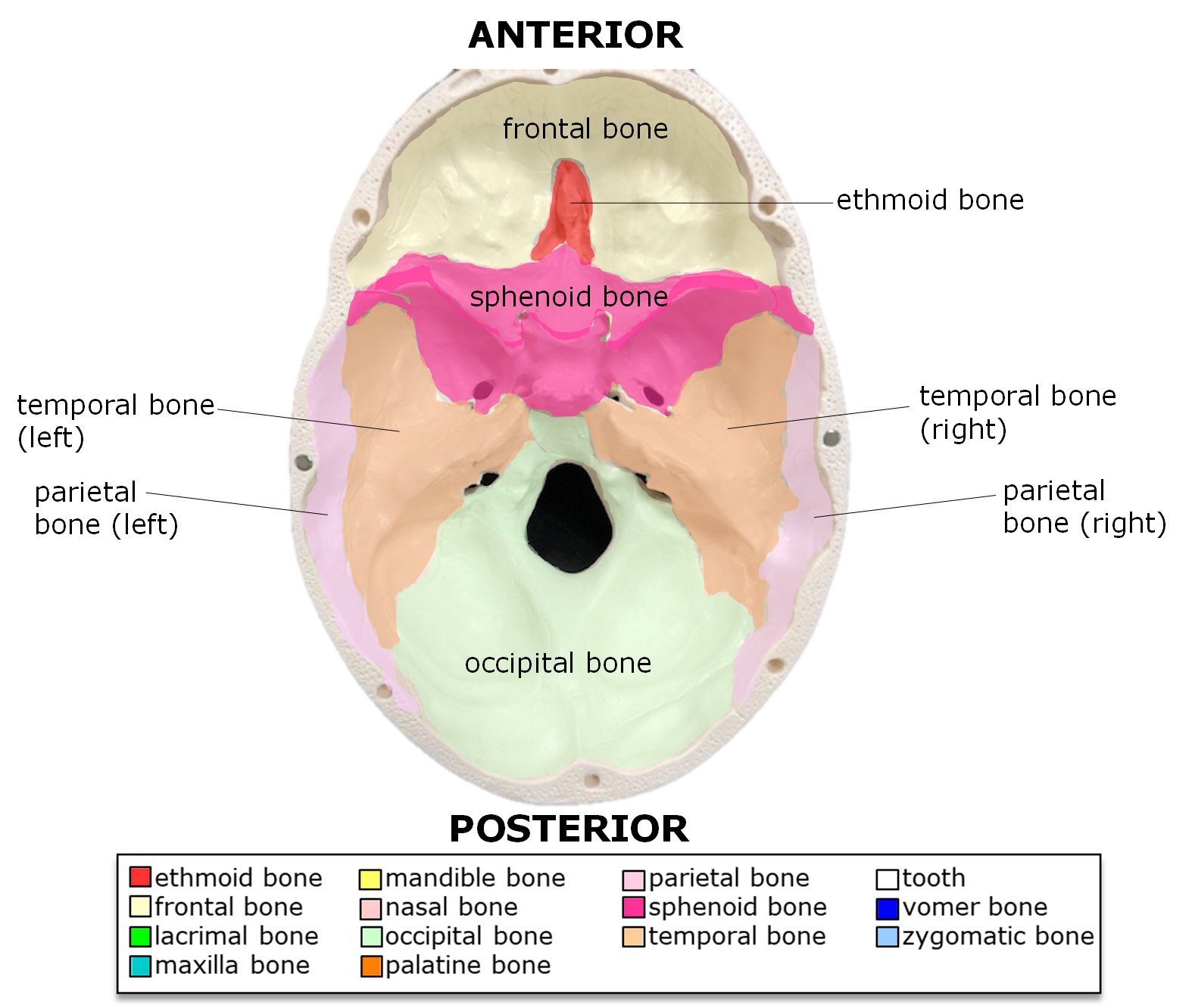
Above: The bones of the cranial cavity.
Attributions (All Skull Sections)
- "Anatomy 204L: Laboratory Manual (Second Edition)" by Ethan Snow, University of North Dakota is licensed under CC BY-NC 4.0
- "Anatomy and Physiology" by J. Gordon Betts et al., OpenStax is licensed under CC BY 4.0
- "Anatomy and Physiology Lab Reference" by Laird C Sheldahl, OpenOregonEducational Resources, Mt. Hood Community College is licensed under CC BY-SA 4.0
- "BIOL 250 Human Anatomy Lab Manual SU 19" by Yancy Aquino, Skyline College is licensed under CC BY-NC-SA 4.0
- "BodyParts3D/Anatomography" by The Database Center for Life Science is licensed under CC BY-SA 2.1
- "Ethmoid.png" by Life Science Databases(LSDB) is licensed under CC BY-SA 2.0
- "Gray151.png" by Henry Vandyke Carter is in the Public Domain
- "Human Skeleton Upper Body Posterior View.jpg" by Andrewmeyerson is licensed under CC BY-SA 3.0
- "Rotation ethmoid.gif" by Life Science Databases(LSDB), animated by was a bee is licensed under CC BY-SA 2.0
- "Sphenoid bone - animation 02.gif" by Database Center for Life Science (DBCLS) is licensed under CC BY-SA 2.1