
8.2.1: Exterior of the Cranium
- Page ID
- 53617

\( \newcommand{\vecs}[1]{\overset { \scriptstyle \rightharpoonup} {\mathbf{#1}} } \)
\( \newcommand{\vecd}[1]{\overset{-\!-\!\rightharpoonup}{\vphantom{a}\smash {#1}}} \)
\( \newcommand{\dsum}{\displaystyle\sum\limits} \)
\( \newcommand{\dint}{\displaystyle\int\limits} \)
\( \newcommand{\dlim}{\displaystyle\lim\limits} \)
\( \newcommand{\id}{\mathrm{id}}\) \( \newcommand{\Span}{\mathrm{span}}\)
( \newcommand{\kernel}{\mathrm{null}\,}\) \( \newcommand{\range}{\mathrm{range}\,}\)
\( \newcommand{\RealPart}{\mathrm{Re}}\) \( \newcommand{\ImaginaryPart}{\mathrm{Im}}\)
\( \newcommand{\Argument}{\mathrm{Arg}}\) \( \newcommand{\norm}[1]{\| #1 \|}\)
\( \newcommand{\inner}[2]{\langle #1, #2 \rangle}\)
\( \newcommand{\Span}{\mathrm{span}}\)
\( \newcommand{\id}{\mathrm{id}}\)
\( \newcommand{\Span}{\mathrm{span}}\)
\( \newcommand{\kernel}{\mathrm{null}\,}\)
\( \newcommand{\range}{\mathrm{range}\,}\)
\( \newcommand{\RealPart}{\mathrm{Re}}\)
\( \newcommand{\ImaginaryPart}{\mathrm{Im}}\)
\( \newcommand{\Argument}{\mathrm{Arg}}\)
\( \newcommand{\norm}[1]{\| #1 \|}\)
\( \newcommand{\inner}[2]{\langle #1, #2 \rangle}\)
\( \newcommand{\Span}{\mathrm{span}}\) \( \newcommand{\AA}{\unicode[.8,0]{x212B}}\)
\( \newcommand{\vectorA}[1]{\vec{#1}} % arrow\)
\( \newcommand{\vectorAt}[1]{\vec{\text{#1}}} % arrow\)
\( \newcommand{\vectorB}[1]{\overset { \scriptstyle \rightharpoonup} {\mathbf{#1}} } \)
\( \newcommand{\vectorC}[1]{\textbf{#1}} \)
\( \newcommand{\vectorD}[1]{\overrightarrow{#1}} \)
\( \newcommand{\vectorDt}[1]{\overrightarrow{\text{#1}}} \)
\( \newcommand{\vectE}[1]{\overset{-\!-\!\rightharpoonup}{\vphantom{a}\smash{\mathbf {#1}}}} \)
\( \newcommand{\vecs}[1]{\overset { \scriptstyle \rightharpoonup} {\mathbf{#1}} } \)
\(\newcommand{\longvect}{\overrightarrow}\)
\( \newcommand{\vecd}[1]{\overset{-\!-\!\rightharpoonup}{\vphantom{a}\smash {#1}}} \)
\(\newcommand{\avec}{\mathbf a}\) \(\newcommand{\bvec}{\mathbf b}\) \(\newcommand{\cvec}{\mathbf c}\) \(\newcommand{\dvec}{\mathbf d}\) \(\newcommand{\dtil}{\widetilde{\mathbf d}}\) \(\newcommand{\evec}{\mathbf e}\) \(\newcommand{\fvec}{\mathbf f}\) \(\newcommand{\nvec}{\mathbf n}\) \(\newcommand{\pvec}{\mathbf p}\) \(\newcommand{\qvec}{\mathbf q}\) \(\newcommand{\svec}{\mathbf s}\) \(\newcommand{\tvec}{\mathbf t}\) \(\newcommand{\uvec}{\mathbf u}\) \(\newcommand{\vvec}{\mathbf v}\) \(\newcommand{\wvec}{\mathbf w}\) \(\newcommand{\xvec}{\mathbf x}\) \(\newcommand{\yvec}{\mathbf y}\) \(\newcommand{\zvec}{\mathbf z}\) \(\newcommand{\rvec}{\mathbf r}\) \(\newcommand{\mvec}{\mathbf m}\) \(\newcommand{\zerovec}{\mathbf 0}\) \(\newcommand{\onevec}{\mathbf 1}\) \(\newcommand{\real}{\mathbb R}\) \(\newcommand{\twovec}[2]{\left[\begin{array}{r}#1 \\ #2 \end{array}\right]}\) \(\newcommand{\ctwovec}[2]{\left[\begin{array}{c}#1 \\ #2 \end{array}\right]}\) \(\newcommand{\threevec}[3]{\left[\begin{array}{r}#1 \\ #2 \\ #3 \end{array}\right]}\) \(\newcommand{\cthreevec}[3]{\left[\begin{array}{c}#1 \\ #2 \\ #3 \end{array}\right]}\) \(\newcommand{\fourvec}[4]{\left[\begin{array}{r}#1 \\ #2 \\ #3 \\ #4 \end{array}\right]}\) \(\newcommand{\cfourvec}[4]{\left[\begin{array}{c}#1 \\ #2 \\ #3 \\ #4 \end{array}\right]}\) \(\newcommand{\fivevec}[5]{\left[\begin{array}{r}#1 \\ #2 \\ #3 \\ #4 \\ #5 \\ \end{array}\right]}\) \(\newcommand{\cfivevec}[5]{\left[\begin{array}{c}#1 \\ #2 \\ #3 \\ #4 \\ #5 \\ \end{array}\right]}\) \(\newcommand{\mattwo}[4]{\left[\begin{array}{rr}#1 \amp #2 \\ #3 \amp #4 \\ \end{array}\right]}\) \(\newcommand{\laspan}[1]{\text{Span}\{#1\}}\) \(\newcommand{\bcal}{\cal B}\) \(\newcommand{\ccal}{\cal C}\) \(\newcommand{\scal}{\cal S}\) \(\newcommand{\wcal}{\cal W}\) \(\newcommand{\ecal}{\cal E}\) \(\newcommand{\coords}[2]{\left\{#1\right\}_{#2}}\) \(\newcommand{\gray}[1]{\color{gray}{#1}}\) \(\newcommand{\lgray}[1]{\color{lightgray}{#1}}\) \(\newcommand{\rank}{\operatorname{rank}}\) \(\newcommand{\row}{\text{Row}}\) \(\newcommand{\col}{\text{Col}}\) \(\renewcommand{\row}{\text{Row}}\) \(\newcommand{\nul}{\text{Nul}}\) \(\newcommand{\var}{\text{Var}}\) \(\newcommand{\corr}{\text{corr}}\) \(\newcommand{\len}[1]{\left|#1\right|}\) \(\newcommand{\bbar}{\overline{\bvec}}\) \(\newcommand{\bhat}{\widehat{\bvec}}\) \(\newcommand{\bperp}{\bvec^\perp}\) \(\newcommand{\xhat}{\widehat{\xvec}}\) \(\newcommand{\vhat}{\widehat{\vvec}}\) \(\newcommand{\uhat}{\widehat{\uvec}}\) \(\newcommand{\what}{\widehat{\wvec}}\) \(\newcommand{\Sighat}{\widehat{\Sigma}}\) \(\newcommand{\lt}{<}\) \(\newcommand{\gt}{>}\) \(\newcommand{\amp}{&}\) \(\definecolor{fillinmathshade}{gray}{0.9}\)Skull
There is only one movable joint in the skull. That is the joint connecting the lower jaw, or mandible, to the rest of the skull. All the other bones in the skull are firmly attached to one another by sutures. Sutures are rigid immovable connections holding bones tightly to one another. Some of the sutures in the skull take a few months-to-years after birth to completely form. The brain is encased in the cranium of the skull. The bones that make up the cranium are called the cranial bones. The remainder of the bones in the skull are the facial bones.
Exterior of the Cranium
The bones of the cranium include the frontal bone (1), parietal bones (2, one right and one left), temporal bones (2, one right and one left), occipital bone (1), sphenoid bone (1), and ethmoid bone (1). These bones enclose and protect the cranial cavity that contains the brain. All of these bones, except for the parietal bones, can also be observed when examining the interior superior view of the cranium (see the Interior of the Cranium section below). Both the sphenoid bone and the ethmoid bone are single, unpaired bones that form deeper parts of the skull and therefore, small portions of these bones can be viewed from the skull exterior (see the Sphenoid Bone and Ethmoid Bone sections below to examine the positions of these bones further).
Bones of the Exterior Cranium
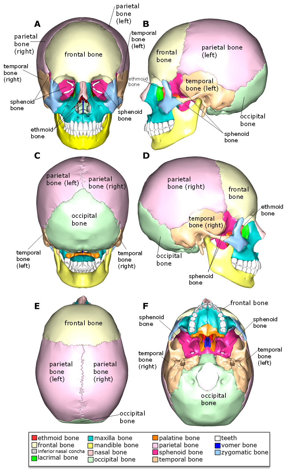
Above: The cranial bones. Figure A is the anterior skull view, figure B is the lateral view of the left side of the skull, figure C is the posterior view of the skull, figure D is the lateral view of the right side of the skull, figure E is the superior view of the skull with the anterior position being at the top of the diagram, and figure F is the inferior view of the skull with mandible removed; the anterior position is at the top of the diagram.
Ethmoid Bone
The ethmoid bone is located deep in the anterior of the skull forming part of the nasal cavity, including the paired superior nasal conchae (right and left) and middle nasal conchae (right and left), bony projections that point medially into the nasal cavities. These conchae form paired canals or passageways (right and left) in the nasal cavities for air passage called nasal meatuses (superior, middle, and inferior nasal meatuses). There are also paired medially-projecting inferior nasal conchae, but these are a part of the maxilla bone, a facial bone. A medial, inferior-pointing projection called the perpendicular plate of the ethmoid bone creates the superior aspect of the septum separating the right and left nasal cavities. The perpendicular plate articulates with part of the vomer bone which form the inferior portion of the nasal septum.
The ethmoid bone also creates separation between the nasal cavity and the cranial cavity. This separation is created by the cribriform plates of the ethmoid bone, two flat regions that house numerous pin-hole foramina called olfactory foramina. At the inferior aspects of the cribriform plates, olfactory epithelia are equipped with olfactory sensory neurons collecting scent information. The olfactory foramina are passageways where axons of these scent-sensing neurons can pass into the cranial cavity and bring olfactory (scent) information to the olfactory bulbs of cranial nerve I (C.N. I olfactory nerve), transmitting scent information to the brain. The two cribriform plates are separated by crista galli, a superior-pointing projection of the ethmoid bone that can be seen inside the cranial cavity. The olfactory bulbs of C.N. I are positioned on either side of crista galli on top of the two cribriform plates.
Small segments of the ethmoid bone also make up medial regions of each of the right and left orbits or eye sockets.
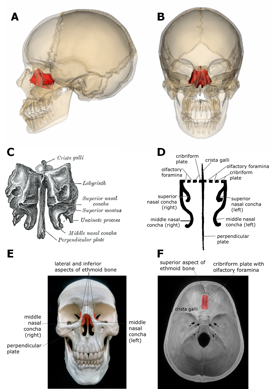

Above: This figure provides images depicting the location of the ethmoid bone and its structures. Image A highlights the ethmoid bone in red with a lateral view. Image B highlights the ethmoid bone in red with an anterior view. Diagram C is an illustration of the ethmoid bone alone with an anterior view. Crista galli, an upward projection of the ethmoid bone, can be observed in the interior of the cranium and the perpendicular plate and middle nasal concha in the nasal cavity. Diagram D shows a simplified version of the ethmoid bone from an anterior view. In this diagram, you can see the cribriform plates, containing olfactory foramina, are located on either side of crista galli. These structures are visible in the cranium interior. Images E and F highlight the ethmoid bone in red and point to important structures of the ethmoid bone. Image E shows the anterior view of the skull and image F shows the superior view of the cranium interior with the anterior of the skull at the top of the image.
Sphenoid Bone
The sphenoid bone is a central bone in the skull located toward the anterior of the skull, but posterior to the ethmoid bone. The sphenoid bone creates some of the exterior of the cranium laterally on both the right and left sides between the temporal, parietal, and frontal bones. Inferiorly, the sphenoid bone is located posterior to the palatine bones (the palatine bone forms the posterior of the roof of the mouth). At this most inferior point of the sphenoid are pterygoid processes where chewing muscles attach.
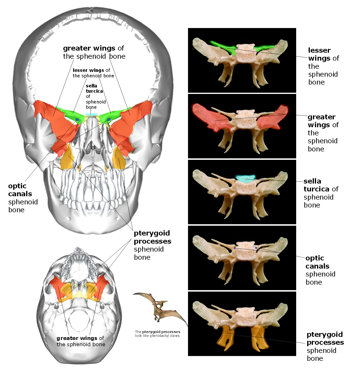
Above: (Left top) Anterior view of the skull with the frontal bone and maxilla bone removed and the sphenoid bone highlighted in yellow. Parts of the sphenoid bone are further colored in green (lesser wings), red (greater wings), blue (optic canals), and orange (pterygoid processes). (Left bottom) Inferior view of the skull with the mandible and bottom teeth removed. The sphenoid bone is highlighted in yellow with the pterygoid processes highlighted orange and the greater wings in red. (Right) The sphenoid bone alone, anterior view. Structures of the sphenoid are highlighted the same colors as the other parts of the figure.
The sphenoid bone also forms the posterior aspect of the orbits (eye sockets) including the right and left pair of superior orbital fissures and the right and left pair of optic canals. These superior orbital fissures allow for the pairs (right and left) of cranial nerves III, IV, part of V, and VI (C.N. III oculomotor, C.N. IV trochlear, C.N. V trigeminal, and C.N. VI abducens) to pass from the brain to the eyes to control movements of the eyes. The optic canals provide a means for the right and left pair of cranial nerve II (C.N. II optic nerve) to pass from the eyes to the cranial cavity carrying sight information to the brain (one through the right optic canal and one through the left optic canal). One side of the optic canals can be see in the posterior aspects of the orbits and the other side of the optic canals can be seen as part of the sphenoid bone in the cranial cavity medial to the lesser wings of the sphenoid.

Above: (Left) Superior view of the interior of the cranium of the skull. (Right) The sphenoid bone alone, superior view. The sphenoid bone is shown highlighted with parts of the bone colored differently to describe its different parts. The lesser wings are colored green, the greater wings are colored red, sella turcica, a saddle-like structure with the hypophyseal fossa at the "seat" of the saddle, is colored light blue, and the optic canals are dark blue.
The sphenoid bone, from the outside, appears to contribute to only a small portion of the cranium, but when the parietal bones are removed and the interior of the cranial cavity (where the brain would be housed) is viewed, you can see the butterfly-like shape of the sphenoid bone makes a large contribution to the floor of the cranial cavity. The sphenoid bone appears to have two sets of wings, one larger, broader, and more inferior (the greater wings), and one smaller, shorter, and more superior (the lesser wings). The lesser wings contribute to the anterior cranial fossa with the frontal bone and the ethmoid bone and the greater wings contribute to the middle cranial fossa with the temporal bone. In the cranial cavity, the sphenoid bone forms a saddle-like structure medial to the greater wings called sella turcica. In the "seat" of the saddle of sella turcica is a basin-like structure called the hypophyseal fossa where the pituitary gland is positioned (note: hypophysis is another term for the pituitary gland).
Sutures of the Cranium
The cranial bones are joined together with sutures, a type of unmoving joint or articulation between bones. Examine the table and figure below listing and showing the cranial sutures.
|
Suture |
Articulating Bones |
|---|---|
|
coronal suture |
Frontal bone and the right/left parietal bones articulate at the coronal suture. This suture occurs in the coronal/frontal plane, hence where its name comes from. |
|
lambdoid suture |
Occipital bone and the right and left parietal bones articulate at the lambdoid suture. The suture shape is similar to the symbol for the uppercase Greek letter lambda (Λ), hence where the "lambd-" of the name comes from. |
|
occipitomastoid suture |
Occipital bone and the right and left temporal bones articulate at the occipitomastoid suture. This suture occurs near the mastoid process of the temporal bone, hence it is called "occipito-" for the occipital bone and "-mastoid" for the mastoid region of the temporal bone. |
|
pterions (2, one right and one left) |
Put your fingers on your temples and you are pointing to the two pterions of your skull. The junction between the parietal bone, temporal bone, frontal bone, and sphenoid bone forms this H-shaped suture configuration called pterion. |
|
sagittal suture |
Right and left parietal bones articulate at the sagittal suture. This suture occurs in the sagittal plane, hence where its name comes from. |
|
sphenofrontal sutures (2, one right and one left) |
Sphenoid bone and frontal bone articulate at these sphenofrontal sutures. The "spheno-" part comes from the sphenoid bone and the "-frontal" part comes from the frontal bone - the two bones articulating at this suture. |
|
sphenoparietal sutures (2, one right and one left) |
Sphenoid bone and one of the parietal bones form each of these paired sutures called sphenoparietal sutures. The "spheno-" part comes from the sphenoid bone and the "-parietal" part comes from the pareital bone - the two bones articulating at this suture. |
|
spheno-squamous suture (2, one right and one left) |
Sphenoid bone and squamous region of the temporal bones articulate at the sphenosquamous sutures. The "spheno-" part comes from the sphenoid bone. "-Squamous" comes from the fact that these regions of the right and left temporal bones are known as "pars squama" regions since they are roughly flat areas. Recall that epithelial tissues with flat cells are called squamous. |
|
squamous sutures (2, one right and one left) |
On the right side, the squamous suture occurs between the right parietal bone and the right temporal bone. On the left side, the squamous suture occurs between the left parietal bone and the left temporal bone. The squamous sutures gets their names because these regions of the right and left temporal bones are known as "pars squama" regions since they are roughly flat areas. Recall that epithelial tissues with flat cells are called squamous. |
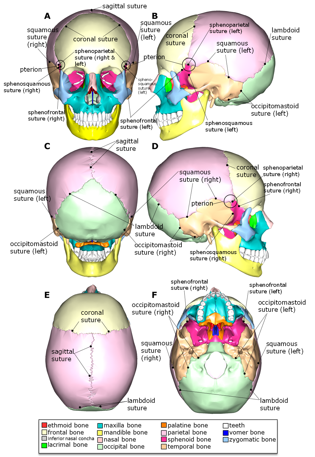
Above: The sutures of the cranial bones. Figure A is the anterior skull view, figure B is the lateral view of the left side of the skull, figure C is the posterior view of the skull, figure D is the lateral view of the right side of the skull, figure E is the superior view of the skull with the anterior position being at the top of the diagram, and figure F is the inferior view of the skull with the mandible removed; the anterior position is at the top of the diagram.
Clinical Application: Fontanelles
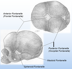
It is common to hear people talking about “soft spots” on the heads of newborn babies. These “soft spots,” otherwise called fontanelles, are areas between skull bones that have not yet fully ossified. The major fontanelles can be found where multiple cranial bones come together. Instead of protective bone, only a membrane and the scalp protect the newborn’s brain at the fontanelles until they ossify. These areas especially must be protected from blunt force until ossification is complete. In the above images, you can see the names fontanelles from a superior view (top right) and a lateral view (bottom left) on a newborn’s skull.
Attributions (All Skull Sections)
- "Anatomy 204L: Laboratory Manual (Second Edition)" by Ethan Snow, University of North Dakota is licensed under CC BY-NC 4.0
- "Anatomy and Physiology" by J. Gordon Betts et al., OpenStax is licensed under CC BY 4.0
- "Anatomy and Physiology Lab Reference" by Laird C Sheldahl, OpenOregonEducational Resources, Mt. Hood Community College is licensed under CC BY-SA 4.0
- "BIOL 250 Human Anatomy Lab Manual SU 19" by Yancy Aquino, Skyline College is licensed under CC BY-NC-SA 4.0
- "BodyParts3D/Anatomography" by The Database Center for Life Science is licensed under CC BY-SA 2.1
- "Ethmoid.png" by Life Science Databases(LSDB) is licensed under CC BY-SA 2.0
- "Gray151.png" by Henry Vandyke Carter is in the Public Domain
- "Human Skeleton Upper Body Posterior View.jpg" by Andrewmeyerson is licensed under CC BY-SA 3.0
- "Rotation ethmoid.gif" by Life Science Databases(LSDB), animated by was a bee is licensed under CC BY-SA 2.0
- "Sphenoid bone - animation 02.gif" by Database Center for Life Science (DBCLS) is licensed under CC BY-SA 2.1
Updated 2025.