
Winter_2024_Bis2A_Facciotti_Reading_07
- Page ID
- 122705
\( \newcommand{\vecs}[1]{\overset { \scriptstyle \rightharpoonup} {\mathbf{#1}} } \)
\( \newcommand{\vecd}[1]{\overset{-\!-\!\rightharpoonup}{\vphantom{a}\smash {#1}}} \)
\( \newcommand{\id}{\mathrm{id}}\) \( \newcommand{\Span}{\mathrm{span}}\)
( \newcommand{\kernel}{\mathrm{null}\,}\) \( \newcommand{\range}{\mathrm{range}\,}\)
\( \newcommand{\RealPart}{\mathrm{Re}}\) \( \newcommand{\ImaginaryPart}{\mathrm{Im}}\)
\( \newcommand{\Argument}{\mathrm{Arg}}\) \( \newcommand{\norm}[1]{\| #1 \|}\)
\( \newcommand{\inner}[2]{\langle #1, #2 \rangle}\)
\( \newcommand{\Span}{\mathrm{span}}\)
\( \newcommand{\id}{\mathrm{id}}\)
\( \newcommand{\Span}{\mathrm{span}}\)
\( \newcommand{\kernel}{\mathrm{null}\,}\)
\( \newcommand{\range}{\mathrm{range}\,}\)
\( \newcommand{\RealPart}{\mathrm{Re}}\)
\( \newcommand{\ImaginaryPart}{\mathrm{Im}}\)
\( \newcommand{\Argument}{\mathrm{Arg}}\)
\( \newcommand{\norm}[1]{\| #1 \|}\)
\( \newcommand{\inner}[2]{\langle #1, #2 \rangle}\)
\( \newcommand{\Span}{\mathrm{span}}\) \( \newcommand{\AA}{\unicode[.8,0]{x212B}}\)
\( \newcommand{\vectorA}[1]{\vec{#1}} % arrow\)
\( \newcommand{\vectorAt}[1]{\vec{\text{#1}}} % arrow\)
\( \newcommand{\vectorB}[1]{\overset { \scriptstyle \rightharpoonup} {\mathbf{#1}} } \)
\( \newcommand{\vectorC}[1]{\textbf{#1}} \)
\( \newcommand{\vectorD}[1]{\overrightarrow{#1}} \)
\( \newcommand{\vectorDt}[1]{\overrightarrow{\text{#1}}} \)
\( \newcommand{\vectE}[1]{\overset{-\!-\!\rightharpoonup}{\vphantom{a}\smash{\mathbf {#1}}}} \)
\( \newcommand{\vecs}[1]{\overset { \scriptstyle \rightharpoonup} {\mathbf{#1}} } \)
\( \newcommand{\vecd}[1]{\overset{-\!-\!\rightharpoonup}{\vphantom{a}\smash {#1}}} \)
Learning Objectives Associated with Winter_2024_Bis2A_Facciotti_Reading_07GC.34 Interpret reaction coordinate diagrams showing either or both catalyzed and uncatalyzed reaction coordinates and identify respective activation energy barriers and relate these to the forward and reverse rates of reaction. GC.36 Describe mechanisms used by enzymes to lower the activation energy and increase rates of reaction. GC.37 Interpret the influence of a regulatory molecule on an enzyme and propose a related functional hypothesis if given kinetic data. MS.18 Hypothesize how binding of small molecules to one or more binding pockets can lead to changes in protein function (i.e. competitive inhibition and/or allostery). MS.19 Describe in general terms the functional link between cofactors, coenzymes, and their associated proteins. MS.17 Draw a rough sketch of an enzyme including its active site and other sites in the enzyme that might impact its function, such as an inhibitor binding site. GC.42 Predict whether two reactions can be theoretically productively coupled by interpreting tables of standard Gibbs enthalpy (energy). GC.50 Describe why an enzyme is critical for coupling an exergonic reaction to an endergonic reaction by applying the concepts from the chemistry and thermodynamics lectures. MS. 14 Form hypotheses about how different types of changes at a molecular interface might influence binding (i.e. proteins-proteins, protein, DNA, protein-lipid, small molecule-lipid, small molecule-protein, etc.).This would include recognizing or inferring different types of chemical bonds from an analysis or knowledge of functional group interactions |
Endergonic and exergonic reactions
Any system of molecules that undergoes a physical transformation/reorganization (aka. reaction) will have an associated change in internal energy and entropy. If we examine a single isolated reaction, in which unique reactants convert into unique products the Gibbs energy of the system will be dependent several factors, key among which are (a) the internal energy and entropy differences associated with the molecular rearrangements and (b) the degree to which the reaction is out-of-equilibrium.
If, for the sake of simplicity we begin by considering only the contribution of the molecular transformations in the system on ∆G, we conclude that reactions with ∆G < 0, the products of the reaction have less Gibbs energy than the reactants. Since ∆G is the difference between the enthalpy and temperature-scaled entropy changes in a reaction, a net negative ∆G can arise in through changes largely of enthalpy, entropy or most often both. The left panel of Figure 1 below shows a common graphical representation of an exergonic reaction. This graph is called a reaction coordinate diagram. It plots Gibbs energy on the y-axis, and the x-axis in arbitrary units shows the progress of a reaction. With an exergonic reaction, the figure on the left shows two key things: (1) the difference between the free energy of the reactants and products is negative and (2) the progress of the reaction requires some input of free energy (shown as an energy "hill" or barrier). This graph does not tell us how the energy in the system redistributed, only that the difference between enthalpy and temperature-scaled entropy is negative. Exergonic reactions are said to occur spontaneously. Understanding which chemical reactions are spontaneous is useful for biologists who are trying to understand whether a reaction is likely to "go" or not.
It is important to note that the term "spontaneous"—in thermodynamics—implies nothing about how fast the reaction proceeds. The change in free energy only describes the difference between beginning and end states, NOT how fast that transition takes place. This is contrary to the everyday use of the term which usually carries the implicit understanding that something happens quickly. As an example, the oxidation/rusting of iron is a spontaneous reaction. However, an iron nail exposed to air does not rust instantly—it may take years.
A chemical reaction with a positive ∆G means that the products of the reaction have a higher free energy than the reactants (see the right panel of Figure 1). These chemical reactions are called endergonic reactions, and they are NOT spontaneous. An endergonic reaction will not take place on its own without transferring energy into the reaction or increase of entropy somewhere else.
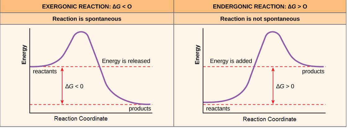
Figure 1. Reaction coordinate diagrams of exergonic and endergonic reactions. Exergonic and endergonic reactions are characterized by changes in Gibbs energy. In the equilibrium state of an exergonic reaction, the Gibbs energy of the products is lower than that of the reactants. Meanwhile, the equilibrium state of an endergonic reaction in, the Gibbs energy of the products is higher than that of the reactants. Attribution: Marc T. Facciotti (own work)
The building of complex molecules, such as sugars, from simpler ones is an anabolic process and is endergonic. On the other hand, the catabolic process, such as the breaking down of sugar into simpler molecules, is exergonic. Like the example of rust above, while the breakdown of biomolecules is generally spontaneous, these reactions don’t occur need to occur instantaneously (quickly). Remember, the terms endergonic and exergonic only refer to the difference in Gibbs energy between the products and reactants; they don't tell you about the rate of the reaction (how fast it happens). Rate will be discussed in later sections.
An important concept in the study of metabolism and energy is that of chemical equilibrium. Most chemical reactions are reversible. They can proceed in both directions, often transferring energy into their environment in one direction and transferring energy in from the environment in the other direction. The same is true for the chemical reactions involved in cell metabolism, such as the breaking down and building up of proteins into and from individual amino acids, respectively. Reactants within a closed system will undergo chemical reactions in both directions until a state of equilibrium is reached. Equilibrium in a chemical reaction is the state in which both reactants and products are present in concentrations that have no further tendency to change with time. Usually, this state results when the forward reaction proceeds at the same rate as the reverse reaction. NOTE THIS LAST STATEMENT! Equilibrium means that the relative concentrations of reactants and products are not changing in time, BUT it does NOT mean that there is no interconversion between substrates and products—it just means that when the reactant(s) are converted to product(s) that product(s) are converted to reactant(s) at an equal rate (see Figure 2). The state of equilibrium is also one of the lowest possible free energy states for the reaction and is a state of maximal entropy.
If a reaction is kept or started far out of equilibrium this state of the system also contributes to the overall Gibbs energy of a reaction. Either a rebalancing of substrate or product concentrations (by adding or removing substrate or product) or a positive change in free energy, typically by the transfer of energy from outside the reaction, can to move a reaction to an out-of-equilibrium state. Note that in a living cell, most chemical reactions do not reach a state of equilibrium—this would require that they reach their lowest free energy state, a state that is almost by definition incompatible with life. Energy is therefore required to keep biological reactions out of their equilibrium state. In this way, living organisms are in a constant, energy-requiring, uphill battle against equilibrium and entropy. This also means that the Gibbs energy of most biological reactions as they occur in the cell must also include a contribution from this out-of-equilibrium state. The Gibbs energy of these reactions, therefore, is often different from that reported under standard conditions.
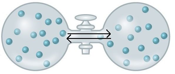
Figure 2. At equilibrium, do not think of a static, unchanging system. Instead, picture molecules moving in equal amounts from one area to another. Here, at equilibrium, molecules are still moving from left to right and right to left. The net movement, however, is equal. There will still be about 15 molecules in each side of this flask once it reaches equilibrium. Source: https://courses.candelalearning.com/...apter/entropy/
Catalysts
For a chemical reaction to happen, the reactants must first find one another in space. Chemicals in solution don't "plan" these collisions; they happen at random. In fact, most times, it's even more complicated. Not only do the reactants need to run into one another, but they also need to come into contact in a specific orientation. If reactants are very dilute, the rate of the reaction will be slow—collisions will happen infrequently. Increasing the concentrations will increase the rate of productive collisions. Another way to change the rate of reaction is to increase the rate of collisions by increasing the rate at which the reactants explore the reaction space—by increasing the velocity of the molecules or their kinetic energy. This can be accomplished by transferring heat into the system or raising the temperature. Those two strategies are often suitable for increasing the rates of chemical reactions that happen in a tube. However, in the cell, transferring heat may not be practical, as it may damage cellular components and lead to death. Cells sometimes use mechanisms to increase concentrations of reactants (we'll see some examples below), but this is rarely enough to drive reaction rates in a biologically relevant regime. This is where catalysts come in.
A catalyst is something that helps increase the rate of a chemical reaction undergoing no change itself. You can think of a catalyst as a chemical change agent.
The most important catalysts in biology are called enzymes. An enzyme is a protein catalyst. Other cellular catalysts include molecules called ribozymes. A ribozyme is a catalyst composed of a ribonucleic acid (RNA). Both will be discussed in more detail later in the course. Like all catalysts, enzymes work by lowering the level of energy that needs to be transferred into a chemical reaction to make it happen. A chemical reaction’s activation energy is the “threshold” level of energy needed to start the reaction.
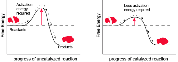
Figure 1. Enzymes and other catalysts decrease the activation energy required to start a given chemical reaction. Without an enzyme (left), the energy input needed for a reaction to begin is high. With the help of an enzyme (right), la reaction needs less energy to begin. Attribution: Marc T. Facciotti (original work)
In the figure above, What do you think the units are on the x-axis? Time would be one guess. However, if you compare the figures, it appears that the products are formed at the same time whether the activation energy barrier is high or low. Wasn't the point of this figure to illustrate that reactions with high activation energy barriers are slower than those with low activation energy barriers? What's going on?
Enzymes Section Overview
Enzymes are biological catalysts that speed up chemical reactions by lowering the activation energy. Enzymes are proteins comprising one or more polypeptide chains. Enzymes have an active site that provides a unique chemical environment made up of certain amino acid R groups (residues). This unique environment is well suited to convert particular chemical reactants for that enzyme, called substrates, into unstable intermediates, called transition states. Enzymes and substrates are thought to bind with an induced fit which means that enzymes and substrates undergo slight conformational adjustments upon substrate contact, leading to binding. Enzymes bind to substrates and catalyze reactions in four different ways: bringing substrates together in an optimal orientation, compromising the bond structures of substrates so that bonds can be more easily broken, providing optimal environmental conditions for a reaction to occur, or taking part directly in their chemical reaction by forming transient covalent bonds with the substrates.
Enzyme action must be regulated so that, in a given cell at a given time, the desired reactions are being catalyzed and the undesired reactions are not. Enzymes are regulated by cellular conditions, such as temperature and pH. They are also regulated through their location within a cell, sometimes being compartmentalized so that they can only catalyze reactions under certain circumstances. Inhibition and activation of enzymes via other molecules are other important ways that enzymes are regulated. Inhibitors can act competitively, noncompetitively, or allosterically; noncompetitive inhibitors are usually allosteric. Activators can also enhance the function of enzymes allosterically. The most common method by which cells regulate the enzymes in metabolic pathways is through feedback inhibition. During feedback inhibition, the products of a metabolic pathway serve as inhibitors (usually allosteric) of one or more of the enzymes (usually the first committed enzyme of the pathway) involved in the pathway that produces them.
Enzymes
A substance that helps a chemical reaction to occur is a catalyst, and the special molecules that catalyze biochemical reactions are called enzymes. Almost all enzymes are proteins, made up of chains of amino acids, and they perform the critical task of lowering the activation energies of chemical reactions inside the cell. Enzymes do this by binding to the reactant molecules and holding them in such a way as to make the chemical bond-breaking and bond-forming processes take place more readily. It is important to remember that enzymes don’t change the ∆G of a reaction. They don’t change whether a reaction is exergonic (spontaneous) or endergonic (not spontaneous). This is because they don’t change the free energy of the reactants or products. They only reduce the activation energy required to reach the transition state.
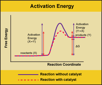
Figure 1. Enzymes lower the activation energy of the reaction but do not change the free energy of the reaction. Here, the solid line in the graph shows the energy required for reactants to turn into products without a catalyst. The dotted line shows the energy required using a catalyst. This figure should say Gibbs Free Energy on the Y-axis and instead of noting ∆H should have ∆G. Attribution: Marc T. Facciotti (own work)
Enzyme active site and substrate specificity
The chemical reactants to which an enzyme binds are the enzyme’s substrates. There may be one or more substrates, depending on the particular chemical reaction. In some reactions, a single-reactant substrate is broken down into multiple products. In others, two substrates may come together to create one larger molecule. Two reactants might also enter a reaction, both become changed, and leave the reaction as two products. The location within the enzyme where the substrate binds is called the enzyme’s active site. The active site is where the “action” happens, so to speak. Since enzymes are proteins, there is a unique combination of amino acid residues (also called side chains, or R groups) within the active site. Each amino acid side chain is characterized by different properties. Amino acids can be classified as large or small, weakly acidic or basic, hydrophilic or hydrophobic, positively or negatively charged, or neutral. The unique combination of amino acids (their positions, sequences, structures, and properties) creates a very specific chemical environment within the active site. This specific environment is suited to bind, albeit briefly, to a specific chemical substrate (or substrates). Due to this jigsaw-puzzle-like match between an enzyme and its substrates (which adapts to find the best fit between the transition state and the active site), enzymes are known for their specificity. The “best fit” between an enzyme and its substrates results from their respective shapes and the chemical complementarity of the functional groups on each binding partner.
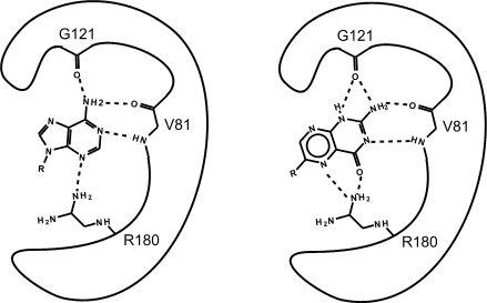
Figure 2. This is an enzyme with two different substrates bound in the active site. The enzymes are represented as blobs, except for the active site, which shows the three R-groups of each of the three amino acids in the active site. These R groups are interacting with the substrates through hydrogen bonding (represented as dashed lines).
At this point in the class, you should be familiar with all the types of bonds as well as the chemical characteristics of all the functional groups. For example, the R group of R180 in the enzyme depicted above is the amino acid Arginine (abbreviated as R) and has an R group that comprises several amino functional groups. Amino functional groups contain a nitrogen (N) and hydrogen (H) atoms. Nitrogen is more electronegative than hydrogen, so the covalent bond between N-H is a polar covalent bond. The hydrogen atoms in this bond will have a positive dipole moment, and the nitrogen atom will have a negative dipole moment. This allows amino groups to form hydrogen bonds with other polar compounds. Likewise, the backbone carbonyl oxygens of valine (V) 81 and glycine (G) 121 the backbone amino hydrogen of V81 are depicted engaged in hydrogen bonds with the small molecule substrate.
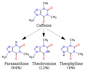
Possible NB Discussion Point: How your body breaks down caffeine
When you drink coffee or other caffeinated beverages like some sodas, you are consuming a molecule called caffeine! Caffeine over time gets metabolized (broken down) via a set of very related "CYP (Cytochrome P450)" enzymes to yield the three products shown in the figure below (Source: Wikipedia). To simplify slightly, you can interpret one arrow to represent a reaction catalyzed by one of the related CYP enzymes to yield paraxanthine, theobromine, or theophylline... all of which themselves get recognized by other enzymes that will further break them down and so on and so forth. Take a moment to examine the four structures below; the general structure should look vaguely familiar to you. Compare the reactant and the three products -- what are the noteworthy functional groups and properties of these molecules? What do you predict to be the key features of the active sites for the enzymes that break down these four molecules? If you were to design an enzyme that would break down caffeine AND theophylline only, how would you design your active site?
Exercise
Look to see which atoms in Figure 2 (above) are involved in the hydrogen bonds between the amino acid R groups and the substrate. You will need to be able to identify these on your own; hydrogen bonds may not be drawn in for you on the test.
If you changed the pH of the solution that this enzyme is located in, would the enzyme still be able to form hydrogen bonds with the substrate?
Which substrate (the left or right one) do you think is more stable in the active site? Why? How?
Figure 3. This is a depiction of an enzyme active site. Only the amino acids in the active site are drawn. The substrate is sitting directly in the center.
Source: created by Marc T. Facciotti (original work)
Exercise
First, identify the type of macromolecule in Figure 3. Second, draw in and label the appropriate interactions between the R groups and the substrate. Explain how these interactions might change if the pH of the solution changed.
Real-Life Connection
A new way of visualizing diseases in the body.
Structural instability of enzymes
The fact that active sites are so well suited to provide specific environmental conditions also means that they are subject to influences by the local environment. It is true that increasing the environmental temperature generally increases reaction rates, enzyme-catalyzed or otherwise. However, increasing or decreasing the temperature outside of an optimal range can affect chemical bonds within the active site in such a way that they are less well suited to bind substrates. High temperatures will eventually cause enzymes, like other biological molecules, to denature, a process that changes the natural properties of a substance. Likewise, the pH of the local environment can also affect enzyme function. Active site amino acid residues have their own acidic or basic properties that are optimal for catalysis. These residues are sensitive to changes in pH that can impair the way substrate molecules bind. Enzymes are suited to function best within a certain pH range, and, as with temperature, extreme pH values (acidic or basic) of the environment can cause enzymes to denature.
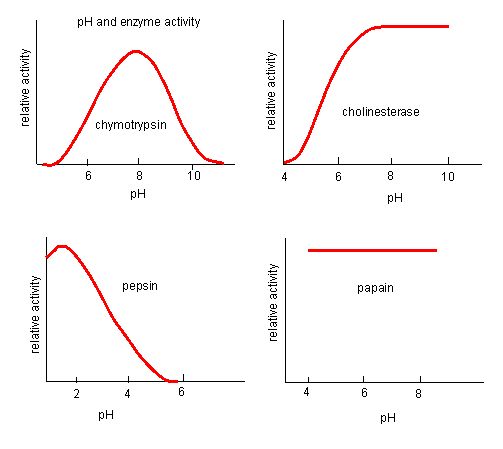
Figure 4. Enzymes have an optimal pH. The pH at which the enzyme is most active will be the pH where the active site R groups are protonated/deprotonated such that the substrate can enter the active site and the initial step in the reaction can begin. Some enzymes require a very low pH (acidic) to be completely active. In the human body, these enzymes are most likely located in the lower stomach, or located in lysosomes (a cellular organelle used to digest large compounds inside the cell).
Source: http://biowiki.ucdavis.edu/Biochemis..._pH_Inhibition
The process where enzymes denature usually starts with the unwinding of the tertiary structure through destabilization of the bonds holding the tertiary structure together. Hydrogen bonds, ionic bonds, and covalent bonds (disulfide bridges and peptide bonds) can all be disrupted by large changes in temperate and pH. Using the chart of enzyme activity and temperature below, make an energy story for the red enzyme. Explain what might be happening from 37 °C to 95 °C.
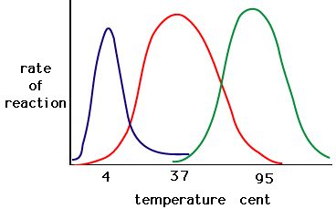
Figure 5. Enzymes have an optimal temperature. The temperature at which the enzyme is most active will usually be the temperature where the structure of the enzyme is stable or uncompromised. Some enzymes require a specific temperature to remain active and not denature. Source: http://academic.brooklyn.cuny.edu/bi...ge/enz_act.htm
Induced fit and enzyme function
For many years, scientists thought that enzyme-substrate binding took place in a simple “lock-and-key” fashion. This model asserted that the enzyme and substrate fit together perfectly in one instantaneous step. However, current research supports a more refined view called induced fit. The induced-fit model expands upon the lock-and-key model by describing a more dynamic interaction between enzyme and substrate. As the enzyme and substrate come together, their interaction causes a mild shift in the enzyme’s structure that confirms a more productive binding arrangement between the enzyme and the transition state of the substrate. This energetically favorable binding maximizes the enzyme’s ability to catalyze its reaction.
When an enzyme binds its substrate, an enzyme-substrate complex is formed. This complex lowers the activation energy of the reaction and promotes its rapid progression in one of many ways. On a basic level, enzymes promote chemical reactions that involve more than one substrate by bringing the substrates together in an optimal orientation. The appropriate region (atoms and bonds) of one molecule is juxtaposed to the appropriate region of the other molecule with which it must react. Another way in which enzymes promote the reaction of their substrates is by creating an energetically favorable environment within the active site for the reaction to occur. Certain chemical reactions might proceed best in a slightly acidic or nonpolar environment. The chemical properties that emerge from the particular arrangement of amino acid residues within an active site create the energetically favorable environment for an enzyme’s specific substrates to react.
The activation energy required for many reactions includes the energy involved in slightly contorting chemical bonds so that they can more easily react. Enzymatic action can aid this process. The enzyme-substrate complex can lower the activation energy by contorting substrate molecules in such a way as to facilitate bond breaking. Finally, enzymes can also lower activation energies by taking part in the chemical reaction itself. The amino acid residues can provide certain ions or chemical groups that actually form covalent bonds with substrate molecules as a necessary step of the reaction process. In these cases, it is important to remember that the enzyme will always return to its original state at the completion of the reaction. One of the hallmark properties of enzymes is that they remain ultimately unchanged by the reactions they catalyze. After an enzyme is done catalyzing a reaction, it releases its product(s).
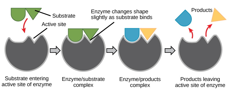
Figure 6. According to the induced-fit model, both enzyme and substrate undergo dynamic conformational changes upon binding. The enzyme contorts the substrate into its transition state, thereby increasing the rate of the reaction.
Creating an energy story for the reaction above
Using Figure 6, answer the questions posed in the energy story.
1. What are the reactants? What are the products?
2. What work was accomplished by the enzyme?
3. What state is the energy in initially? What state is the energy transformed into in the final state? This one might be tricky still, but try to identify where the energy is in the initial state and the final state.
Enzyme regulation
Why regulate enzymes?
Cellular needs and conditions vary from cell to cell and change within individual cells over time. The required enzymes and energetic demands of stomach cells are different from those of fat storage cells, skin cells, blood cells, and nerve cells. Furthermore, a digestive cell works much harder to process and break down nutrients during the time closely following a meal compared with many hours after a meal. As these cellular demands and conditions vary, so do the needed amounts and functionality of different enzymes.
Regulation of enzymes by molecules
Enzymes can be regulated in ways that either promote or reduce their activity. There are many kinds of molecules that inhibit or promote enzyme function, and various mechanisms exist for doing so. In some cases of enzyme inhibition, for example, an inhibitor molecule is similar enough to a substrate that it can bind to the active site and simply block the substrate from binding. When this happens, the enzyme is inhibited through competitive inhibition, because an inhibitor molecule competes with the substrate for active site binding. On the other hand, in noncompetitive inhibition, an inhibitor molecule binds to the enzyme in a location other than an active site and still manages to block substrate binding to the active site.
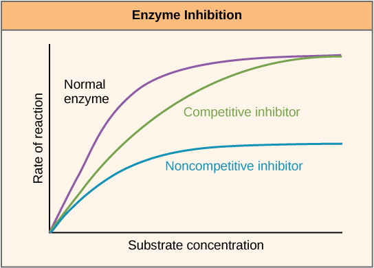
Figure 7. Competitive and noncompetitive inhibition affect the rate of reaction differently. Competitive inhibitors affect the initial rate but do not affect the maximal rate, whereas noncompetitive inhibitors affect the maximal rate.
Some inhibitor molecules bind to enzymes in a location where their binding induces a conformational change that reduces the affinity of the enzyme for its substrate. This type of inhibition is called allosteric inhibition. Most allosterically regulated enzymes are made up of more than one polypeptide, meaning that they have more than one protein subunit. When an allosteric inhibitor binds to an enzyme, all active sites on the protein subunits are changed slightly such that they bind their substrates with less efficiency. There are allosteric activators as well as inhibitors. Allosteric activators bind to locations on an enzyme away from the active site, inducing a conformational change that increases the affinity of the enzyme’s active site(s) for its substrate(s).
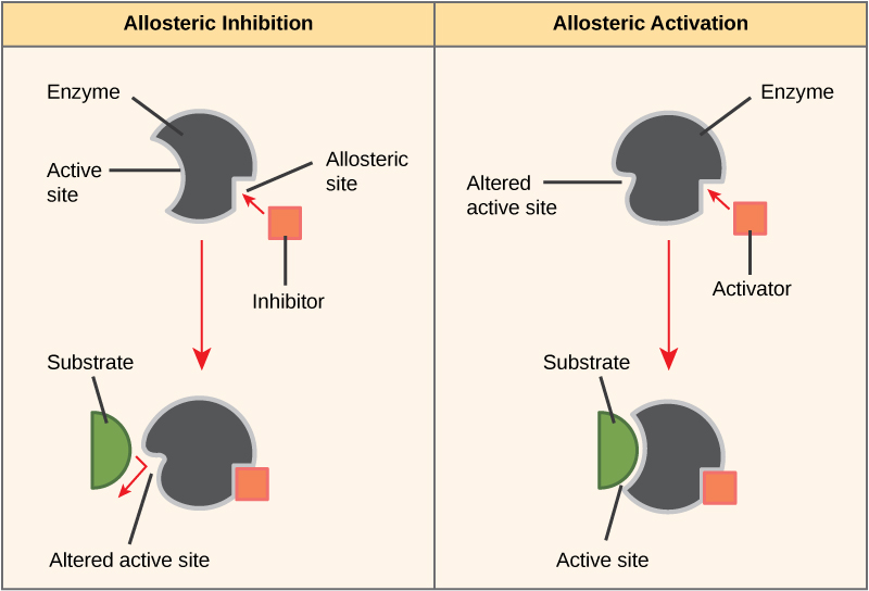
Figure 8. Allosteric inhibitors modify the active site of the enzyme so that substrate binding is reduced or prevented. In contrast, allosteric activators modify the active site of the enzyme so that the affinity for the substrate increases.
Video link
Check out this short (one-minute) video on competitive vs. noncompetitive enzymatic inhibition. Also, take a look at this video (1.2 minutes) on feedback inhibition.
Many enzymes don’t work optimally, or even at all, unless bound to other specific non-protein helper molecules, either temporarily through ionic or hydrogen bonds or permanently through stronger covalent bonds. Two types of helper molecules are cofactors and coenzymes. Binding to these molecules promotes optimal conformation and function for their respective enzymes. Cofactors are inorganic ions such as iron(II) (Fe2+) and magnesium(II) (Mg2+). One example of an enzyme that requires a metal ion as a cofactor is the enzyme that builds DNA molecules, DNA polymerase, which requires a bound zinc(II) ion (Zn2+) to function. Coenzymes are organic helper molecules, with a basic atomic structure made up of carbon and hydrogen, that are required for enzyme action. The most common sources of coenzymes are dietary vitamins. Some vitamins are precursors to coenzymes, and others act directly as coenzymes. Vitamin C is a coenzyme for multiple enzymes that take part in building the important connective tissue component, collagen. An important step in the breakdown of glucose to yield energy is catalysis by a multi-enzyme complex called pyruvate dehydrogenase. Pyruvate dehydrogenase is a complex of several enzymes that actually requires one cofactor (a magnesium ion) and five different organic coenzymes to catalyze its specific chemical reaction. Therefore, enzyme function is, in part, regulated by an abundance of various cofactors and coenzymes, which are supplied primarily by the diets of most organisms.
Enzyme compartmentalization
In eukaryotic cells, molecules such as enzymes are usually compartmentalized into different organelles. This allows for yet another level of regulation of enzyme activity. Enzymes required only for certain cellular processes can be housed separately along with their substrates, allowing for more efficient chemical reactions. Examples of this sort of enzyme regulation based on location and proximity include the enzymes involved in the latter stages of cellular respiration, which take place exclusively in the mitochondria, and the enzymes involved in the digestion of cellular debris and foreign materials, located within lysosomes.
Possible NB Discussion Point: Reversing the Effects of Caffeine
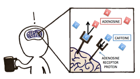
Previously, we discussed caffeine and its metabolism. Let’s now think about caffeine’s pharmacology (mode of action). Were you able to identify, compare, and contrast the molecule that caffeine had a similar structure to? Because of caffeine's structural similarity to the molecule adenosine, it is actually able to bind to the adenosine-specific receptor protein in the brain. However, because the exact lock-and-key-fit is unsatisfied, caffeine will not "activate" the adenosine receptors upon binding as adenosine would. Normally, when adenosine binds to and thereby activates its specific receptor protein in the brain, the physiological effect is increased drowsiness and muscle relaxation. It makes sense that we get tired at night because we accumulate adenosine over the day -- that's a lot of receptor activation! But back to caffeine -- when caffeine is present, it can bind to the adenosine receptor protein, thereby blocking adenosine from binding/activating the receptor. The lack of adenosine action is what leads to suppressed sleepiness and increased alertness. The inhibition seen with this receptor protein and caffeine is similar to some of the inhibition we see with enzymes. What type of inhibition would you classify this as? Follow up question: If you were hired by a company to design a solution to reverse the effect of caffeine post-ingestion, what strategies would you try to test? Explain!
Additional links
Khan Academy
The following links will take you to a series of videos on kinetics. The first link contains four videos on reaction rates, and the second link contains nine videos related to the relationship between reaction rates and concentration. These videos are supplemental and are provided to give you an outside resource to further explore enzyme kinetics.
PRACTICE POST GUIDE
General Practice
3. Why: The reaction coordinate diagram is a type of figure/abstraction commonly used to represent the energetics of a reaction. Not only is it a piece of “common language” in the sciences, once you learn how to use it, it’s a useful construct that can help you interpret a number of “how does this go” questions.
How to practice: Draw a free energy reaction coordinate diagram for both a generic endergonic and a generic exergonic reaction (label each). Make sure to label the x- and y-axes. Now label where reactants and products should go on the figure. Draw something that illustrates the free energy change for each reaction. Now write something like the following next to your figures: “reactants <—> products”.
- What do the reaction coordinate diagrams have to do with the text you just wrote?
- What is represented by the graphs that is not represented in the text?
- Which of the two reaction coordinate diagrams represents a thermodynamically spontaneous reaction?
This exercise is good practice for learning goals: “GC.39 Interpret reaction coordinate diagrams and associate changes in Gibbs enthalpy and activation energy with relative rates of reactions, equilibrium conditions, and whether a reaction is endergonic or exergonic.”
4. Why: More on reaction coordinate diagrams.
How to practice: Return to the diagram you drew in #3 and do the following. Start with the endergonic reaction. Add the term “Keq" (equilibrium constant) above the arrow in the text “reactants <—> products”. This constant is defined as: [products]/[reactants], or the ratio of the product concentration to reactant concentration at equilibrium. Do the same for the exergonic reaction. Now for each reaction write whether Keq is greater than, less than, or equal to one. If you don’t know the answers, look them up, ask TAs, or ask the instructor.
This exercise helps you practice learning objectives: “GC.38 Describe the concept of equilibrium in the context of reaction coordinate diagrams.”
5. Why: reaction coordinate diagrams continued . . .
How to practice: Use your figure from exercise #4 to make predictions about what would happen to both endergonic and exergonic reactions if (a) they were to start at equilibrium and (b) either reactants or products are added to or removed from them. We’ll return to these figures for the next lecture so don’t trash them.
This is an extension of practicing the learning objectives above and making a link to a previous learning objectives: “GC.35 Apply the concept of chemical equilibrium and the equilibrium constant to describe the progress of a chemical reaction, initially out of equilibrium, towards equilibrium, and finally at equilibrium in terms of "forward" and "reverse" reaction rates and concentrations of chemical reactants.” (From lecture 3 - review)
6. Why: We use reaction coordinates and the concepts of equilibrium in our discussion and to describe our understanding of how enzymes help reactions “go”. So here we’re going to integrate what you practiced above with the concepts of activation energies and energetic “barriers” that we use in the discussion of catalysts and enzymes.
How to practice: Using your sketchpad, draw a Gibbs enthalpy (energy) reaction coordinate diagram for an exergonic reaction and include the concept of an activation energy barrier. Label the diagram with ∆G and ∆G‡ (the double dagger notation is sometimes used to indicate the activation energy barrier). Now draw a curve for the same reaction that represents the reaction progress for a catalyzed reaction. What changed? What didn’t? Do the same for an endergonic reaction. Make sure to label reactants and products in both cases.
This is an extension of practicing the learning objectives above and making a link to a previous learning objectives: “GC.34 Interpret reaction coordinate diagrams showing either or both catalyzed and uncatalyzed reaction coordinates and identify respective activation energy barriers and relate these to the forward and reverse rates of reaction.”
7. Why: The likelihood and “strength” of molecular scale interactions and reactions in biology are associated with factors like the balance between energetically favorable and unfavorable bonds and non-covalent interactions and the entropy of a system, “before” and “after” a reaction. The interactions (bonds, etc.) and entropy (ways to spread energy in the system) contribute to ∆G of a reaction. Here I want you to start explicitly relating/thinking about how bonds and entropy contribute to ∆G by looking at a “simple” example - the dissolution of NaCl in H2O. It is intended to help you link the molecular scale happenings in the reaction to the more population-level thermodynamic quantities like ∆G, ∆H, and ∆S. Think about what kinds of bonds are lost and what kinds of new interactions are formed when NaCl dissolves in water. How do those contribute to ∆H? How does the reorganization of molecules contribute to ∆S?
How to practice: In your sketchbook, draw a free energy reaction coordinate diagram for an energy story associated with the dissociation of NaCl (table salt) in water. Recall that this reaction is spontaneous. Be sure to label the axes and the other components of the diagram (reactants, products, etc). Use each figure to tell yourself a convincing energy story for the reaction and make sure that you include the concepts of bonds forming and breaking and a change of entropy in your story.
While I don’t have an explicit learning objective, yet, for the skill practiced here (one will come), this exercise wants you to relate the skills of using reaction coordinate diagrams and the associated thermodynamics concepts together with your understanding of molecular interactions. You’re bringing together various learning objective here and will build the mental picture to help with similar ideas involving the role of enzymes.
8. Why: This practice asks you to extend on the practice you did in exercise #7 by getting you to think about the connection between the molecular events that happen in an enzyme and the associated thermodynamic quantities that describe the reaction and are represented in a reaction coordinate diagram.
How to practice: In class, we often talk about an enzyme lowering the activation energy barrier and we often draw a smooth curve that has a smaller ∆G‡ than that of the uncatalyzed reaction. However, sometimes you will see figures like the at right. Can you think of a reason why the red curve is depicted with so many bumps?
This exercise is practice for the integration of some of the learning objectives practiced above with those that describe concepts of how enzymes work. For instance: “GC.34 Interpret reaction coordinate diagrams showing either or both catalyzed and uncatalyzed reaction coordinates and identify respective activation energy barriers and relate these to the forward and reverse rates of reaction.”; & “GC.36 Describe mechanisms used by enzymes to lower the activation energy and increase rates of reaction.”
9. Why: Enzymes work to increase the rates of reactions and their activity can be modulated. Both of these processes occur because of interactions between molecules that happen at the molecular level. This encourages you to think about the link between what happens (allosteric regulation) and how it happens (the kinds of molecular interactions that might take place).

How to practice: The figure below (“For question 9”) depicts the rate of an enzyme catalyzed reaction with respect to substrate concentration. The black curve in the center is a curve showing enzyme activity when the experiment is carried out with only enzyme and substrate in the tube. The blue and red curves depict the influence of two different molecules on the rates of reactions. What is the influence of the “blue” compound? The “red” compound? What are each of these generic types of compounds called and how are they influencing the rate of reaction? (This can be found in the reading). Use a drawing of an enzyme and relevant binding sites to help explain your interpretation.
This is practice for learning objectives: “MS.18 Hypothesize how binding of small molecules to one or more binding pockets can lead to changes in protein function (i.e. competitive inhibition and/or allostery).” & “MS.17 Draw a rough sketch of an enzyme including its active site and other sites in the enzyme that might impact its function, such as an inhibitor binding site.”
10. Why: The exercises above ask you to think about and picture the molecular basis for interactions between molecules and how this can be extended to understanding/picturing how enzymes work. Here we extend this idea and ask you to imagine “What if we changed some of those interactions? How would that impact function?”
How to practice: Based on what you know about the structure of proteins, the types of bonds that hold them together, the role of amino acids in binding substrate and catalysis, try to enumerate some different ways in which specific changes in amino acid composition could influence the behavior of a protein. I suggest looking back at a table of amino acids and asking something like: “Here’s a valine. What might be a consequence of changing this to a glutamate?” You need to think about what role a valine might play in a structure and figure out how changing that might change the role. How might that role be enhanced or disrupted by a change in amino acids?
This is a revisiting of the following learning objective in the specific context of proteins: “MS. 14 Form hypotheses about how different types of changes at a molecular interface might influence binding (i.e. proteins-proteins, protein, DNA, protein-lipid, small molecule-lipid, small molecule-protein, etc.).This would include recognizing or inferring different types of chemical bonds from an analysis or knowledge of functional group interactions.”
PRACTICE EXAM QUESTIONS
Question Q7.1
Q7.1 Dissolving potassium hydroxide in water is an exothermic process (it gives up energy to the solution and surroundings) and spontaneous at room temperature. Note that exothermic and exergonic are different terms. Meanwhile, dissolving ammonium nitrate is an endothermic process and spontaneous at room temperature (the solution absorbs energy from the environment). Which of the following explains this?
- While ΔH(potassium hydroxide/water) is positive, TΔS(ammonium nitrate/water) is larger.
- While ΔH(ammonium nitrate/water) is positive, TΔS(ammonium nitrate/water) is larger.
- While ΔH(ammonium nitrate/water) is negative, TΔS(ammonium nitrate/water) is larger.
- Keq of the potassium hydroxide reaction is >1 while it is <1 for ammonium nitrate.
- Dissolving ammonium nitrate is slower than dissolving potassium hydroxide.
Question Q7.2
Q7.2 Exergonic reactions:
- are necessarily exothermic.
- are spontaneous reactions.
- create products with greater free energy than the substrates.
- All statements are true.
Question Q7.3
Q7.3 A reaction with ΔH > 0 and ΔS < 0 (and room temperature) will be:
- spontaneous
- favorable at high temperatures
- non-spontaneous
- favorable at cold temperatures
- There is insufficient data to answer the question.
Question Q7.4
Q7.4 The reaction coordinate diagram depicts the free energy profile for a reaction both catalyzed and uncatalyzed. S = substrates, P = products. The bar graphs represent hypothetical equilibrium concentrations for S and P of the same reaction. Which bar chart below best represents the equilibrium conditions of the catalyzed and uncatalyzed reaction?

Question Q7.5
Q7.5 A reaction coordinate diagram for a reaction converting S to P is shown on the right. Which combination of differences in free energy describes forward and reverse rates for the catalyzed reaction, respectively?

- 1 only
- 1 and 3
- 2 and 5
- 3 only
- 3 and 4