
8.3: Introduction to Bacterial Identification using Genotypic methods
- Page ID
- 52312
\( \newcommand{\vecs}[1]{\overset { \scriptstyle \rightharpoonup} {\mathbf{#1}} } \)
\( \newcommand{\vecd}[1]{\overset{-\!-\!\rightharpoonup}{\vphantom{a}\smash {#1}}} \)
\( \newcommand{\id}{\mathrm{id}}\) \( \newcommand{\Span}{\mathrm{span}}\)
( \newcommand{\kernel}{\mathrm{null}\,}\) \( \newcommand{\range}{\mathrm{range}\,}\)
\( \newcommand{\RealPart}{\mathrm{Re}}\) \( \newcommand{\ImaginaryPart}{\mathrm{Im}}\)
\( \newcommand{\Argument}{\mathrm{Arg}}\) \( \newcommand{\norm}[1]{\| #1 \|}\)
\( \newcommand{\inner}[2]{\langle #1, #2 \rangle}\)
\( \newcommand{\Span}{\mathrm{span}}\)
\( \newcommand{\id}{\mathrm{id}}\)
\( \newcommand{\Span}{\mathrm{span}}\)
\( \newcommand{\kernel}{\mathrm{null}\,}\)
\( \newcommand{\range}{\mathrm{range}\,}\)
\( \newcommand{\RealPart}{\mathrm{Re}}\)
\( \newcommand{\ImaginaryPart}{\mathrm{Im}}\)
\( \newcommand{\Argument}{\mathrm{Arg}}\)
\( \newcommand{\norm}[1]{\| #1 \|}\)
\( \newcommand{\inner}[2]{\langle #1, #2 \rangle}\)
\( \newcommand{\Span}{\mathrm{span}}\) \( \newcommand{\AA}{\unicode[.8,0]{x212B}}\)
\( \newcommand{\vectorA}[1]{\vec{#1}} % arrow\)
\( \newcommand{\vectorAt}[1]{\vec{\text{#1}}} % arrow\)
\( \newcommand{\vectorB}[1]{\overset { \scriptstyle \rightharpoonup} {\mathbf{#1}} } \)
\( \newcommand{\vectorC}[1]{\textbf{#1}} \)
\( \newcommand{\vectorD}[1]{\overrightarrow{#1}} \)
\( \newcommand{\vectorDt}[1]{\overrightarrow{\text{#1}}} \)
\( \newcommand{\vectE}[1]{\overset{-\!-\!\rightharpoonup}{\vphantom{a}\smash{\mathbf {#1}}}} \)
\( \newcommand{\vecs}[1]{\overset { \scriptstyle \rightharpoonup} {\mathbf{#1}} } \)
\( \newcommand{\vecd}[1]{\overset{-\!-\!\rightharpoonup}{\vphantom{a}\smash {#1}}} \)
\(\newcommand{\avec}{\mathbf a}\) \(\newcommand{\bvec}{\mathbf b}\) \(\newcommand{\cvec}{\mathbf c}\) \(\newcommand{\dvec}{\mathbf d}\) \(\newcommand{\dtil}{\widetilde{\mathbf d}}\) \(\newcommand{\evec}{\mathbf e}\) \(\newcommand{\fvec}{\mathbf f}\) \(\newcommand{\nvec}{\mathbf n}\) \(\newcommand{\pvec}{\mathbf p}\) \(\newcommand{\qvec}{\mathbf q}\) \(\newcommand{\svec}{\mathbf s}\) \(\newcommand{\tvec}{\mathbf t}\) \(\newcommand{\uvec}{\mathbf u}\) \(\newcommand{\vvec}{\mathbf v}\) \(\newcommand{\wvec}{\mathbf w}\) \(\newcommand{\xvec}{\mathbf x}\) \(\newcommand{\yvec}{\mathbf y}\) \(\newcommand{\zvec}{\mathbf z}\) \(\newcommand{\rvec}{\mathbf r}\) \(\newcommand{\mvec}{\mathbf m}\) \(\newcommand{\zerovec}{\mathbf 0}\) \(\newcommand{\onevec}{\mathbf 1}\) \(\newcommand{\real}{\mathbb R}\) \(\newcommand{\twovec}[2]{\left[\begin{array}{r}#1 \\ #2 \end{array}\right]}\) \(\newcommand{\ctwovec}[2]{\left[\begin{array}{c}#1 \\ #2 \end{array}\right]}\) \(\newcommand{\threevec}[3]{\left[\begin{array}{r}#1 \\ #2 \\ #3 \end{array}\right]}\) \(\newcommand{\cthreevec}[3]{\left[\begin{array}{c}#1 \\ #2 \\ #3 \end{array}\right]}\) \(\newcommand{\fourvec}[4]{\left[\begin{array}{r}#1 \\ #2 \\ #3 \\ #4 \end{array}\right]}\) \(\newcommand{\cfourvec}[4]{\left[\begin{array}{c}#1 \\ #2 \\ #3 \\ #4 \end{array}\right]}\) \(\newcommand{\fivevec}[5]{\left[\begin{array}{r}#1 \\ #2 \\ #3 \\ #4 \\ #5 \\ \end{array}\right]}\) \(\newcommand{\cfivevec}[5]{\left[\begin{array}{c}#1 \\ #2 \\ #3 \\ #4 \\ #5 \\ \end{array}\right]}\) \(\newcommand{\mattwo}[4]{\left[\begin{array}{rr}#1 \amp #2 \\ #3 \amp #4 \\ \end{array}\right]}\) \(\newcommand{\laspan}[1]{\text{Span}\{#1\}}\) \(\newcommand{\bcal}{\cal B}\) \(\newcommand{\ccal}{\cal C}\) \(\newcommand{\scal}{\cal S}\) \(\newcommand{\wcal}{\cal W}\) \(\newcommand{\ecal}{\cal E}\) \(\newcommand{\coords}[2]{\left\{#1\right\}_{#2}}\) \(\newcommand{\gray}[1]{\color{gray}{#1}}\) \(\newcommand{\lgray}[1]{\color{lightgray}{#1}}\) \(\newcommand{\rank}{\operatorname{rank}}\) \(\newcommand{\row}{\text{Row}}\) \(\newcommand{\col}{\text{Col}}\) \(\renewcommand{\row}{\text{Row}}\) \(\newcommand{\nul}{\text{Nul}}\) \(\newcommand{\var}{\text{Var}}\) \(\newcommand{\corr}{\text{corr}}\) \(\newcommand{\len}[1]{\left|#1\right|}\) \(\newcommand{\bbar}{\overline{\bvec}}\) \(\newcommand{\bhat}{\widehat{\bvec}}\) \(\newcommand{\bperp}{\bvec^\perp}\) \(\newcommand{\xhat}{\widehat{\xvec}}\) \(\newcommand{\vhat}{\widehat{\vvec}}\) \(\newcommand{\uhat}{\widehat{\uvec}}\) \(\newcommand{\what}{\widehat{\wvec}}\) \(\newcommand{\Sighat}{\widehat{\Sigma}}\) \(\newcommand{\lt}{<}\) \(\newcommand{\gt}{>}\) \(\newcommand{\amp}{&}\) \(\definecolor{fillinmathshade}{gray}{0.9}\)Learning Outcomes
- Discuss the characterization of microbes based on phenotypic and genotypic methods
- Discuss how PCR is used to identify bacterial species. Describe the process of PCR.
- Explain the theory of PCR, its purpose, and applications
- Discuss how to visualize an agarose gel
- Interpret a DNA gel
Bacterial identification and characterization
Phenotypic methods
Throughout this semester, we have learned about many methods to characterize and identify bacteria. These methods include characterizing cell shape (cellular morphology), identifying Gram status or specialized cellular features through staining, growth requirements (oxygen, pH, temperature, etc), appearance of colonies (colony morphology), and through biochemical reactions (enterotubes, selective and/or differential media types, etc.). All of these are primarily Phenotypic methods of analysis and characterization, that is, the results are a product of the expression of their genes.
-
Cellular morphology: cell shape--through microscopy
-
Staining characteristics: Gram status, cell structures such as flagella, endospores---microscopy
-
Growth characteristics: culturing requirements such as oxygen, osmotic pressure, temperature, colony morphology----culturing techniques
- Biochemical characteristics: biochemical tests such as enterotubes, oxidase, catalase tests, selective/differential media.
Genotypic methods
The last method used for the identification of bacteria is Genetic analysis through the use of nucleic acid probes or other molecular techniques.
The application of molecular techniques for detecting and identifying pathogens is widely used. In particular, in surveillance studies these methods provide reliable epidemiological data for tracing the source of human infections, such as a foodborne illness outbreak. A wide range of molecular techniques (including pulsed field gel electrophoresis, multilocus sequence typing, random amplified polymorphism deoxyribonucleic acid, repetitive extragenic palindromic, deoxyribonucleic acid sequencing, multiplex polymerase chain reaction and many more) have been used for detecting, speciating, typing, classifying and/or characterizing pathogens of great significance to humans.
The advent of the “molecular biology age” has provided a plethora of tools and techniques for the detection, identification, characterization, and typing of bacteria for a range of clinical and research purposes. Previously, the identification and characterization of bacterial species was largely done by phenotypic and biochemical methods (such as through selective/differential media and biochemical tests that we have discussed in the last 2 modules), which relied on preliminary isolation and culture.
While these methods continue to hold place in certain settings, molecular-based techniques have provided unprecedented insights into bacterial identification and typing. To name a few examples, genotypic methods have enabled the identification of a large diversity of previously unknown taxa, the characterization of uncultivable bacteria, and facilitated metagenomics studies on large and diverse bacterial communities. Both clinical and research setting have provided in depth insights into bacterial virulence, pathogenesis, antibiotic resistance, and epidemiological typing, as well as identification of novel, emerging, and re-emerging species. In addition, the widespread use and availability of molecular tools for bacterial genotyping has resulted in high throughput analysis, more sensitive and discriminatory results, and rapid turn-around-times, which are only likely to get better with automated tools and data analysis pipelines.
Most molecular methods for bacterial identification are based on some variation of DNA analysis, either amplification or sequencing based. These methods range from relatively simple DNA amplification-based approaches (PCR, real-time PCR, RAPD-PCR) towards more complex methods based on restriction fragment analysis, targeted gene and whole-genome sequencing, and mass spectrometry. In addition to this, approaches based on unique protein signatures such as matrix-assisted laser desorption/ionization time-of-flight mass spectrometry (MALDI-TOF-MS) and similar variations have also been explored.
Polymerase Chain Reaction
Many of these molecular techniques utilize the polymerase chain reaction or PCR. A technique you have probably heard of in other classes. Polymerase chain reaction (PCR) enables researchers to produce millions of copies of a specific DNA sequence in approximately two hours. This automated process bypasses the need to use bacteria for amplifying DNA. During the course of a bacterial infection, the rapid identification of the causative agent(s) is necessary for the determination of effective treatment options, thus molecular methods such as PCR is a popular option due to its speed. You may already know that for detection of the SARS-CoV2 virus, RT-PCR is widely used.
PCR allows DNA amplification so that specific segments of DNA can be copied numerous times, and then separated and analyzed by gel electrophoresis. The presence of a specific fragment of amplified DNA can be used to either identify an organisms or a particular characteristic such as antibiotic resistance. We often use the 16S rRNA gene for bacterial identification purposes because it is present in all bacteria, it is highly conserved sequences (large regions of nucleotide similarity) which are interspersed with variable regions that are genus- or species-specific. Bacteria can be identified by nucleotide sequence analysis of the 16S rRNA PCR product and comparing it to a database with known sequences.

Figure 1: Schematic PCR primers binding to the 16S rRNA gene for amplification from a bacterial chromosome.
In a PCR reaction, the is a series of steps that occur. Usually the dsDNA is denatured to ssDNA. At 55-58degC, a pair of synthesized oligonucleotide primers anneal to the ssDNA that flank the sequence of interest. At 72degC, a thermostable DNA polymerase will replicate the ssDNA to dsDNA sequences. The cycle repeats itself 20-40x to amplify the DNA.

Figure 2: PCR reaction. Image by Erica Suchman, Colorado State University, Ft. Collins, CO.
The sequence of events in the PCR is as follows. The temperature is raised to 92-98oC, causing the DNA strands to separate. Two primer sequences of approximately 20 nucleotides each are annealed to opposite strands of DNA. (RNA requires an initial reverse transcription step to create a double-stranded cDNA template.) The temperature is raised to the optimum for a polymerase from a thermophylic bacterium; usually Thermus aquaticus (Taq) is used at 72oC. Replication continues from the 3' OH of the primers, producing two copies of the DNA. The temperature is again raised to 92-98oC, causing the DNA strands to separate. Then the temperature is lowered to allow new primers to attach to each of the four strands created in the previous reaction. The temperature used during the annealing of primers must be optimized for each individual primer set. The Taq polymerase fortunately is stable during the DNA melting step and is able to begin a new cycle of synthesis. The process is repeated for 20 to 40 cycles so that additional copies arise exponentially, i.e., in a chain reaction.
Watch Video 1: how to set up a PCR reaction
Watch Video 1: how to set up a PCR reaction. Video by Hands-on DNA. (4:38) URL:https://youtu.be/95qOSslefMM
Visualizing PCR products
After amplification, the PCR product, sometimes called an amplicon, is visualized and analyzed on an agarose gel and is abundant enough to be detected with an ethidium bromide or SYBR safe stain (see image below). The amplicon is compared to known sized molecular markers for production of bands of the correct size.
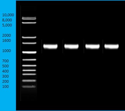
Image 1: Agarose gel electrophoresis of 16S rRNA PCR products. Note the 4 bands at the 1600 bp marker.
The 16S rRNA gene is frequently a target for those who are interested in identifying the genera or species of a bacterium. If you amplify the entire gene (~1500 bp), that is more than sufficient for a sequencing results that will tell you the identity of the bacterium.
Watch video 2: how to run an agarose gel
Watch video 2: how to load and run an agarose gel. Video by Bio-Rad Explorer. (4:06) URL: https://youtu.be/uAttNVEEEwY
Targeting other genes
In some cases, you don’t need to identify the genus or species of the bacterium, but rather some other characteristics, such as antibiotic resistance. In which case, you design your primers to target a specific antibiotic gene. In the gel below, the mecA gene for methicillin resistance was targeted.
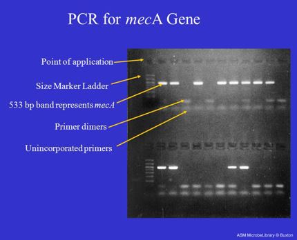
Image 2: Ethidium bromide-stained agarose mini-gel visualizing the products of multiple PCR reactions amplifying a portion of the mecA gene that encodes for methicillin resistance in Staphylococcus aureus ("MRSA"). Distinct 533 base-pair bands are clearly visible in the samples containing the resistance gene. Less distinct bands, indicating smaller primer dimers and unincorporated primers, are also visible . (Labeled view) (Rebecca Buxton, University of Utah, Salt Lake City, UT)
Watch video 3: how to interpret a DNA gel
Watch video 3: how to interpret a DNA gel. Video by Nicole Lantz. (2:30) URL: https://youtu.be/eDmaBtxym30