
8.2: Introduction to Bacterial Identification using Enterotube test
- Page ID
- 52310
Learning Outcomes
- Observe, interpret, and identify bacteria using the Enterotubes/Enteropluri test
Bacterial Identification
The identification of microorganisms is an important part of what many microbiologists do. As you can imagine, clinical microbiologists would need to identify the pathogen that is causing disease in patients. A microbial ecologist is interested in what microbes are contributing to environmental change or they might want to identify new species in the field. Perhaps you are working in a lab and have a contaminant and want to know where it is coming from and what it is. We have already learned some ways to identify microbes, in the last module, you learned about the many biochemical tests available to microbiologists that aid in their identification of bacteria. We’ll continue with some of these biochemical tests, focusing on ones that are capable of combining multiple biochemical tests into one, and the use of selective and differential media to isolate and identify bacteria. Then we’ll move into bacterial genetics and how we can use molecular methods to identify bacteria.
Enteropluri/Enterotubes
A number of techniques can be used for the identification of specific species and subspecies of Enterobacteriaceae. Speciation is important because it provides data regarding patterns of susceptibility to antimicrobial agents and changes that occur over a period of time. It is also essential for epidemiological studies such as determination of nosocomial infections and their spread.
In an effort to simplify the speciation of the Enterobacteriaceae and reduce the amount of prepared media and incubation space needed by the clinical lab, a number of self-contained multi-test systems have been commercially marketed. Some of these multi-test systems have been combined with a computer-prepared manual to provide identification based on the overall probability of occurrence for each of the biochemical reactions. In this way, a large number of biochemical tests can economically be performed in a short period of time, and the results can be accurately interpreted with relative ease and assurance.
The EnteroPluri-Test /Enterotube is a self-contained, compartmented plastic tube containing 12 different agars (enabling the performance of a total of 15 standard biochemical tests) and an enclosed inoculating wire. After inoculation and incubation, the resulting combination of reactions, together with a Computer Coding and Identification System (CCIS), allows for easy identification. The various biochemical reactions of the EnteroPluri-Test and their correct interpretation are discussed below. Although it is designed to identify members of the bacterial family Enterobacteriaceae, it will sometimes also identify common biotypes of Pseudomonas and other non-fermentative Gram-negative bacilli. It does not identify Pseudomonas aeruginosa.
IDENTIFYING MEMBERS OF THE ENTEROBACTERIACEAE WITH THE ENTEROPLURI-TEST
The EnteroPluri-Test contains 12 different agars that can be used to carry out 15 standard biochemical test. Interpret the results of your EnteroPluri-Test is based on a coding chart included with the test.
The enterotube is a self-contained, compartmented plastic tube containing twelve different media that allow determination of 15 biochemical reactions (glucose, gas production from glucose, lysine decarboxylase, ornithine decarboxylase, hydrogen sulfide (H2S), indole, adonitol, lactose, arabinose, sorbitol, Voges-Proskauer (VP), dulcitol, phenylalanine deaminase (PA), urea, and citrate). The enclosed inoculating wire allows inoculation of all compartments in one step from one or a few single colonies of your unknown microorganism. The resulting combination of enterotube reactions, together with other metabolism tests can help you to identify unknown organisms.

Image 1: Four Enterotubes shown. Uninoculated control tube and 3 tubes inoculated with various bacteria. 24 hr incubation.

Image 2: Enterotube chambers
Interpretation of Enterotube chamber Results:
A. Fermentation of Sugars
Among the common products of carbohydrate breakdown by microorganisms using fermentative pathways are organic acids (acetic, lactic, etc.), alcohols, and gases (carbon dioxide and hydrogen). The types of product formed, and the proportion of each, depends on the species of microorganism as well as the particular carbohydrate being fermented.
The ability to ferment different sugar compounds will be tested by inoculating a single enterotube. The 5 carbohydrates tested in 1 enterotube are: glucose, adonitol, lactose, arabinose, and sorbitol. The formation of acids can be readily detected by including a pH indicator in the microbial growth medium. Acid production will lower the pH of the medium, resulting in a color change of the indicator, creosol red, from red (alkaline) to yellow (acidic).
End products of bacterial fermentation of glucose are either acid, or acid and gas. Gas production will be indicated by the definite and complete separation of the wax overlay from the surface of the glucose chamber. The glucose chamber medium is covered with wax to provide anaerobic conditions to allow detection of gas formation. Fermentation of adonitol, lactose, arabinose, and sorbitol will also result in formation of acidic end products indicated by a change in color of indicator present in the medium from red to yellow. Any sign of yellow is interpreted as a positive reaction; red should be considered negative. Some strains will give slightly variable reactions, refer to the manufacturer's chart for more details.
B. Urease
Many bacteria are able to use urea as a nitrogen source by splitting it into ammonia and carbon dioxide through the hydrolysis reaction catalyzed by the enzyme urease:
O
ǁ urease
NH2 — C—NH2 + H20 ------------------ ► 2NH3 + CO2
urea Ammonia Carbon dioxide
The ammonia reacts in solution to form ammonium carbonate, which results in an increase in the pH of the medium. Urease activity is detected by inoculating a medium containing urea and a pH indicator, phenol red (yellow/beige/light amber at acid pH and red-purple at alkaline pH). Initially the urea media chamber is mostly yellow. After incubation, a red-purple color throughout the medium indicates a rapid urea splitter, a positive urease result. No color change, (the agar remains yellow/beige/light amber) is a negative urease result. The urease test is included in the enterotube, see Image 1, chamber 11. Escherichia coli and Enterobacter aerogenes does not normally have the enzyme urease. Proteus vulgaris is a rapid urea splitter, as indicated by the bright purple chamber 11.
C. Lysine and Ornithine Decarboxylation
Decarboxylase tests are useful for differentiating bacteria. Microorganisms that have the enzyme decarboxylase can remove the carboxyl group from an amino acid. Certain bacteria can decarboxylate the amino acid lysine using the enzyme lysine decarboxylase, which results in the formation of the alkaline end product, cadaverine. Some bacteria can decarboxylate ornithine (a product of arginine hydrolysis) using the enzyme ornithine decarboxylase (ODC), which results in the formation of the alkaline end product putrescine. The presence of these decarboxylation byproducts, cadaverine and putrescine, are indicated by a change in the color of bromcresol purple, the pH indicator in the enterotube medium, from pale yellow (acidic) to purple (alkaline). The medium is covered with wax to provide anaerobic conditions. Any degree of purple should be interpreted as a positive reaction. The medium remains yellow, a negative reaction, if decarboxylation of lysine or ornithine does not occur. These tests are included in the enterotube
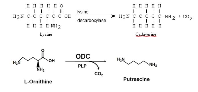
Image 3 Lysine and ornithine decarboxylation reactions. The enzymatic reaction catalyzed by ornithine decarboxylase.The pyridoxal phosphate (PLP)-dependent ODC enzyme catalyzes decarboxylation of ornithine and produces putrescine.
D. Voges-Proskauer
Different bacteria convert dextrose and glucose to pyruvate using different metabolic pathways. Some of these pathways produce unstable acidic products which quickly convert to neutral compounds. Some organisms use the butylene glycol pathway, which produces neutral end products, including acetylmethylcarbinol (acetoin) and 2,3-butanediol. The Voges-Proskauer (VP) test detects organisms that utilize the butylene glycol pathway and produce acetoin during glucose metabolism. After inoculation and incubation of the enterotube, the production of acetoin is detected using Barritt’s reagent (potassium hydroxide, and alpha-naphthol) in the VP chamber. The acetoin product is oxidized in the presence of KOH to diacetyl. The diacetyl then reacts to produce a red color. The presence of acetoin, a positive result, is indicated by the development of a red color within 20 minutes, a negative reaction will appear colorless or light amber. The use of Barritt’s reagent in the VP chamber will be performed after all the results are read for the other chambers in the enterotube.
E. Citrate Utilization
This test detects those organisms which are capable of utilizing citrate, in the form of its sodium salt, as the sole source of carbon. Organisms capable of utilizing citrate produce alkaline metabolites which change the color of the indicator from green (acidic) to deep blue (alkaline). Any degree of blue should be considered positive. Certain microorganisms will not always produce the ideal “strong” positive color change. Lighter shades of the same basic color should be considered positive here. The citrate utilization is tested in the enterotube.
How to Inoculate & Interpret an Enterotube
Watch video 1: how to inoculate an Enterotube with your bacterial sample
Watch Video 1: how to inoculate an enterotube performed in Microbiology labs at NC State. (5:08) URL: https://youtu.be/3pmaDdZPLJg
Watch Video 2: how to interpret Enterotube results
Watch Video 2: how to interpret Enterotube results. Video by Professor B (5:23). URL: https://youtu.be/CwxvUq4lTZ4