
18.4: Microfungi
- Page ID
- 123998

\( \newcommand{\vecs}[1]{\overset { \scriptstyle \rightharpoonup} {\mathbf{#1}} } \)
\( \newcommand{\vecd}[1]{\overset{-\!-\!\rightharpoonup}{\vphantom{a}\smash {#1}}} \)
\( \newcommand{\dsum}{\displaystyle\sum\limits} \)
\( \newcommand{\dint}{\displaystyle\int\limits} \)
\( \newcommand{\dlim}{\displaystyle\lim\limits} \)
\( \newcommand{\id}{\mathrm{id}}\) \( \newcommand{\Span}{\mathrm{span}}\)
( \newcommand{\kernel}{\mathrm{null}\,}\) \( \newcommand{\range}{\mathrm{range}\,}\)
\( \newcommand{\RealPart}{\mathrm{Re}}\) \( \newcommand{\ImaginaryPart}{\mathrm{Im}}\)
\( \newcommand{\Argument}{\mathrm{Arg}}\) \( \newcommand{\norm}[1]{\| #1 \|}\)
\( \newcommand{\inner}[2]{\langle #1, #2 \rangle}\)
\( \newcommand{\Span}{\mathrm{span}}\)
\( \newcommand{\id}{\mathrm{id}}\)
\( \newcommand{\Span}{\mathrm{span}}\)
\( \newcommand{\kernel}{\mathrm{null}\,}\)
\( \newcommand{\range}{\mathrm{range}\,}\)
\( \newcommand{\RealPart}{\mathrm{Re}}\)
\( \newcommand{\ImaginaryPart}{\mathrm{Im}}\)
\( \newcommand{\Argument}{\mathrm{Arg}}\)
\( \newcommand{\norm}[1]{\| #1 \|}\)
\( \newcommand{\inner}[2]{\langle #1, #2 \rangle}\)
\( \newcommand{\Span}{\mathrm{span}}\) \( \newcommand{\AA}{\unicode[.8,0]{x212B}}\)
\( \newcommand{\vectorA}[1]{\vec{#1}} % arrow\)
\( \newcommand{\vectorAt}[1]{\vec{\text{#1}}} % arrow\)
\( \newcommand{\vectorB}[1]{\overset { \scriptstyle \rightharpoonup} {\mathbf{#1}} } \)
\( \newcommand{\vectorC}[1]{\textbf{#1}} \)
\( \newcommand{\vectorD}[1]{\overrightarrow{#1}} \)
\( \newcommand{\vectorDt}[1]{\overrightarrow{\text{#1}}} \)
\( \newcommand{\vectE}[1]{\overset{-\!-\!\rightharpoonup}{\vphantom{a}\smash{\mathbf {#1}}}} \)
\( \newcommand{\vecs}[1]{\overset { \scriptstyle \rightharpoonup} {\mathbf{#1}} } \)
\(\newcommand{\longvect}{\overrightarrow}\)
\( \newcommand{\vecd}[1]{\overset{-\!-\!\rightharpoonup}{\vphantom{a}\smash {#1}}} \)
\(\newcommand{\avec}{\mathbf a}\) \(\newcommand{\bvec}{\mathbf b}\) \(\newcommand{\cvec}{\mathbf c}\) \(\newcommand{\dvec}{\mathbf d}\) \(\newcommand{\dtil}{\widetilde{\mathbf d}}\) \(\newcommand{\evec}{\mathbf e}\) \(\newcommand{\fvec}{\mathbf f}\) \(\newcommand{\nvec}{\mathbf n}\) \(\newcommand{\pvec}{\mathbf p}\) \(\newcommand{\qvec}{\mathbf q}\) \(\newcommand{\svec}{\mathbf s}\) \(\newcommand{\tvec}{\mathbf t}\) \(\newcommand{\uvec}{\mathbf u}\) \(\newcommand{\vvec}{\mathbf v}\) \(\newcommand{\wvec}{\mathbf w}\) \(\newcommand{\xvec}{\mathbf x}\) \(\newcommand{\yvec}{\mathbf y}\) \(\newcommand{\zvec}{\mathbf z}\) \(\newcommand{\rvec}{\mathbf r}\) \(\newcommand{\mvec}{\mathbf m}\) \(\newcommand{\zerovec}{\mathbf 0}\) \(\newcommand{\onevec}{\mathbf 1}\) \(\newcommand{\real}{\mathbb R}\) \(\newcommand{\twovec}[2]{\left[\begin{array}{r}#1 \\ #2 \end{array}\right]}\) \(\newcommand{\ctwovec}[2]{\left[\begin{array}{c}#1 \\ #2 \end{array}\right]}\) \(\newcommand{\threevec}[3]{\left[\begin{array}{r}#1 \\ #2 \\ #3 \end{array}\right]}\) \(\newcommand{\cthreevec}[3]{\left[\begin{array}{c}#1 \\ #2 \\ #3 \end{array}\right]}\) \(\newcommand{\fourvec}[4]{\left[\begin{array}{r}#1 \\ #2 \\ #3 \\ #4 \end{array}\right]}\) \(\newcommand{\cfourvec}[4]{\left[\begin{array}{c}#1 \\ #2 \\ #3 \\ #4 \end{array}\right]}\) \(\newcommand{\fivevec}[5]{\left[\begin{array}{r}#1 \\ #2 \\ #3 \\ #4 \\ #5 \\ \end{array}\right]}\) \(\newcommand{\cfivevec}[5]{\left[\begin{array}{c}#1 \\ #2 \\ #3 \\ #4 \\ #5 \\ \end{array}\right]}\) \(\newcommand{\mattwo}[4]{\left[\begin{array}{rr}#1 \amp #2 \\ #3 \amp #4 \\ \end{array}\right]}\) \(\newcommand{\laspan}[1]{\text{Span}\{#1\}}\) \(\newcommand{\bcal}{\cal B}\) \(\newcommand{\ccal}{\cal C}\) \(\newcommand{\scal}{\cal S}\) \(\newcommand{\wcal}{\cal W}\) \(\newcommand{\ecal}{\cal E}\) \(\newcommand{\coords}[2]{\left\{#1\right\}_{#2}}\) \(\newcommand{\gray}[1]{\color{gray}{#1}}\) \(\newcommand{\lgray}[1]{\color{lightgray}{#1}}\) \(\newcommand{\rank}{\operatorname{rank}}\) \(\newcommand{\row}{\text{Row}}\) \(\newcommand{\col}{\text{Col}}\) \(\renewcommand{\row}{\text{Row}}\) \(\newcommand{\nul}{\text{Nul}}\) \(\newcommand{\var}{\text{Var}}\) \(\newcommand{\corr}{\text{corr}}\) \(\newcommand{\len}[1]{\left|#1\right|}\) \(\newcommand{\bbar}{\overline{\bvec}}\) \(\newcommand{\bhat}{\widehat{\bvec}}\) \(\newcommand{\bperp}{\bvec^\perp}\) \(\newcommand{\xhat}{\widehat{\xvec}}\) \(\newcommand{\vhat}{\widehat{\vvec}}\) \(\newcommand{\uhat}{\widehat{\uvec}}\) \(\newcommand{\what}{\widehat{\wvec}}\) \(\newcommand{\Sighat}{\widehat{\Sigma}}\) \(\newcommand{\lt}{<}\) \(\newcommand{\gt}{>}\) \(\newcommand{\amp}{&}\) \(\definecolor{fillinmathshade}{gray}{0.9}\)- Use characteristics to distinguish between groups of microfungi.
- Describe some of the roles microfungi have in ecosystems.
- Explain the relationship between plants and members of Glomeromycota.
- Identify structures in the "zygomycete" life cycle and know their ploidy.
Chytrids
This small group (~1000 species) is thought to be the most primitive of the fungi. Once all classified as Chytridiomycota, these early, aquatic fungi are now grouped into Blastocladiomycota, Chytridiomycota, and Neocallimastigomycota. Unlike all the other fungi, its members produce flagellated gametes (for sexual reproduction) and flagellated zoospores (for asexual reproduction). They are mostly aquatic.
The evolutionary record shows that the first recognizable chytrids appeared during the late pre-Cambrian period, more than 500 million years ago. Like all fungi, chytrids have chitin in their cell walls, but one group of chytrids has both cellulose and chitin in the cell wall. Most chytrids are unicellular; a few form multicellular thalli with filamentous hyphae, which have no septa between cells (coenocytic). They produce gametes and diploid zoospores that swim with the help of a single flagellum.
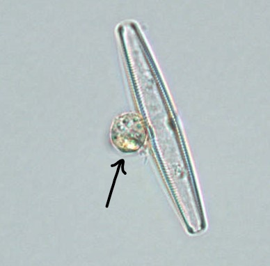
Chytrids usually live in aquatic environments, although some species live on land or as mutualists in the rumens of ungulates. Some species thrive as parasites on plants, insects, or amphibians (Figure \(\PageIndex{3}\)), while others are saprotrophs. The chytrid genus Allomyces (Figure \(\PageIndex{2}\)) is well characterized as an experimental organism; fungal sex horomones (specifically sirenin) were first discovered in this group. Its reproductive cycle includes both asexual and sexual phases. Allomyces produces diploid or haploid flagellated zoospores in a sporangium.
.jpeg?revision=1&size=bestfit&width=462&height=391)
.jpeg?revision=1&size=bestfit&width=363&height=388)
Parasitic Chytrids
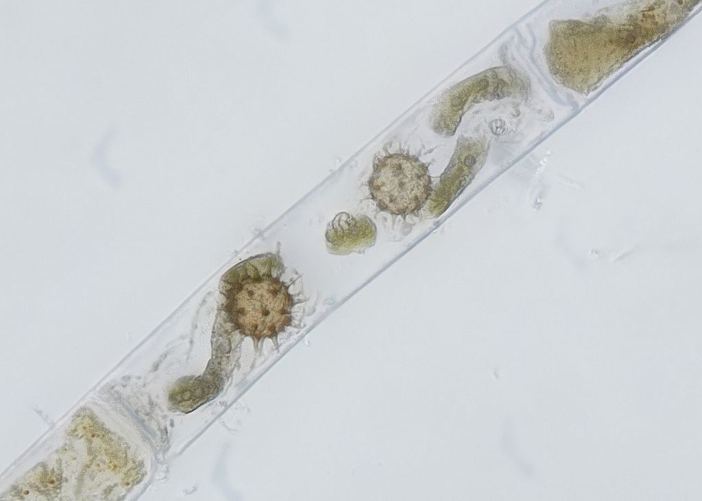
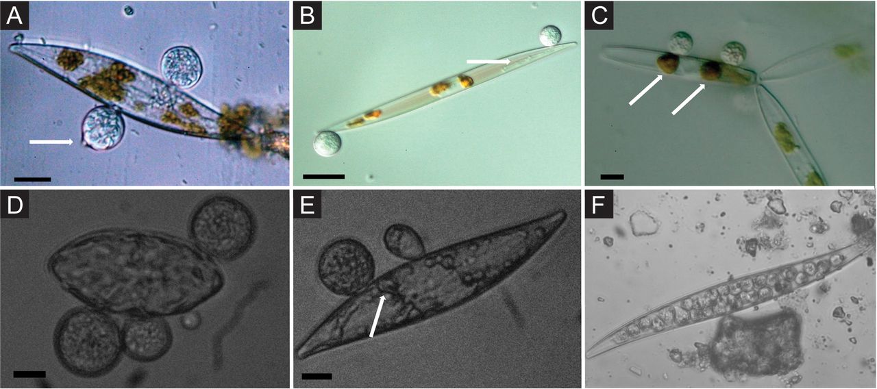
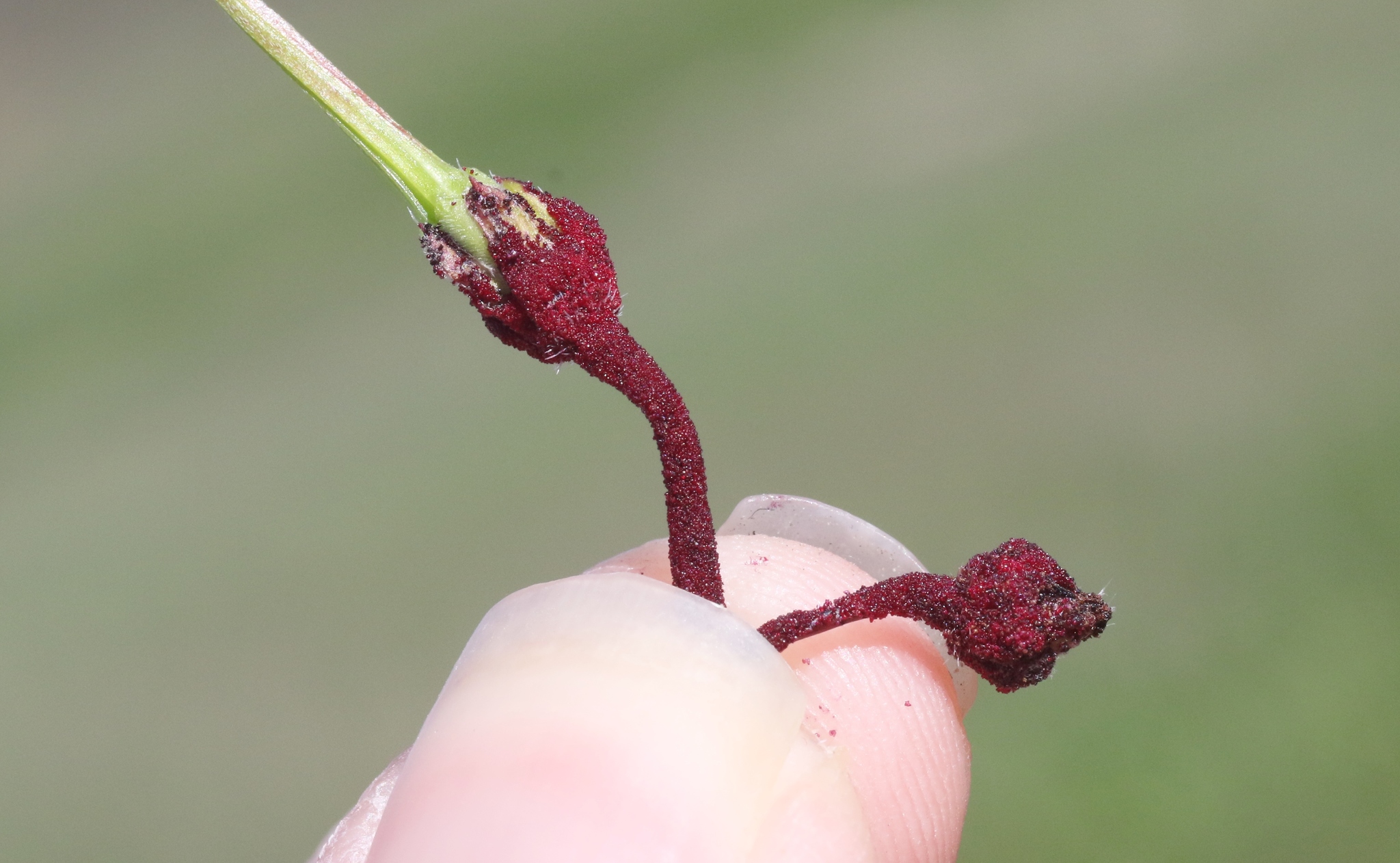
Many chytrids are parasites, perhaps due to their need for an aqueous environment. Some chytrid species are parasites of other chytrids (Rozella allomycis is a parasite that lives within Allomyces), some are parasites of algal species (Figure \(\PageIndex{3}\) and Figure \(\PageIndex{4}\)), some are parasites of plants (Figure \(\PageIndex{5}\), while still others are parasites of animals (Figure \(\PageIndex{6}\). The most infamous chytrid species is a parasite on amphibians, discussed in the following section.
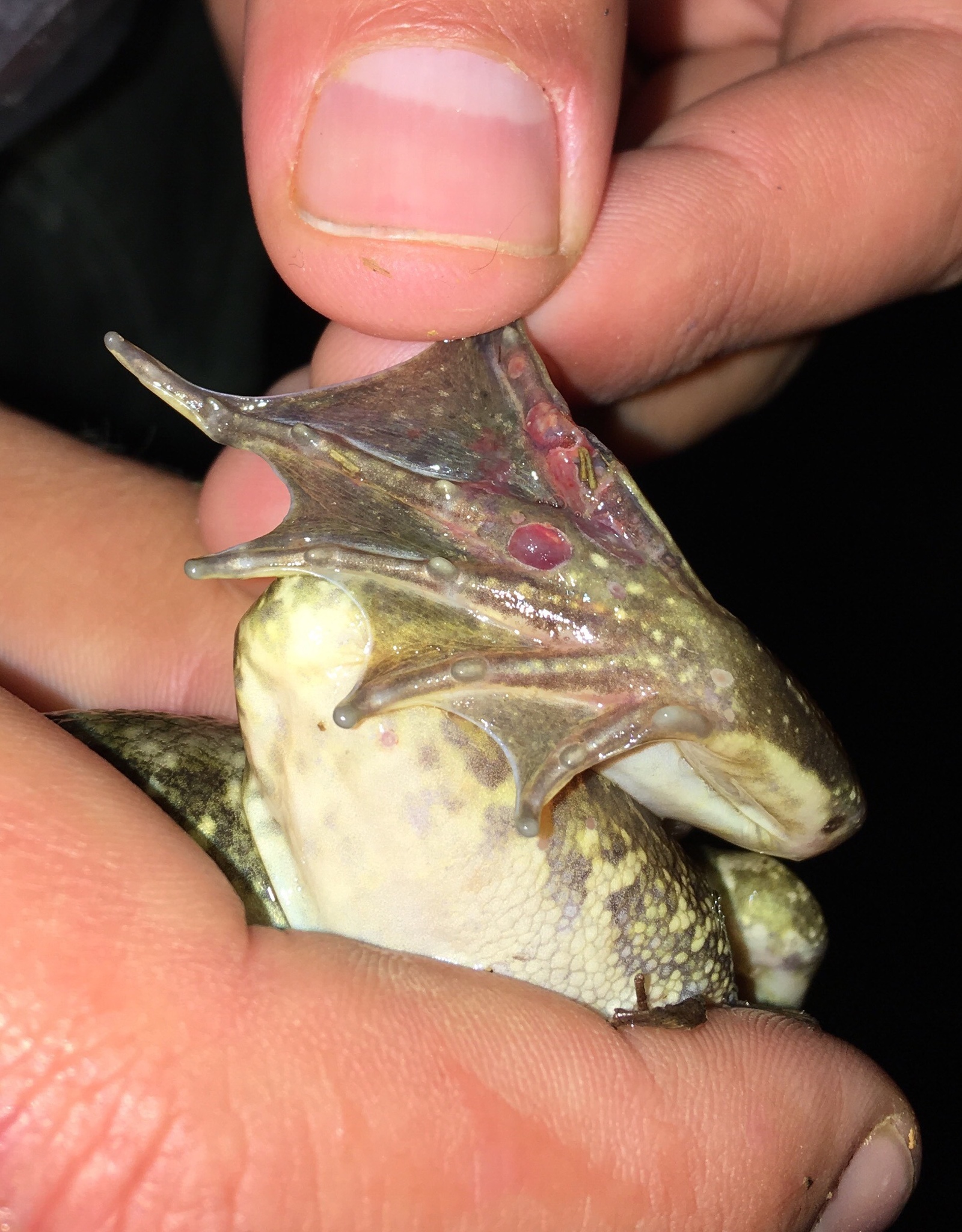
Bd (Batrachochytrium dendrobatidis)
Batrachochytrium dendrobatidis, commonly called Bd, is a chytrid fungus that infects the skin of amphibians. The infection causes hardening of the permeable skin, making it difficult for the animal to breathe. Swimming spores are produced in lesions on the skin and are easily spread through aquatic environments. Amphibians, who must spend part of their life cycle in water, are highly susceptible to this parasite, particularly as other stressors like climate change and pollution lower their resistance. Bd is contributing to a worldwide decline in many amphibian species (though there are numerous other contributing factors), particularly frogs.
Neocallimastix
Neocallimastix is an anaerobic chytrid that lives in the rumen of some ungulates. Cows and other grazing animals can't actually break down the cellulose in the plants they eat. Instead, this plant material first travels to a pre-stomach called a rumen. A community of anaerobic organisms breaks down the cellulose, producing methane. When the pre-digested plant matter has been transformed into compounds that the cow can break down, it then moves to the stomach of the cow. The methane released by cows is due to this anaerobic activity in the rumen.
Zygospore-forming Fungi
Characteristics
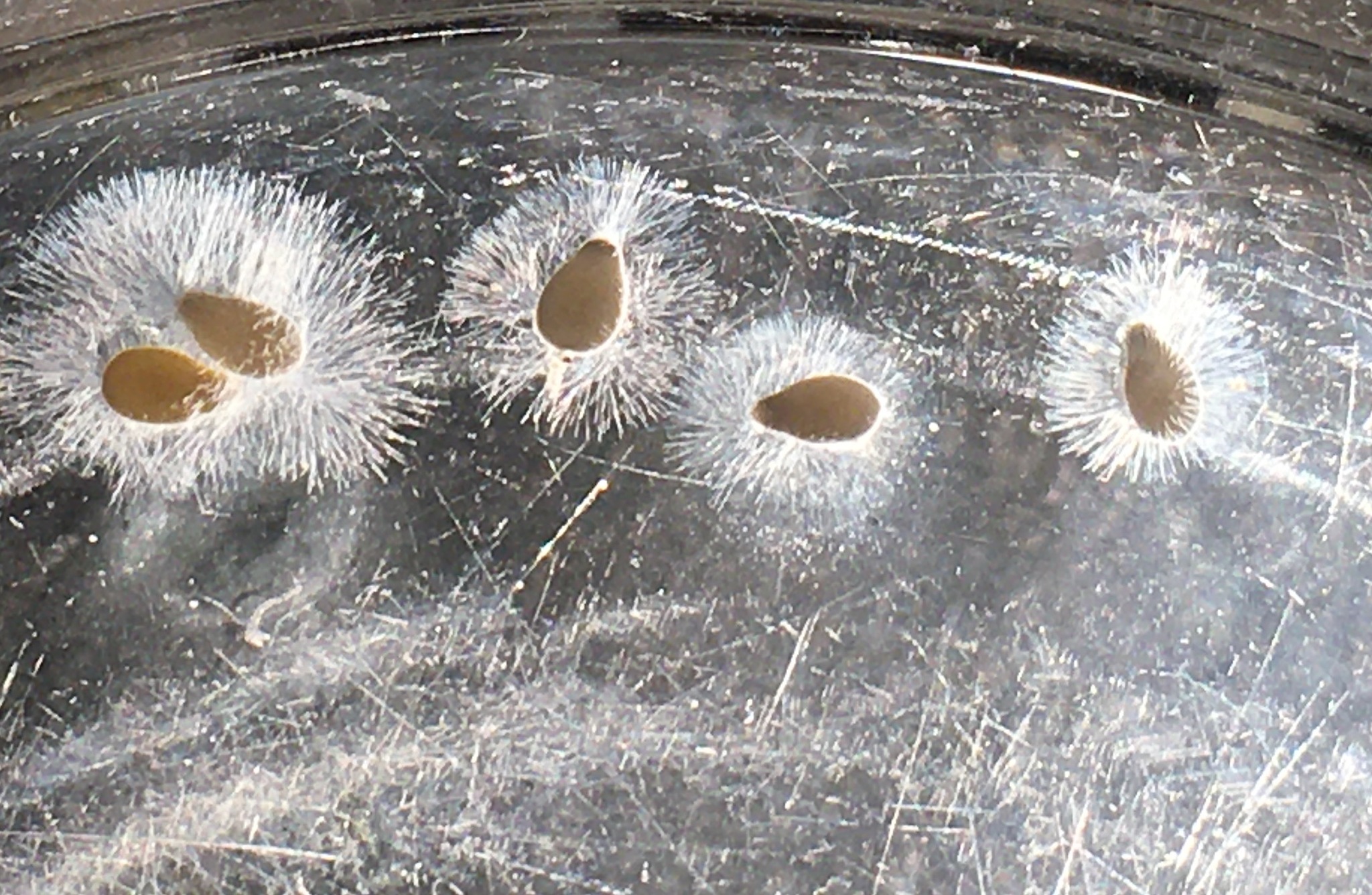
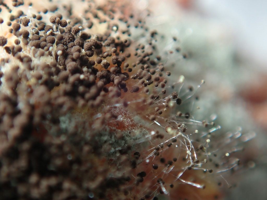
Fungi in the former Zygomycota were recently split into several lineages because they are a paraphyletic group. These fungi produce a large sexual structure called a zygospore and so are called "zygomycetes". They are mainly saprotrophs with coenocytic hyphae and haploid nuclei. They produce haploid sporangiospores by mitosis during asexual reproduction. The group name comes from the zygospores that they use for sexual reproduction (Figure \(\PageIndex{8}\)), which have hard walls formed from the fusion of reproductive cells from two individuals. Zygomycetes are important for food science and as crop pathogens. One example is Rhizopus stolonifer (Figure \(\PageIndex{8}\)), an important bread mold that also causes rice seedling blight. Mucor is a genus of fungi that can potentially cause necrotizing infections in humans, although most species are intolerant of temperatures found in mammalian bodies.
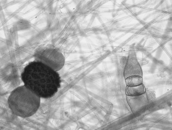
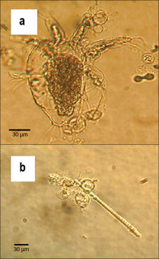
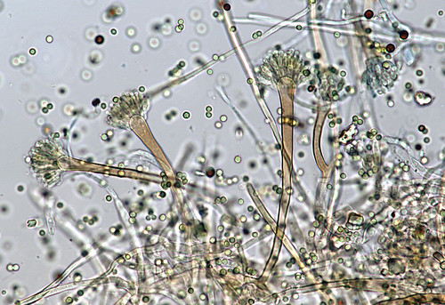
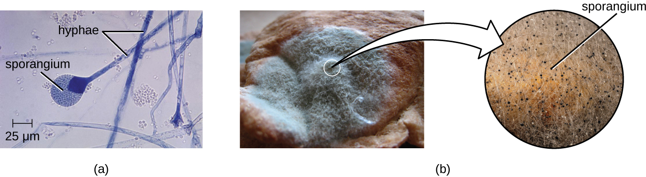 Figure \(\PageIndex{9}\): These images show asexually produced spores. (a) This brightfield micrograph shows the release of spores from a sporangium at the end of a hypha called a sporangiophore. The organism is a Mucor sp. fungus, a mold often found indoors. (b) Sporangia grow at the ends of stalks, which appear as the white fuzz seen on this bread mold, Rhizopus stolonifer. The tips of bread mold are the dark, spore-containing sporangia. (credit a: modification of work by Centers for Disease Control and Prevention; credit b right: modification of work by “Andrew”/Flickr)
Life Cycle
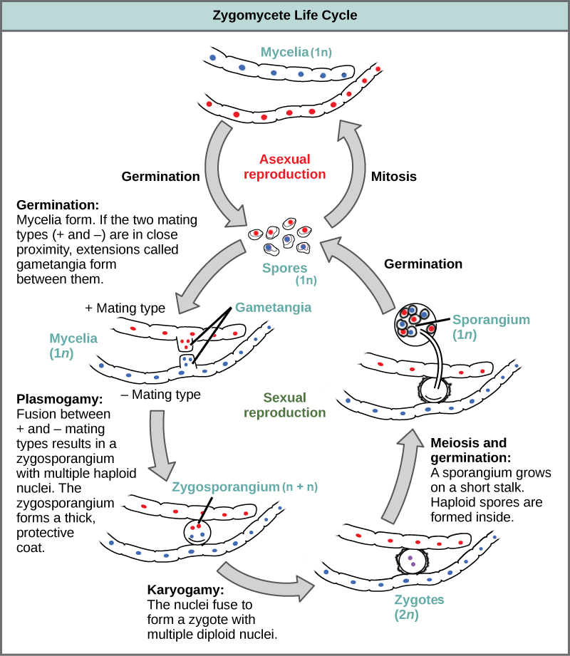
Zygomycetes have a thallus of coenocytic hyphae in which the nuclei are haploid when the organism is in the vegetative stage. Asexual reproduction occurs through mitosporangia that produce sporangiospores by mitosis. These can germinate and develop into a new haploid mycelium that is genetically identical to the parent. The black tips of bread mold are the swollen sporangia packed with black spores (Figure \(\PageIndex{8}\)). Sexual reproduction starts when conditions become unfavorable. Two different mating strains (type + and type –) must be in close proximity for gametangia from the hyphae to be produced and fuse (plasmogamy), forming a multinucleate zygosporangium. Many sets of haploid nuclei fuse (karyogamy) to form diploid nuclei. The developing diploid zygospores have thick coats that protect them from desiccation and other hazards. They may remain dormant until environmental conditions are favorable. When the zygospore germinates, it undergoes meiosis and produces haploid spores from a sporangium. To differentiate between the sporangia produced in sexual and asexual reproduction, you must trace them back to the base. Did it emerge from a zygospore or from a hypha? Both types of spores are haploid and can germinate to form a new haploid mycelium. Spores that germinate from the sporangium of the zygospore will be genetically distinct from the parent mycelia.
Glomeromycota
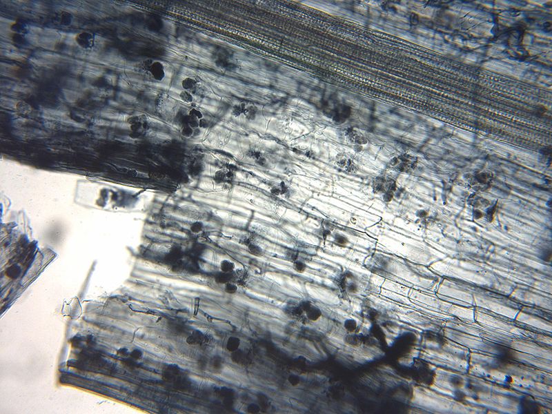
Glomeromycota represent the arbuscular endomycorrhizal fungi. These fungi form a mutualistic relationship with plant roots, forming hyphal structures within the plant cell walls (Figure \(\PageIndex{12}\)), branching to maximize contact with the plasma membrane. Outside of the direct interaction between these fungi and plants, the fungi themselves are not well understood.
Molds
Molds are fungi that are asexually reproducing or sometimes growing vegetatively. If a mold is coenocytic, it is likely from the zygospore-forming lineages. If the mold is septate, it could be a member of the Ascomycota or Basidiomycota. Spores produced by molds are called conidia (sing. conidium) and the structures they are produced on are called conidiophores (Figure \(\PageIndex{13}\)). Molds are important decomposers and pathogens. Commercially, molds are important for a variety of uses, such as food production (e.g. cheese making, soy sauce, or the production of citric acid), food spoilage, and the production of antiobiotics.
Summary
Microfungi is a term used to refer to groups of fungi that form microscopic reproductive structures. The chytrids are early diverging lineages of fungi that have swimming spores with a single flagellum. These lineages are primarily aquatic, but some have evolved to live terrestrially. They have important roles as decomposers, mutualists, and parasites, including one causing worldwide declines in amphibians.
The zygospore-forming fungi, formerly classified as zygomycetes, have a haplontic life cycle and some can reproduce asexually by mitosis. When they sexually reproduce, they form a multinucleate zygosporangium that is often thick-walled and highly ornamented. These fungi are commonly found on sugary substrates like fruits or nutrient-dense substrates like dung. Some lineages are pathogens of arthropods.
The Glomeromycota are a strange group of fungi that form endomycorrhizal relationships with plants. These fungi penetrate the cells within plant roots and form highly branched structures called arbuscules to increase surface area for nutrient exchange. The plants transfer sugars to the fungus, while the fungus supplies nutrients like phosphorus and nitrogen, as well as water, from the soil substrate.
Molds are fungi reproducing asexually and can be from the lineages of microfungi (coenocytic hyphae) or higher fungi (septate hyphae).
Attribution
Curated and authored by Maria Morrow, CC-BY-NC, using the following sources:
- 19.1.7 Fungi from Biology by John. W. Kimball (licensed CC-BY)
- 24 Fungi from Biology 2e by OpenStax (licensed CC-BY). Access for free at openstax.org.