
16.9: The Skeletal System
- Page ID
- 46306
\( \newcommand{\vecs}[1]{\overset { \scriptstyle \rightharpoonup} {\mathbf{#1}} } \)
\( \newcommand{\vecd}[1]{\overset{-\!-\!\rightharpoonup}{\vphantom{a}\smash {#1}}} \)
\( \newcommand{\id}{\mathrm{id}}\) \( \newcommand{\Span}{\mathrm{span}}\)
( \newcommand{\kernel}{\mathrm{null}\,}\) \( \newcommand{\range}{\mathrm{range}\,}\)
\( \newcommand{\RealPart}{\mathrm{Re}}\) \( \newcommand{\ImaginaryPart}{\mathrm{Im}}\)
\( \newcommand{\Argument}{\mathrm{Arg}}\) \( \newcommand{\norm}[1]{\| #1 \|}\)
\( \newcommand{\inner}[2]{\langle #1, #2 \rangle}\)
\( \newcommand{\Span}{\mathrm{span}}\)
\( \newcommand{\id}{\mathrm{id}}\)
\( \newcommand{\Span}{\mathrm{span}}\)
\( \newcommand{\kernel}{\mathrm{null}\,}\)
\( \newcommand{\range}{\mathrm{range}\,}\)
\( \newcommand{\RealPart}{\mathrm{Re}}\)
\( \newcommand{\ImaginaryPart}{\mathrm{Im}}\)
\( \newcommand{\Argument}{\mathrm{Arg}}\)
\( \newcommand{\norm}[1]{\| #1 \|}\)
\( \newcommand{\inner}[2]{\langle #1, #2 \rangle}\)
\( \newcommand{\Span}{\mathrm{span}}\) \( \newcommand{\AA}{\unicode[.8,0]{x212B}}\)
\( \newcommand{\vectorA}[1]{\vec{#1}} % arrow\)
\( \newcommand{\vectorAt}[1]{\vec{\text{#1}}} % arrow\)
\( \newcommand{\vectorB}[1]{\overset { \scriptstyle \rightharpoonup} {\mathbf{#1}} } \)
\( \newcommand{\vectorC}[1]{\textbf{#1}} \)
\( \newcommand{\vectorD}[1]{\overrightarrow{#1}} \)
\( \newcommand{\vectorDt}[1]{\overrightarrow{\text{#1}}} \)
\( \newcommand{\vectE}[1]{\overset{-\!-\!\rightharpoonup}{\vphantom{a}\smash{\mathbf {#1}}}} \)
\( \newcommand{\vecs}[1]{\overset { \scriptstyle \rightharpoonup} {\mathbf{#1}} } \)
\( \newcommand{\vecd}[1]{\overset{-\!-\!\rightharpoonup}{\vphantom{a}\smash {#1}}} \)
\(\newcommand{\avec}{\mathbf a}\) \(\newcommand{\bvec}{\mathbf b}\) \(\newcommand{\cvec}{\mathbf c}\) \(\newcommand{\dvec}{\mathbf d}\) \(\newcommand{\dtil}{\widetilde{\mathbf d}}\) \(\newcommand{\evec}{\mathbf e}\) \(\newcommand{\fvec}{\mathbf f}\) \(\newcommand{\nvec}{\mathbf n}\) \(\newcommand{\pvec}{\mathbf p}\) \(\newcommand{\qvec}{\mathbf q}\) \(\newcommand{\svec}{\mathbf s}\) \(\newcommand{\tvec}{\mathbf t}\) \(\newcommand{\uvec}{\mathbf u}\) \(\newcommand{\vvec}{\mathbf v}\) \(\newcommand{\wvec}{\mathbf w}\) \(\newcommand{\xvec}{\mathbf x}\) \(\newcommand{\yvec}{\mathbf y}\) \(\newcommand{\zvec}{\mathbf z}\) \(\newcommand{\rvec}{\mathbf r}\) \(\newcommand{\mvec}{\mathbf m}\) \(\newcommand{\zerovec}{\mathbf 0}\) \(\newcommand{\onevec}{\mathbf 1}\) \(\newcommand{\real}{\mathbb R}\) \(\newcommand{\twovec}[2]{\left[\begin{array}{r}#1 \\ #2 \end{array}\right]}\) \(\newcommand{\ctwovec}[2]{\left[\begin{array}{c}#1 \\ #2 \end{array}\right]}\) \(\newcommand{\threevec}[3]{\left[\begin{array}{r}#1 \\ #2 \\ #3 \end{array}\right]}\) \(\newcommand{\cthreevec}[3]{\left[\begin{array}{c}#1 \\ #2 \\ #3 \end{array}\right]}\) \(\newcommand{\fourvec}[4]{\left[\begin{array}{r}#1 \\ #2 \\ #3 \\ #4 \end{array}\right]}\) \(\newcommand{\cfourvec}[4]{\left[\begin{array}{c}#1 \\ #2 \\ #3 \\ #4 \end{array}\right]}\) \(\newcommand{\fivevec}[5]{\left[\begin{array}{r}#1 \\ #2 \\ #3 \\ #4 \\ #5 \\ \end{array}\right]}\) \(\newcommand{\cfivevec}[5]{\left[\begin{array}{c}#1 \\ #2 \\ #3 \\ #4 \\ #5 \\ \end{array}\right]}\) \(\newcommand{\mattwo}[4]{\left[\begin{array}{rr}#1 \amp #2 \\ #3 \amp #4 \\ \end{array}\right]}\) \(\newcommand{\laspan}[1]{\text{Span}\{#1\}}\) \(\newcommand{\bcal}{\cal B}\) \(\newcommand{\ccal}{\cal C}\) \(\newcommand{\scal}{\cal S}\) \(\newcommand{\wcal}{\cal W}\) \(\newcommand{\ecal}{\cal E}\) \(\newcommand{\coords}[2]{\left\{#1\right\}_{#2}}\) \(\newcommand{\gray}[1]{\color{gray}{#1}}\) \(\newcommand{\lgray}[1]{\color{lightgray}{#1}}\) \(\newcommand{\rank}{\operatorname{rank}}\) \(\newcommand{\row}{\text{Row}}\) \(\newcommand{\col}{\text{Col}}\) \(\renewcommand{\row}{\text{Row}}\) \(\newcommand{\nul}{\text{Nul}}\) \(\newcommand{\var}{\text{Var}}\) \(\newcommand{\corr}{\text{corr}}\) \(\newcommand{\len}[1]{\left|#1\right|}\) \(\newcommand{\bbar}{\overline{\bvec}}\) \(\newcommand{\bhat}{\widehat{\bvec}}\) \(\newcommand{\bperp}{\bvec^\perp}\) \(\newcommand{\xhat}{\widehat{\xvec}}\) \(\newcommand{\vhat}{\widehat{\vvec}}\) \(\newcommand{\uhat}{\widehat{\uvec}}\) \(\newcommand{\what}{\widehat{\wvec}}\) \(\newcommand{\Sighat}{\widehat{\Sigma}}\) \(\newcommand{\lt}{<}\) \(\newcommand{\gt}{>}\) \(\newcommand{\amp}{&}\) \(\definecolor{fillinmathshade}{gray}{0.9}\)- Identify the structure and function of the skeletal system
The skeletal system not only helps to provide movement and support but also serves as a storage area for calcium and inorganic salts and a source of blood cells. The adult human body has 206 bones in a variety of shapes and sizes. Basically there are 4 types of bones categorized according to shape:
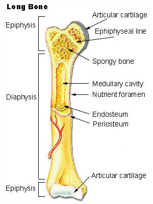
- Long bones have a long longitudinal axis (Figure 1).
- Short bones have a short longitudinal axis and are more cube-like.
- Flat bones are thin and curved such as some of the bones of the skull.
- Irregular bones are often found in groups and have a variety of shapes and sizes.
Notice the long shaft or diaphysis in the middle of the bone. The diaphysis contains compact bone surrounding a medullary cavity containing bone marrow On either end is an epiphysis containing cancellous or spongy bone. The epiphyseal line is a remnant of the growth plate. The epiphyses also contain hyaline cartilage for forming joints with other bones. Surrounding the bone is a membrane called the periosteum. The periosteum contains blood vessels and cells that help to repair and restore bone.
There are also 2 types of bone tissue in different amounts in bones. Compact bone (sometimes called cortical bone) is very dense. Cancellous bone (sometimes called spongy bone) looks more like a trabeculated matrix (Figure 2). It is found in the central regions of some of the skull bones or at ends (epiphyses) of long bones. The bone forming cells (osteocytes) get their nutrients by diffusion.
Notice the spongy appearance of the trabeculated bone. The cortical bone is located near the margins of the bone and is more dense.

Bone Structure
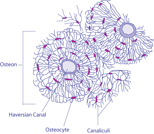
Compact bone is organized according to structural units called Haversian systems or osteons (Figure 3). These are located along the lines of force and line up along the long axis of the bone. The Haversian systems are connected together and form an interconnected structure that provides support and strength to bones.
Haversian systems contain a central canal (Haversian canal) that serves as a pathway for blood vessels and nerves. The bone is deposited along concentric rings called lamellae. Along the lamellae are small openings called lacunae. The lacunae contain fluid and bone cells called osteocytes. Radiating out in all directions from lacunae are small canals called canaliculi. Haversian systems are interconnected by a series of larger canals called Volksmann’s canals (perforating canals).
Bone Cells
There are 3 basic types of cells in bone. Osteoblasts undergo mitosis and secrete a substance that acts as the framework for bone. Once this substance (called osteoid) is secreted minerals can deposit and form hardened bone. Osteoblasts respond to certain bone forming hormones as well as from physical stress. Osteocytes are mature osteoblasts that cannot divide by mitosis (Figure 4).
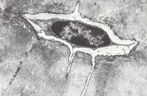
Osteocytes reside in lacunae. Osteoclasts are capable of demineralizing bone. They free up calcium from bone to make it available to the body depending on the body’s needs.
Bone Marrow
Bone marrow is located in the medullary (marrow) cavity of long bones and in some spongy bones. There are 2 kinds of marrow. Red marrow exists in the bones of infants and children. It is called red because it contains a large number of red blood cells. In adults the red marrow is replaced by yellow marrow. It is called yellow because it contains a large proportion of fat cells. Yellow marrow decreases its ability to form new red blood cells. However, not all adult bones contain yellow marrow. The following bones continue to contain red marrow and produce red blood cells:
- Proximal end of humerus
- Ribs
- Bodies of vertebrae
- Pelvis
- Femur
The Skeleton
The skeleton is divided into 2 sections: the axial and appendicular sections (Figure 5). The axial skeleton includes the skull, spine, ribcage, and sacrum and is indicated in blue in the figure below. The appendicular skeleton is indicated with red labels.

This video provides another introduction to the skeletal system:

A YouTube element has been excluded from this version of the text. You can view it online here: pb.libretexts.org/fob1/?p=496
Contributors and Attributions
- An eText of Human Anatomy and Physiology. Authored by: Dr. Bruce Forciea. Located at: www.bruceforciea.com/etextchapters/etexthumananatrevmay12.pdf. License: CC BY: Attribution
- Transverse Section Of Bone. Authored by: BDB. Located at: https://commons.wikimedia.org/wiki/File:Transverse_Section_Of_Bone.png. License: CC BY: Attribution
- The Skeletal System. Authored by: Crash Course. Located at: https://youtu.be/rDGqkMHPDqE. Project: Crash Course A&P. License: All Rights Reserved. License Terms: Standard YouTube License
- Illu long bone. Provided by: Cancer.gov. Located at: https://commons.wikimedia.org/wiki/File:Illu_long_bone.jpg. License: Public Domain: No Known Copyright
- Osteocyte. Provided by: Wikimedia Commons. Located at: commons.wikimedia.org/wiki/File:Osteocyte_2.jpg. License: Public Domain: No Known Copyright
- Human skeleton front. Authored by: LadyofHats. Located at: https://commons.wikimedia.org/wiki/File:Human_skeleton_front_en.svg. License: Public Domain: No Known Copyright