
4.2: Cell Theory
- Page ID
- 35677
What you’ll learn to do: State the basic principles of the unified cell theory
A cell is the smallest unit of a living thing. A living thing, whether made of one cell (like bacteria) or many cells (like a human), is called an organism. Thus, cells are the basic building blocks of all organisms. In this outcome we will learn about the discovery and origin of cells.
State the basic principles of the unified cell theory
A cell is the smallest unit of life. Most cells are so tiny that they cannot be seen with the naked eye. Therefore, scientists use microscopes to study cells. The first microscopes were used in the 1600s by Antony van Leeuwenhoek, a Dutch shopkeeper who had great skill in crafting lenses. Despite the limitations of his now-ancient lenses, van Leeuwenhoek observed the movements of protista (a type of single-celled organism) and sperm, which he collectively termed “animalcules.”
In a 1665 publication called Micrographia, experimental scientist Robert Hooke coined the term “cell” for the box-like structures he observed when viewing cork tissue through a lens. In the 1670s, van Leeuwenhoek discovered bacteria and protozoa. Later advances in lenses, microscope construction, and staining techniques enabled other scientists to see some components inside cells.
By the late 1830s, botanist Matthias Schleiden and zoologist Theodor Schwann were studying tissues and proposed the unified cell theory, which states that all living things are composed of one or more cells, the cell is the basic unit of life, and new cells arise from existing cells. Rudolf Virchow later made important contributions to this theory.
Microscopes
The microscopes we use today are far more complex than those used in the 1600s and 1800s. There are two primary types of modern microscopes used: light microscopes and electron microscopes. Electron microscopes provide higher magnification, higher resolution, and more detail than light microscopes. However, a light microscope is required to study living cells as the method used to prepare the specimen for viewing with an electron microscope kills the specimen.
Cytotechnologist
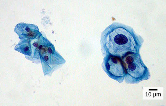
Have you ever heard of a medical test called a Pap smear (shown in Figure 1)? In this test, a doctor takes a small sample of cells from the uterine cervix of a patient and sends it to a medical lab where a cytotechnologist stains the cells and examines them for any changes that could indicate cervical cancer or a microbial infection.
Cytotechnologists (cyto = “cell”) are professionals who study cells via microscopic examinations and other laboratory tests. They are trained to determine which cellular changes are within normal limits and which are abnormal. Their focus is not limited to cervical cells; they study cellular specimens that come from all organs. When they notice abnormalities, they consult a pathologist, who is a medical doctor who can make a clinical diagnosis.
Cytotechnologists play a vital role in saving people’s lives. When abnormalities are discovered early, a patient’s treatment can begin sooner, which usually increases the chances of a successful outcome.
Check Your Understanding
Answer the question(s) below to see how well you understand the topics covered in the previous section. This short quiz does not count toward your grade in the class, and you can retake it an unlimited number of times.
Use this quiz to check your understanding and decide whether to (1) study the previous section further or (2) move on to the next section.
Contributors and Attributions
- Introduction to Cell Theory. Provided by: Lumen Learning. License: CC BY: Attribution
- Biology. Provided by: OpenStax CNX. Located at: http://cnx.org/contents/185cbf87-c72e-48f5-b51e-f14f21b5eabd@10.8. License: CC BY: Attribution. License Terms: Download for free at http://cnx.org/contents/185cbf87-c72...f21b5eabd@10.8
- Cell theory. Provided by: Khan Academy. Located at: www.khanacademy.org/science/biology/structure-of-a-cell/introduction-to-cells/v/cell-theory. License: CC BY-NC-SA: Attribution-NonCommercial-ShareAlike