
9.5: Marchantiophyta- The Liverworts
- Page ID
- 134633

\( \newcommand{\vecs}[1]{\overset { \scriptstyle \rightharpoonup} {\mathbf{#1}} } \)
\( \newcommand{\vecd}[1]{\overset{-\!-\!\rightharpoonup}{\vphantom{a}\smash {#1}}} \)
\( \newcommand{\dsum}{\displaystyle\sum\limits} \)
\( \newcommand{\dint}{\displaystyle\int\limits} \)
\( \newcommand{\dlim}{\displaystyle\lim\limits} \)
\( \newcommand{\id}{\mathrm{id}}\) \( \newcommand{\Span}{\mathrm{span}}\)
( \newcommand{\kernel}{\mathrm{null}\,}\) \( \newcommand{\range}{\mathrm{range}\,}\)
\( \newcommand{\RealPart}{\mathrm{Re}}\) \( \newcommand{\ImaginaryPart}{\mathrm{Im}}\)
\( \newcommand{\Argument}{\mathrm{Arg}}\) \( \newcommand{\norm}[1]{\| #1 \|}\)
\( \newcommand{\inner}[2]{\langle #1, #2 \rangle}\)
\( \newcommand{\Span}{\mathrm{span}}\)
\( \newcommand{\id}{\mathrm{id}}\)
\( \newcommand{\Span}{\mathrm{span}}\)
\( \newcommand{\kernel}{\mathrm{null}\,}\)
\( \newcommand{\range}{\mathrm{range}\,}\)
\( \newcommand{\RealPart}{\mathrm{Re}}\)
\( \newcommand{\ImaginaryPart}{\mathrm{Im}}\)
\( \newcommand{\Argument}{\mathrm{Arg}}\)
\( \newcommand{\norm}[1]{\| #1 \|}\)
\( \newcommand{\inner}[2]{\langle #1, #2 \rangle}\)
\( \newcommand{\Span}{\mathrm{span}}\) \( \newcommand{\AA}{\unicode[.8,0]{x212B}}\)
\( \newcommand{\vectorA}[1]{\vec{#1}} % arrow\)
\( \newcommand{\vectorAt}[1]{\vec{\text{#1}}} % arrow\)
\( \newcommand{\vectorB}[1]{\overset { \scriptstyle \rightharpoonup} {\mathbf{#1}} } \)
\( \newcommand{\vectorC}[1]{\textbf{#1}} \)
\( \newcommand{\vectorD}[1]{\overrightarrow{#1}} \)
\( \newcommand{\vectorDt}[1]{\overrightarrow{\text{#1}}} \)
\( \newcommand{\vectE}[1]{\overset{-\!-\!\rightharpoonup}{\vphantom{a}\smash{\mathbf {#1}}}} \)
\( \newcommand{\vecs}[1]{\overset { \scriptstyle \rightharpoonup} {\mathbf{#1}} } \)
\(\newcommand{\longvect}{\overrightarrow}\)
\( \newcommand{\vecd}[1]{\overset{-\!-\!\rightharpoonup}{\vphantom{a}\smash {#1}}} \)
\(\newcommand{\avec}{\mathbf a}\) \(\newcommand{\bvec}{\mathbf b}\) \(\newcommand{\cvec}{\mathbf c}\) \(\newcommand{\dvec}{\mathbf d}\) \(\newcommand{\dtil}{\widetilde{\mathbf d}}\) \(\newcommand{\evec}{\mathbf e}\) \(\newcommand{\fvec}{\mathbf f}\) \(\newcommand{\nvec}{\mathbf n}\) \(\newcommand{\pvec}{\mathbf p}\) \(\newcommand{\qvec}{\mathbf q}\) \(\newcommand{\svec}{\mathbf s}\) \(\newcommand{\tvec}{\mathbf t}\) \(\newcommand{\uvec}{\mathbf u}\) \(\newcommand{\vvec}{\mathbf v}\) \(\newcommand{\wvec}{\mathbf w}\) \(\newcommand{\xvec}{\mathbf x}\) \(\newcommand{\yvec}{\mathbf y}\) \(\newcommand{\zvec}{\mathbf z}\) \(\newcommand{\rvec}{\mathbf r}\) \(\newcommand{\mvec}{\mathbf m}\) \(\newcommand{\zerovec}{\mathbf 0}\) \(\newcommand{\onevec}{\mathbf 1}\) \(\newcommand{\real}{\mathbb R}\) \(\newcommand{\twovec}[2]{\left[\begin{array}{r}#1 \\ #2 \end{array}\right]}\) \(\newcommand{\ctwovec}[2]{\left[\begin{array}{c}#1 \\ #2 \end{array}\right]}\) \(\newcommand{\threevec}[3]{\left[\begin{array}{r}#1 \\ #2 \\ #3 \end{array}\right]}\) \(\newcommand{\cthreevec}[3]{\left[\begin{array}{c}#1 \\ #2 \\ #3 \end{array}\right]}\) \(\newcommand{\fourvec}[4]{\left[\begin{array}{r}#1 \\ #2 \\ #3 \\ #4 \end{array}\right]}\) \(\newcommand{\cfourvec}[4]{\left[\begin{array}{c}#1 \\ #2 \\ #3 \\ #4 \end{array}\right]}\) \(\newcommand{\fivevec}[5]{\left[\begin{array}{r}#1 \\ #2 \\ #3 \\ #4 \\ #5 \\ \end{array}\right]}\) \(\newcommand{\cfivevec}[5]{\left[\begin{array}{c}#1 \\ #2 \\ #3 \\ #4 \\ #5 \\ \end{array}\right]}\) \(\newcommand{\mattwo}[4]{\left[\begin{array}{rr}#1 \amp #2 \\ #3 \amp #4 \\ \end{array}\right]}\) \(\newcommand{\laspan}[1]{\text{Span}\{#1\}}\) \(\newcommand{\bcal}{\cal B}\) \(\newcommand{\ccal}{\cal C}\) \(\newcommand{\scal}{\cal S}\) \(\newcommand{\wcal}{\cal W}\) \(\newcommand{\ecal}{\cal E}\) \(\newcommand{\coords}[2]{\left\{#1\right\}_{#2}}\) \(\newcommand{\gray}[1]{\color{gray}{#1}}\) \(\newcommand{\lgray}[1]{\color{lightgray}{#1}}\) \(\newcommand{\rank}{\operatorname{rank}}\) \(\newcommand{\row}{\text{Row}}\) \(\newcommand{\col}{\text{Col}}\) \(\renewcommand{\row}{\text{Row}}\) \(\newcommand{\nul}{\text{Nul}}\) \(\newcommand{\var}{\text{Var}}\) \(\newcommand{\corr}{\text{corr}}\) \(\newcommand{\len}[1]{\left|#1\right|}\) \(\newcommand{\bbar}{\overline{\bvec}}\) \(\newcommand{\bhat}{\widehat{\bvec}}\) \(\newcommand{\bperp}{\bvec^\perp}\) \(\newcommand{\xhat}{\widehat{\xvec}}\) \(\newcommand{\vhat}{\widehat{\vvec}}\) \(\newcommand{\uhat}{\widehat{\uvec}}\) \(\newcommand{\what}{\widehat{\wvec}}\) \(\newcommand{\Sighat}{\widehat{\Sigma}}\) \(\newcommand{\lt}{<}\) \(\newcommand{\gt}{>}\) \(\newcommand{\amp}{&}\) \(\definecolor{fillinmathshade}{gray}{0.9}\)- Gametophyte morphology
- Leafy liverworts have leaves arranged in a flat plane with a set of smaller underleaves
- Thalloid liverworts have no leaves
- Liverwort cells have multiple chloroplasts
- Simple pores allow for gas exchange (no guard cells, meaning pores are permanently open)
- Asexual clones, called gemmae, are sometimes produced
- Sporophyte morphology
- Leafy liverworts produce single sporangium at the end of a seta (often fragile, transparent)
- Marchantia, a thalloid liverwort, develops complex structures called where gametangia are produced
Leafy Liverworts
Cut off the end of a leafy liverwort and make a wet mount or obtain a prepared slide to view under the compound microscope. Draw the specimen below and indicate the three different rows of leaves (two in one plane, and a row of smaller underleaves running beneath the stem).
Still, under the compound microscope, observe one of the leaf cells. Can you tell how many chloroplasts are in it? Draw this cell in the space below and label the chloroplasts and any other features you recognize.
Thalloid Liverworts: Marchantia
If available, observe a Marchantia polymorpha gametophyte under the dissecting scope. Look for simple pores, rhizoids, archegoniophores, antheridiophores, and gemmae cups containing asexual clones of the gametophyte, called gemmae. Label the bolded features in the life cycle diagram.
Marchantia life cycle:

Figure \(\PageIndex{1}\): This is the life cycle diagram for the thalloid liverwort Marchantia. It includes all of the structures described in the following pages.
In the diagram above, indicate where meiosis and fertilization occur. Color the haploid and diploid tissue differently, and draw arrows to show when mitosis is happening.
Obtain a prepared slide of a Marchantia antheridiophore. The male gametangia, antheridia, are produced on the top of this structure. Each antheridium produces haploid, swimming sperm by mitosis. Label the bolded features in the life cycle diagram.

Figure \(\PageIndex{2}\): Antheridia are visible as dark elongated sacs, shaped almost like footballs on the top of the antheridiophore
Obtain a prepared slide of an unfertilized Marchantia archegoniophore. This is the structure that produces the female gametangia, archegonia. Each archegonium produces a single haploid egg by mitosis.
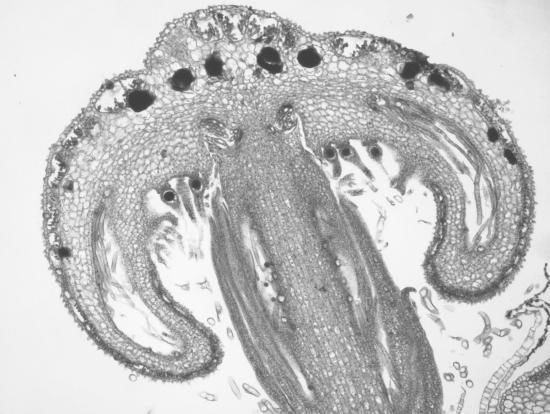
Figure \(\PageIndex{3}\): Archegonia are visible emerging from the underside of the palm tree-like branches of the archegoniophore. An egg is present at the base of each upside down, vase-like archegonium.
A sperm will be transported by water to the archegoniophore, travel down the venter of the archegonium, and fertilize the egg. This forms a diploid zygote. Label the bolded features in the life cycle diagram.

Figure \(\PageIndex{4}\): Archegonia are visible emerging from the underside of the archegoniophore. An egg is present at the base of each upside down, vase-like archegonium.
Obtain a prepared slide of a fertilized Marchantia archegoniophore with sporophytes. The zygote will be retained within the archegonium and nourished through the placenta, an area of gametophyte tissue adjacent to the foot of the sporophyte.
The mature sporophyte produces haploid spores via meiosis, which will grow into gametophytes. The sporophyte has a seta (the stalk), sporangium (also called a capsule), spores, and elaters, which aid in spore dispersal. Label the bolded features in the life cycle diagram.

Figure \(\PageIndex{5}\): The sporophyte emerges upside down from the archegonium. Spores are visible in chains within the sporangium, surrounded by elaters. There is a small fleshy area at the base of the sporangium. This is the seta.