
27.3: Lab Report
- Page ID
- 105950
\( \newcommand{\vecs}[1]{\overset { \scriptstyle \rightharpoonup} {\mathbf{#1}} } \)
\( \newcommand{\vecd}[1]{\overset{-\!-\!\rightharpoonup}{\vphantom{a}\smash {#1}}} \)
\( \newcommand{\id}{\mathrm{id}}\) \( \newcommand{\Span}{\mathrm{span}}\)
( \newcommand{\kernel}{\mathrm{null}\,}\) \( \newcommand{\range}{\mathrm{range}\,}\)
\( \newcommand{\RealPart}{\mathrm{Re}}\) \( \newcommand{\ImaginaryPart}{\mathrm{Im}}\)
\( \newcommand{\Argument}{\mathrm{Arg}}\) \( \newcommand{\norm}[1]{\| #1 \|}\)
\( \newcommand{\inner}[2]{\langle #1, #2 \rangle}\)
\( \newcommand{\Span}{\mathrm{span}}\)
\( \newcommand{\id}{\mathrm{id}}\)
\( \newcommand{\Span}{\mathrm{span}}\)
\( \newcommand{\kernel}{\mathrm{null}\,}\)
\( \newcommand{\range}{\mathrm{range}\,}\)
\( \newcommand{\RealPart}{\mathrm{Re}}\)
\( \newcommand{\ImaginaryPart}{\mathrm{Im}}\)
\( \newcommand{\Argument}{\mathrm{Arg}}\)
\( \newcommand{\norm}[1]{\| #1 \|}\)
\( \newcommand{\inner}[2]{\langle #1, #2 \rangle}\)
\( \newcommand{\Span}{\mathrm{span}}\) \( \newcommand{\AA}{\unicode[.8,0]{x212B}}\)
\( \newcommand{\vectorA}[1]{\vec{#1}} % arrow\)
\( \newcommand{\vectorAt}[1]{\vec{\text{#1}}} % arrow\)
\( \newcommand{\vectorB}[1]{\overset { \scriptstyle \rightharpoonup} {\mathbf{#1}} } \)
\( \newcommand{\vectorC}[1]{\textbf{#1}} \)
\( \newcommand{\vectorD}[1]{\overrightarrow{#1}} \)
\( \newcommand{\vectorDt}[1]{\overrightarrow{\text{#1}}} \)
\( \newcommand{\vectE}[1]{\overset{-\!-\!\rightharpoonup}{\vphantom{a}\smash{\mathbf {#1}}}} \)
\( \newcommand{\vecs}[1]{\overset { \scriptstyle \rightharpoonup} {\mathbf{#1}} } \)
\( \newcommand{\vecd}[1]{\overset{-\!-\!\rightharpoonup}{\vphantom{a}\smash {#1}}} \)
A1. Draw a cross section of the celery petiole, labeling parenchyma in the epidermis, collenchyma in the cortex, and sclerenchyma in the vascular tissue. Make notes about the differences in the cell wall for your future study.
A2. The central region of the celery petiole is called the pith. What type of cells are present in this region?
A3. What other cellular changes might occur to signal that a pear is ripe?
A4. Is this sclereid alive or dead? What about the parenchyma cells around it?
A5. What is the compound in the secondary wall that stains differently from the primary wall?
B1. How does the location of the trichomes relate to prevention of water loss?
B2. View a leaf under the dissecting scope. Can you find trichomes, guard cells, or other specialized epidermal cells?
B3. What cell type (-enchyma) are these cells most similar to?
B4. View a prepared slide of a leaf cross section. Draw what you see below. Identify and label as many tissues, cell types, and specialized cells as you can.
Summary Table of Cells and Tissues in the Leaf Organ
A simple tissue contains only a single cell type, while a complex tissue contains multiple cell types. The leaf organ is composed of both simple and complex tissues. In the table below under Tissue Type, try to identify whether it is a simple or complex tissue. If it is a simple tissue, identify which cell type it is composed of.
|
Tissue |
Specialized Cells |
Tissue Type |
Function |
|---|---|---|---|
|
Epidermis |
1. Guard cell 2. Trichome |
||
|
Mesophyll |
Mesophyll cells |
||
|
Xylem |
Tracheids & vessel elements |
||
|
Phloem |
1. Sieve tube elements 2. Companion cells 3. Phloem fibers |
Why didn’t I include a stoma among the specialized cells in the epidermis?
The image below is a specialized cell called a tracheid. What tissue would you find this cell in? Is that tissue simple or complex?
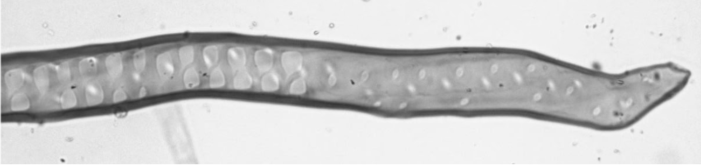
Based on your knowledge of root words, what does the term tracheophytes mean?
Early plants have tracheids, while later groups of plants have an additional type of water conducting cell: vessel elements.
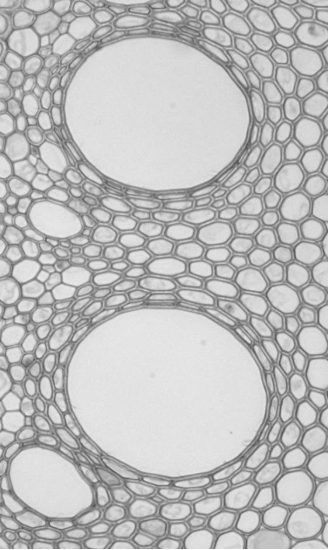
The image above is a cross section through the xylem of a corn root. You can see large open areas (vessel elements) surrounded by smaller, more densely packed cells (tracheids).
How would these two cell types differ in the ability to take up and transport water?
Transpiration in Action
Water moves through the dead water-conducting cells in the xylem much like it moves through a tube. Transpiration acts like suction from the top of the tube, but as you saw in the previous experiment, other forces aid in the movement of the water: cohesion, adhesion, tension, and capillary action. All of these forces work to pull water into the plant through the root hairs, into the xylem, and out through the stomata.
You set up four plants at the start of lab. What were the conditions for each plant?
Plant 1 -
Plant 2 -
Plant 3 -
Plant 4 -
Check on the plants and, before doing anything, simply observe the appearance of the bags. Describe your observations below.
Turn each plant on its side and carefully remove the bags. Try not to let any condensation in the bag escape. Use a scale to obtain the mass of each bag. Note: if you used different types of bags, adjust your end mass measurements by subtracting the initial mass.
|
Mass of bag (adjusted) |
1: |
2: |
3: |
4: |
|---|
C1. Fill in the diagram below with the following terms: cortex, ground meristem, pericycle, primary phloem, and protoderm. Choose a different color to represent meristems and tissues, then color the boxes accordingly.
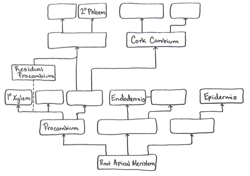
C2. How would this flowchart be different in a monocot? Draw a flowchart of monocot root development in the space below.
C3. Observe a cross section of the zone of maturation in a Zea mays root. Draw and label your microscope slide.
C4. Observe a cross section of a Salix (willow) root producing lateral roots. Locate the primary xylem, primary phloem, pericycle, cortex, and epidermis. Draw and label these from your microscope slide.
C5. Observe a cross section of a Ranunculus (buttercup) root. Locate the pericycle, endodermis, casparian strip, and passage cells. The suberin in the casparian strip often stains similarly to the secondary walls in the xylem. Draw and label the cross section.

In the figure above, label any adventitious roots, prop roots, and storage roots. Label each system as either netted or taproot (except the topmost root system, which is an underground stem).
C6. Observe a prepared slide of a Coleus shoot tip long section. Locate the meristems, tissues, and specialized cells. Draw what you see and label any important features.
Flowchart of Shoot Development
Below is a flowchart of eudicot shoot development in primary growth. There are two lines that will later lead to the secondary meristems. With this information and the filled in boxes, you should be able to determine which meristems and tissues go in the empty boxes.
Fill in the diagram below with the following terms: cortex, epidermis, fascicular cambium, ground meristem, pith rays, procambium and primary xylem.. Choose a different color to represent meristems and tissues, then color the boxes accordingly.

Figure \(\PageIndex{5}\): Flowchart of eudicot shoot development in primary growth
C7. Using a slide of a monocot stem, draw and label the xylem, phloem, a vessel element, sieve tube element, and companion cell.
C8. Observe a cross section of a young Helianthus (or other eudicot) stem. Draw and label the epidermis, cortex, primary phloem, primary xylem, pith, and pith rays.
What cell types (-enchymas) can you identify in the Tilia cross section? Which tissue(s) do you find each cell type in?
C9. Draw a cross section of a mesophytic leaf, labeling each structure or tissue with its name and function. Consider simplifying the image to use as an easy reference.
In the leaf you are viewing, are there more stomata on the upper or lower epidermis? Can you think of any reasons why this might be?
How does the structure of the spongy mesophyll contribute to its function?